Transcription
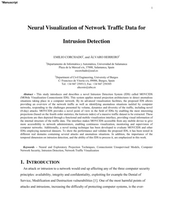
Medical Hypotheses (2003) 61(2), 282–291ª 2003 Elsevier Science Ltd. All rights reserved.doi:10.1016/S0306-9877(03)00175-0The neural basis of the complexmental task of meditation:neurotransmitter and neurochemicalconsiderationsA. B. Newberg,1 J. Iversen21University of Pennsylvania, Philadelphia, PA, USA; 2Stanford University, Stanford, CA 94309, USASummary Meditation is a complex mental process involving changes in cognition, sensory perception, affect,hormones, and autonomic activity. Meditation has also become widely used in psychological and medicalpractices for stress management as well as a variety of physical and mental disorders. However, until now, therehas been limited understanding of the overall biological mechanism of these practices in terms of the effectsin both the brain and body. We have previously described a rudimentary neuropsychological model to explain thebrain mechanisms underlying meditative experiences. This paper provides a substantial development byintegrating neurotransmitter systems and the results of recent brain imaging advances into the model. Thefollowing is a review and synthesis of the current literature regarding the various neurophysiological mechanismsand neurochemical substrates that underlie the complex processes of meditation. It is hoped that this modelwill provide hypotheses for future biological and clinical studies of meditation.ª 2003 Elsevier Science Ltd. All rights reserved.INTRODUCTIONThe complex mental task of meditation is potentially oneof the most important areas of research that may bepursued by science in the next decade. Meditation offersa fascinating window into human consciousness, psychology, and experience; the relationship betweenmental states and body physiology; emotional and cognitive processing; and the biological correlates of religious experience. In the past thirty years, scientists haveexplored the biological effects and mechanisms of meditation in great detail. Initial studies measured changes inautonomic activity such as heart rate and blood pressure,as well as electroencephalographic (EEG) changes. MoreReceived 1 November 2002Accepted 26 March 2003Correspondence to: Andrew B. Newberg MD, Division of Nuclear Medicine,110 Donner Building, H.U.P., 3400 Spruce Street, Philadelphia, PA 19104,USA. Phone: 215-662-3092; Fax: 215-349-5843;E-mail: newberg@rad.upenn.edu282recent studies have explored changes in hormonal andimmunological function associated with meditation.Studies have also explored the clinical effects of meditation in both physical and psychological disorders.Functional neuroimaging has opened a new windowinto the investigation of meditative states by exploringthe neurological correlates of these experiences. Fivestudies, currently available in the literature (see Table 1),measured cerebral function during meditation techniques. These neuroimaging techniques include positronemission tomography [PET; (1–3)], single photon emission computed tomography [SPECT; (4)], and functionalmagnetic resonance imaging [fMRI; (5)]. Each of thesetechniques provides different advantages and disadvantages in the study of meditation. Although functional MRIhas the ability of immediate anatomic correlation and hasimproved resolution over SPECT, it would be very difficultto utilize for the study of meditation because of the noisefrom the machine and the problem of requiring the subject to lie prone in the machine, an atypical posture formany forms of meditation. In fact, we attempted the use of
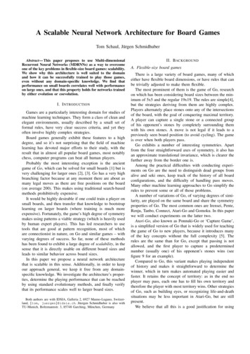
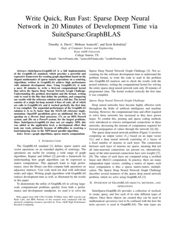
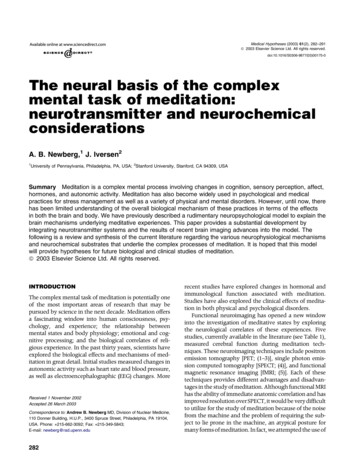
The neural basis of the complex mental task of meditation283Table 1 Summary of current neuroimaging studies of meditationaStudyType ueHerzog et al. (1)YogaPET8CBF"Frontal lobe, #parietallobeLou et al. (2)Tantric YogaPET9CBF"Parietal during focus onself, "hippocampus, #PFCNewberg et al.(4)TibetanSPECT8CBF"PFC, thalamus, brainstem,#PSPLLazar et al. (5)KundaliniMRI5CBF"PFC, parietal, hippocampus, temporal lobe cingulate gyrus, hypothalamusKjaer et al. (3)Yoga NidraPET5Dopamine"Dopamine in the striatumMeditation in the scanner,limited regional analysis,one time pointSubjects listened to tapeguiding the meditation,different forms of meditation,one time pointNo cross correlation withother modalities, SPECToffers lowest resolution,one time pointAttempted to factor in MRInoise to prevent distraction,smallest N, limited abilityto detect decreased CBF,multiple time pointsSubjects listened to tapeguiding the meditation,one time point, firstneurotransmitter studyN ¼ number of subjects.fMRI with one of our initial meditation subjects in orderto determine feasibility, but the subject found it extremelydifficult to carry out the meditation practice. WhilePET imaging also provides better resolution than SPECT,if one strives to make the environment relativelydistraction free to maximize the chances of having astrong meditative experience, then it is sometimes beneficial to perform these studies during non-clinical times,which may make PET radiopharmaceuticals such asfluorodeoxyglucose difficult to obtain. Finally, the functional imaging techniques of PET and SPECT offerthe opportunity to study changes in a variety of neurotransmitter systems in the brain associated with meditation practices. Overall, functional brain imagingoffers important techniques for studying meditation, although the best approach may depend on factors relatedto each technique as well as specific parameters to bemeasured.In this paper, we review existing data on the neurophysiology and physiology of meditation practices, andwe attempt to integrate this data into a comprehensiveneurochemical model of such practices. However, thereare many possible neurochemical changes that may occur during meditation, even though they may not occurin every type of practice or in each individual. This modelis designed to provide a framework of the neurologicaland physiological correlates of meditative experiences,and to create a springboard for future research.TYPES OF MEDITATIONAlthough there are many specific approaches to meditation, we have typically divided such practices into twoª 2003 Elsevier Science Ltd. All rights reserved.basic categories. The first category is one in which thesubjects simply attempt to clear all thought from theirsphere of attention. This form of meditation is one inwhich the practitioner attempts to reach a subjective statecharacterized by a sense of no space, no time, and nothought. Further, this state is cognitively experienced asfully integrated and unified, such that there is no sense ofa self and other. This includes practices such as thoseassociated with traditions such as Theravada Buddhism(6). The second category is one in which the subjectsfocus their attention on a particular object, image, phrase,or word, and it includes practices such as transcendentalmeditation and various forms of Tibetan Buddhism. Thisform of meditation is designed to lead one to a subjectiveexperience of absorption with the object of focus. There isanother distinction in which some meditation is guidedby following along with a leader, either in person or ontape, who is verbally directing the practitioner. Otherspractice the meditation on their own volition. We mightexpect that this difference between volitional and guidedmeditation should also be reflected in specific differencesin cerebral activation.Phenomenological analysis suggests that the end results of many practices of meditation are similar, although these results might be described using differentcharacteristics depending on the culture and individual.Therefore, it seems reasonable that while the initialneurophysiological activation occurring during any given practice may differ, there should eventually be aconvergence. We present a description of volitionalmeditation, which will hopefully provide an overallframework from which any given type of meditation canbe considered (see Fig. 1).Medical Hypotheses (2003) 61(2), 282–291
284 Newberg and IversenFig. 1 Schematic overview of the neurophysiological network possibly associated with meditative states. The circuits and activationsgenerally apply to both hemispheres even though they may be ascribed to only one hemisphere on the diagram below.ACTIVATION OF THE PREFRONTAL ANDCINGULATE CORTEXBrain imaging studies suggest that willful acts and tasksthat require sustained attention are initiated via activityin the prefrontal cortex (PFC), particularly in the righthemisphere (7–10). The cingulate gyrus appears to beinvolved in focusing attention, probably in conjunctionwith the PFC (11). Since meditation requires intense focus of attention, it seems appropriate that a model formeditation begin with activation of the PFC, particularlyin the right hemisphere, as well as the cingulate gyrus.This notion is supported by the increased activity observed in these regions on several of the brain imagingstudies of volitional types of meditation, including thosefrom our laboratory (1,4,5). In our study of eight TibetanBuddhist meditators, subjects had an intravenous lineMedical Hypotheses (2003) 61(2), 282–291placed in the arm, and were injected with a cerebralblood flow tracer while at rest in order to acquire a‘baseline’ image. They then meditated for approximately1 h when they were again injected with the tracer whilethey continued to meditate. At the time of injection, thetracer was fixed in the brain so that when the imageswere acquired approximately 20 min later, they reflectedthe cerebral blood flow during the meditation state.Quantitative analysis demonstrated increased activity inthe PFC bilaterally (greater on the right) and the cingulate gyrus during meditation. Therefore, meditation appears to begin by activating the prefrontal and cingulatecortex associated with the will or intent to clear one’smind of thoughts or to focus on an object. One PETstudy of a guided type of meditation did not demonstrate increased prefrontal activity. However, a recentª 2003 Elsevier Science Ltd. All rights reserved.
The neural basis of the complex mental task of meditationstudy showed decreased frontal activity during externally guided word generation compared to internal orvolitional word generation (12). Thus, prefrontal andcingulate activation may be associated with the volitional aspects of meditation.THALAMIC ACTIVATIONSeveral animal studies have shown that the PFC, whenactivated, innervates the reticular nucleus of the thalamus (13), particularly as part of a more global attentionalnetwork (14). Such activation may be accomplished bythe PFC’s production and distribution of the excitatoryneurotransmitter glutamate, which the PFC neurons useto communicate among themselves and to innervateother brain structures (15). The thalamus itself governsthe flow of sensory information to cortical processingareas via its interactions with the lateral geniculate andlateral posterior nuclei and also likely uses the glutamatesystem in order to activate neurons in other structures(16). The lateral geniculate nucleus receives raw visualdata from the optic tract and routes it to the striatecortex for processing (17). The lateral posterior nucleusof the thalamus provides the posterior superior parietallobule (PSPL) with the sensory information it needs todetermine the body’s spatial orientation (18).When excited, the reticular nucleus secretes the inhibitory neurotransmitter c-aminobutyric acid (GABA)onto the lateral posterior and geniculate nuclei, cuttingoff input to the PSPL and visual centers in proportion tothe reticular activation (19). During meditation, due tothe increased activity in the PFC, particularly in the righthemisphere, there should be a concomitant increase inthe activity in the reticular nucleus of the thalamus.While brain imaging studies of meditation have not yethad the resolution to distinguish the reticular nuclei, ourrecent SPECT study did demonstrate a general increasein thalamic activity that was proportional to the activitylevels in the PFC. This finding is consistent with, butdoes not confirm, the specific interaction between thePFC and the reticular nuclei. If the activation of the rightPFC causes activity to increase in the reticular nucleusduring meditation, the result may be a decrease in sen-285sory input entering into the PSPL. Several studies havedemonstrated an increase in serum GABA during meditation (see Table 2 for an overview of neurochemicalchanges during meditation), possibly reflecting increased central GABA activity (20). This functional deafferentation related to increased GABA would meanthat fewer distracting outside stimuli would arrive at thevisual cortex and PSPL enhancing the sense of focus.It should also be noted that the dopaminergic system,via the basal ganglia, is believed to participate in regulating the glutamatergic system and the interactionsbetween the prefrontal cortex and subcortical structures.A recent PET study utilizing 11C-Raclopride to measurethe dopaminergic tone during Yoga Nidra meditationdemonstrated a significant increase in dopamine levelsduring the meditation practice (3). The experimentershypothesized that this increase may be associated withthe gating of cortical–subcortical interactions, leading toan overall decrease in readiness for action that is associated with this particular type of meditation. Futurestudies will be necessary to elaborate on the role of dopamine during meditative practices, as well as on theinteractions between dopamine and other neurotransmitter systems.PSPL DEAFFERENTATIONThe PSPL is heavily involved in the analysis and integration of higher-order visual, auditory, and somaesthetic information (21). It is also involved in a complexattentional network that includes the PFC and thalamus(22). Through the reception of auditory and visual inputfrom the thalamus, the PSPL is able to help generate athree-dimensional image of the body in space, provide asense of spatial coordinates in which the body is oriented,
The neural basis of the complex mental task of meditation: neurotransmitter and neurochemical considerations A. B. Newberg,1 J. Iversen2 1University of Pennsylvania, Philadelphia, PA, USA; 2Stanford University, Stanford, CA 94309, USA Summary Meditation is a complex mental process involving changes in cognition, sensory perception, affect,