Transcription
NeuroImage 136 (2016) 186–196Contents lists available at ScienceDirectNeuroImagejournal homepage: www.elsevier.com/locate/ynimgDynamics of neural recruitment surrounding the spontaneous arising ofthoughts in experienced mindfulness practitionersMelissa Ellamil a, Kieran C.R. Fox a, Matthew L. Dixon a, Sean Pritchard b, Rebecca M. Todd a,c,Evan Thompson d, Kalina Christoff a,c,⁎aDepartment of Psychology, University of British Columbia, 2136 West Mall, Vancouver, British Columbia V6T 1Z4, CanadaSchool of Psychology, Fielding Graduate University, 2020 De la Vina Street, Santa Barbara, CA 93105, United StatesCentre for Brain Health, University of British Columbia, 2215 Wesbrook Mall, Vancouver, British Columbia V6T 1Z3, CanadadDepartment of Philosophy, University of British Columbia, 1866 Main Mall, Vancouver, British Columbia V6T 1Z1, Canadabca r t i c l ei n f oArticle history:Received 22 December 2015Revised 12 April 2016Accepted 14 April 2016Available online 23 April 2016Keywords:Spontaneous thoughtDefault mode networkMedial temporal lobeNeural antecedentsfMRIa b s t r a c tThoughts arise spontaneously in our minds with remarkable frequency, but tracking the brain systems associatedwith the early inception of a thought has proved challenging. Here we addressed this issue by taking advantage ofthe heightened introspective ability of experienced mindfulness practitioners to observe the onset of their spontaneously arising thoughts. We found subtle differences in timing among the many regions typically recruited byspontaneous thought. In some of these regions, fMRI signal peaked prior to the spontaneous arising of a thought— most notably in the medial temporal lobe and inferior parietal lobule. In contrast, activation in the medial prefrontal, temporopolar, mid-insular, lateral prefrontal, and dorsal anterior cingulate cortices peaked together withor immediately following the arising of spontaneous thought. We propose that brain regions that show antecedent recruitment may be preferentially involved in the initial inception of spontaneous thoughts, while those thatshow later recruitment may be preferentially involved in the subsequent elaboration and metacognitive processing of spontaneous thoughts. Our findings highlight the temporal dynamics of neural recruitment surroundingthe emergence of spontaneous thoughts and may help account for some of spontaneous thought's peculiar qualities, including its wild diversity of content and its links to memory and attention. 2016 The Authors. Published by Elsevier Inc. This is an open access article under the CC BY /).1. IntroductionWhere do thoughts come from? One of the most intriguing yet leastunderstood aspects of the human mind is its tendency to spontaneouslygive rise to words, images, and emotions that flow effortlessly from onetopic to another. There is a stunning ubiquity of such spontaneouslyarising mental content in people's lives: thoughts that occur withoutour deliberate control take up as much as one-third of our mental experience (Klinger and Cox 1987).The last decade marked an upsurge of investigations into a numberof closely related mental phenomena, often investigated using termssuch as ‘mind-wandering’, ‘stimulus-independent thought’, or ‘task-unrelated thought’ (Christoff, 2012). Psychological research has focused onthe conditions that facilitate these kinds of thought processes, showingthat their frequency increases when task demands are low or when external sensory stimulation is weak (Smallwood and Schooler, 2006). Inparallel, neuroscientific investigations have focused on identifying the⁎ Corresponding author at: Department of Psychology, University of British Columbia,2136 West Mall, Vancouver, British Columbia V6T 1Z4, Canada.E-mail address: kchristoff@psych.ubc.ca (K. Christoff).brain regions that are consistently recruited during such conditions.The majority of investigations have placed particular emphasis on recruitment of the default mode network (for review, see Christoff,2012), although a number of executive control network regions are recruited with equal consistency (Christoff et al., 2009a; Fox et al., 2015).While these investigations have greatly improved our understanding ofwhich brain regions are engaged when spontaneous thought is ongoing,we still have virtually no knowledge of how these various brain regionscontribute to the different components of a spontaneous thought experience and, in particular, which brain regions support the early inception of spontaneous thoughts (Smallwood, 2013).The goal of the present study, therefore, was to investigate key brainregions associated with the arising of spontaneous thoughts. To do this,we set out to examine brain recruitment that occurs immediately priorto the onset of a spontaneous thought. The central difficulty in investigating this question lies in identifying the precise temporal onset ofspontaneously arising mental content. There are currently no thirdperson behavioral measures that provide such information reliably,making it necessary for scientists to use first-person introspective reports from participants. Although most individuals can provide fairly reliable descriptions of the basic contents of their thoughts (Hurlburt 0341053-8119/ 2016 The Authors. Published by Elsevier Inc. This is an open access article under the CC BY license (http://creativecommons.org/licenses/by/4.0/).
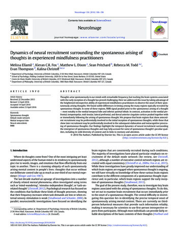
M. Ellamil et al. / NeuroImage 136 (2016) 186–196Schwitzgebel, 2007), their ability to report on the mental processes thatgive rise to thought content is generally considered to be poor (Nisbettand Wilson, 1977). This apparent lack of introspective ability, combinedwith the need for reliance on introspective measures, has created amethodological knot that has tied the hands of researchers interestedin differentiating the neural systems associated with the early formationof spontaneous thoughts.The ability to detect the arising of spontaneous thoughts, however,can vary across individuals (Seli et al., 2015) as well as within thesame individual (Zedelius et al., 2015). This raises the possibilitythat the methodological difficulty described earlier can be tackled byemploying participants with heightened introspective skills, a methodknown as neurophenomenology (Fazelpour and Thompson, 2015;Varela, 1996). In this approach, participants with introspective traininggive first-person reports about their mental processes that are then related to measures of neural activity (Lutz et al., 2002). One form of mental training that specifically focuses on observing the spontaneousarising of one's own thoughts is mindfulness practice (Lutz et al.,2008; Tang et al., 2015). Mindfulness training has been shown to enhance attention in numerous ways, including a greater ability to noticesubtle or rapid events, such as increased line length discrimination at visual threshold during a vigilance task (MacLean et al., 2010; Sahdraet al., 2011; Zanesco et al., 2013) and improved second target detectionduring an attentional blink task (Slagter et al., 2007). Specifically, introspective ability appears to be enhanced through mindfulness training,as shown by increased metacognitive memory confidence judgments(Baird et al., 2014) and greater accuracy in emotional self-awareness relative to no training (Sze et al., 2010). Thus, we employed a group of highlyexperienced mindfulness practitioners, who tracked the arising of spontaneous thoughts during fMRI scanning, to capitalize on this potentiallyheightened introspective ability and more specifically examine the neuralantecedents of spontaneous mental content as well as the full suite ofneural recruitment before, during, and after spontaneous thought reports.We hypothesized that the medial temporal lobe would show preferential recruitment during the initial generation of spontaneous thoughtas it is often recruited during rest (Buckner et al., 2008; Christoff et al.,2004; Stark and Squire, 2001) and has been associated with memory retrieval (Squire et al., 2004), future thinking (Addis et al., 2009), and mental simulation (Hassabis et al., 2007), which form a large portion ofspontaneous thought content (Klinger, 2009; Klinger and Cox, 1987).We also hypothesized that the medial and lateral prefrontal cortices,also consistently recruited during rest (Christoff et al., 2009a; Fox et al.,2015), would show increased recruitment after the initial generationof spontaneous thought as they have been associated more with theevaluation and monitoring of self-generated thought content (Buckneret al., 2008; Dixon and Christoff, 2012; Dixon and Christoff, 2014; Dixonet al., 2014; Fox and Christoff, 2014; Miller and Cohen, 2001).2. Materials and methods2.1. ParticipantsEighteen participants (8 male and 10 female; M 49.91 years old,SD 11.17, range 29.39–68.42) took part in the experiment. The participants were long-term, expert mindfulness meditators with morethan 3000 h of lifetime meditation experience and at least 1 h of dailypractice in the Mahasi Vipassana tradition (M 8338.60 h, SD 5989.98, range 3174–23,700). The number of hours reported didnot include practice in other traditions (e.g., Zen meditation, Transcendental meditation, Goenka or body scanning meditation, Yoga, Tai Chi,Qi Gong). Eligibility screening consisted of the administration of a meditation experience questionnaire and a phone interview by S.P., a formerMahasi monk. The screening ensured consistency of the meditationtechnique across participants and attainment of extensive knowledgeof and experience with the meditation technique through regular practice and attendance of several long-term intensive retreats.187The participants were recruited from Vipassana meditation communities in Vancouver (BC, Canada), Vancouver Island (BC, Canada),Boulder (CO, USA), San Francisco (CA, USA), and Seattle (WA, USA).All participants had normal or corrected-to-normal vision with no MRIcontraindications and no current psychiatric medication use. Fourteenwere right-handed and 4 were left-handed, but all used their righthand to respond during the experiment. From an original sample of22 participants, three participants were excluded from the analysesdue to excessive motion (2 with N5 pitch rotation, and 1 with N5 pitch rotation and N 5 mm in the z-direction). One participant was excluded from the analyses due to technical problems with behavioraldata recording. All protocols were approved by the University of BritishColumbia (UBC; Vancouver, BC, Canada) Clinical Research Ethics Boardand the UBC MRI Research Center. All participants gave informed writtenconsent prior to participating and received payment as compensation.2.2. ProcedureOne to two days prior to the actual scanning session, participants engaged in a practice session identical to the actual scanning procedure ina mock scanner environment in order to become acclimatized to thetask, button pressing, and scanner noises. Participants alternated between 30 s blocks of monitoring thoughts that arose spontaneously(thought condition, Fig. 1A) and 30 s blocks of monitoring words thatappeared onscreen (word condition, Fig. 1B). During both blocks, participants attended to the rising and falling of the abdomen (i.e., breathing).Participants reported with a first button press (index finger) to indicatewhen a thought arose or when a word appeared onscreen (see Stimuli),and with a second button press to indicate what type of thought or wordit was (index finger image or symbol, middle finger narrative orinner speech, ring finger emotion, pinky finger body sensation).Each word stayed onscreen until the first button press. The first buttonpress was followed by one asterisk (*) onscreen for 250 ms and the second button press was followed by two asterisks (**) onscreen for250 ms to signal successful button presses. Each participant completedthree 9-min task runs. Each run consisted of 8 thought blocks and 8word blocks; each participant therefore completed 24 thought blocksand 24 word blocks in total. Blocks were separated by interstimulus intervals (ISIs) with jittered durations (randomly chosen from 250 ms,500 ms, or 750 ms), with each ISI appearing an equal number of times.The instructions were designed to be consistent with a typical mindfulness practice session (Sayadaw, 1985, 2002): the first and secondbutton presses roughly corresponded to the standard steps of (i) notingthe occurrence of thought and then (ii) labeling its content. In thethought condition, participants were instructed to report on the occurrence of mental events that took attention away from one's breathing,or became more prominent in attention than breathing. This enabledthe assessment of mental content arising without being explicitly elicitedby external cues in the environment. In the word condition, participantswere instructed to briefly think about the definition of each word presented on the screen before responding. This enabled the assessment ofmental content elicited by external cues in the environment.The onsets and categories of words presented within each wordblock were selected in real-time by a computer algorithm that matchedthe timing onsets and categories of words to the timing onsets and categories of spontaneous thoughts reported in the immediately precedingthought block. Thus, each pair of thought and word blocks had the samenumber of events, and these events had the same content categoriesand appeared at the same time within the block.2.3. StimuliThe words presented during word monitoring blocks were randomlychosen from four lists that corresponded to the categories of thoughtsroutinely identified during mindfulness meditation (Sayadaw, 1985,2002) and covered a large portion of spontaneous thought content
188M. Ellamil et al. / NeuroImage 136 (2016) 186–196Fig. 1. Experimental procedure. Participants alternated between (A) spontaneous thought detection blocks (30 s) and (B) matched word detection blocks (30 s). Participants indicatedwith a first button press as soon as they noticed a thought spontaneously arising, which served as the experimental measure of the thought's onset, and with a second button presswhat category of thought it was, which allowed online matching of the subsequent word detection block. Each word block was constructed in real-time to correspond to the timingand categories of spontaneous thoughts reported during the immediately preceding thought block, allowing a closely matched comparison between neural recruitment associatedwith spontaneously arising and perceptually triggered mental content.(Fox et al., 2013): (i) image or symbol, (ii) narrative or inner speech, (iii)emotion, or (iv) body sensation. Words in the image list consisted of 30nouns (e.g., mountain, beach, rain, sun, pet) selected from the MedicalResearch Council (MRC) Psycholinguistics Database (Wilson, 1988),which had imageability, concreteness, and familiarity ratings of500–700 (on scales of 100 very low to 700 very high) to ensureease of visualization. Words in the narrative list consisted of 30 nouns(e.g., work, money, family, goals, health) selected from the EdinburghAssociative Thesaurus (EAT) (Kiss et al., 1973), which were associatedwith the types of current concerns that people tend to have and thatare thought to be major determinants of spontaneous thought content(Klinger, 2009; Klinger and Cox, 1987). These included home andhousehold matters; employment and finance; partner, family, and relatives; friends and acquaintances; love, intimacy, and sexual matters;self-changes; education and training; health and medical matters; spiritual matters; and hobbies, pastimes, and recreation (Klinger and Cox,2004). Words in the emotion list consisted of 30 adjectives (e.g., calm,happy, sad, afraid, worried) also selected from the EAT that were associated with various emotions (e.g., happiness, sadness, anger, disgust,fear, surprise). Words in the body sensation list consisted of 30 nounsand adjectives similarly selected from the EAT that were associatedwith various body sensations (e.g., warmth, tickle, vibration, pressure,pain). Each word contained 3–10 letters and 1–3 syllables. The wordsand fixation cross appeared as gray text on a black background. Thetask and stimuli were implemented and presented using E-Prime 2.0(Psychology Software Tools, Sharpsburg, PA, USA).2.4. Data acquisitionFunctional and structural MRI data were collected using a 3.0 TeslaPhilips Intera MRI scanner (Best, Netherlands) with a standard headcoil. Head movement was restricted using foam padding around thehead. T2*-weighted functional images were acquired parallel to theanterior commissure/posterior commissure (AC/PC) line using asingle-shot gradient echo-planar sequence (EPI; repetition time[TR] 2 s, echo time [TE] 30 ms, flip angle [FA] 90 , field of view[FOV] 240 240 143 mm, matrix size 80 80, SENSE factor 1.0). A total of 265 functional volumes were acquired for each taskrun, each including 36 interleaved axial slices (3 mm thick with 1 mmskip) covering the entire brain. Before functional imaging, an inversionrecovery prepared T1-weighted structural volume was acquired in thesame slice locations and orientation as the functional images using afast spin-echo sequence (TR 2 s, TE 10 ms, FA 90 , FOV 224 224 143 mm, acquisition matrix size 240 235, reconstructed matrix size 480 480, inversion delay [IR] 800 ms, spin-echoturbo factor 5).2.5. Data preprocessingfMRI data for each participant were preprocessed and analyzedusing SPM8 (Statistical Parametric Mapping, Wellcome Department ofImaging Neuroscience, London, UK). Slice timing correction was performed using sinc interpolation and resampling with the middle(18th) slice as a reference point. All functional volumes were realignedto the first volume to correct for between-scan motion. The structuralvolume was coregistered to the mean functional image and segmentedto extract a gray matter image. The segmented structural volumewas then spatially normalized to a gray matter image of the MontrealNeurological Institute (MNI) template and resliced to a voxel size of2 2 2 mm. The derived spatial transformations were applied tothe realigned functional volumes to bring them into standardized MNIspace. Finally, the functional volumes were smoothed with an 8-mmfull-width at half-maximum (FWHM) isotropic Gaussian kernel tocompensate for residual between-subject variability after spatial normalization and to permit application of Gaussian random field theoryfor corrected statistical inference (Friston et al., 1994). To ensure that
M. Ellamil et al. / NeuroImage 136 (2016) 186–196189statistical analysis was performed for all brain regions, including thosewhere the signal might have been low due to susceptibility artifacts, amask was created by averaging and thresholding at N.80 the firstpreprocessed functional volume from all participants and was explicitlyspecified during model estimation at the individual level. To removelow-frequency drift in the blood oxygen-level dependent (BOLD) signal,the data were high-pass filtered using an upper cut-off period of 128 s.No global scaling was performed.significance of the results at p b .05 was computed using permutationtesting with 500 permutations. Correction for multiple comparisonswas not necessary as the whole spatiotemporal pattern was tested inone analytical step instead of in a series of voxel-wise statistical tests.The reliability of the results was computed using bootstrap estimationof standard errors with 300 iterations. Reliable voxels were signifiedby bootstrap ratios greater than 2.576, which is approximately equalto a Z-score with p b .01.2.6. General linear model3. ResultsCondition effects at each voxel were estimated according to the general linear model for the whole-brain analyses. The model included (a)the observed tim
Dynamics of neural recruitment surrounding the spontaneous arising of thoughts in experienced mindfulness practitioners Melissa Ellamila, Kieran C.R. Foxa, Matthew L. Dixona, Sean Pritchardb, Rebecca M. Todda,c, Evan Thompsond, Kalina Christoffa,c,⁎ a Department of Psychology, University of British Columbia, 21