Transcription
Article published online: 2021-01-20Original ArticleMechanical Alignment in Knee ReplacementHomogenizes Postoperative Coronal Hip–Knee–AnkleAngle in Varus Knees: A Navigation-Based Study1 Department of Orthopadic and Trauma, Hospital UniversitarioInfanta Leonor, Madrid, Spain2 Departamento Cirugía, Facultad de Medicina, UniversidadComplutense, Madrid, Spain3 Departamento de Estadística e Investigación Operativa, UnidadDepartamental de Bioestadística, Facultad de Medicina, UniversidadComplutense, Madrid SpainAddress for correspondence Ricardo Larrainzar-Garijo, MD, PhD,Department of Orthopadic and Trauma, Hospital Universitario InfantaLeonor, C/Gran Via Este 80, 28031, Madrid, Spain(e-mail: rlarrainzar@gmail.com).J Knee SurgAbstractKeywords computer hanical angle dynamic alignmentreceivedJuly 30, 2020accepted after revisionNovember 29, 2020After knee replacement, postoperative lower limb alignment is influenced by thegeometry of the prosthesis position and surrounding soft tissue that contributes to thehip–knee–ankle (HKA) angle. The purpose of this study is to determine the dynamiccoronal HKA angle after mechanical alignment in total knee replacement usingcomputer navigation. We conducted a pre–post design study of 71 patients withvarus osteoarthritic knees on which total knee arthroplasty was performed. The HKAwas measured before and at the end of the surgical procedure with the patient in thesupine position using a navigation system at 30, 60, and 90 degrees of knee flexion.Postoperative implant position and flexion and extension gaps were assessed. HKA wasclustered in three preoperative dynamic patterns (PDPs; Varus-Neutral, Varus-Valgus,and Varus-Varus). There were statistically significant differences in the dynamic coronalHKA between the preoperative and postoperative statuses after mechanically alignedknee replacement (with p 0.0001) Before the surgical procedure, statistically significant differences were found between patterns at any angle of flexion confirming a welldifferentiated preoperative dynamic behavior between the three groups. Postoperatively, 98.6% (71 out of 72) of the knees were within 3 degrees of the HKA at fullextension. Fifty-eight knees (80.6%) were assessed to a “within-range” postoperativedynamic alignment at any grade of flexion considered. There are differences betweenthe preoperative and postoperative status of the dynamic coronal HKA angle aftermechanically aligned knee replacement. We proposed that an excellent dynamic HKAalignment is achieved not only at full extension within the range of 0 3 degrees butalso when this alignment is maintained at 30, 60, and 90 degrees. 2021. Thieme. All rights reserved.Thieme Medical Publishers, Inc.,333 Seventh Avenue, 18th Floor,New York, NY 10001, USADOI https://doi.org/10.1055/s-0040-1722694.ISSN 1538-8506.This document was downloaded for personal use only. Unauthorized distribution is strictly prohibited.Ricardo Larrainzar-Garijo, MD, PhD1,2Elisa M. Molanes-Lopez, PhD3 David Murillo-Vizuete, MD11Raul Garcia-Bogalo, MD David Escobar-Anton, MD1 Jesus Lopez-Rodriguez, MD1Angel Diez-Fernandez, MD1 Fernando Corella-Montoya, MD, PhD1,2
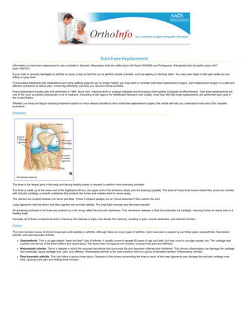
After total knee arthroplasty (TKA), the postoperative lowerlimb alignment is influenced by the geometry of the prosthesis position1 and surrounding soft tissue that contributeto the hip–knee–ankle (HKA) angle.2–4 It is not clearlyestablished what the optimal TKA alignment is due towide variations between individual patients, sexes, andraces.5–9The HKA angle within 3 degrees of neutral has been usedas the essential outcome measure in TKA, and mechanicalalignment (MA) has been associated with implant survival.However, the relationship between the survival of a primaryTKA and mechanical axis alignment may be weaker thandescribed in several previous reports. Parratte et al foundthat a postoperative mechanical axis of 0 3 degrees did notimprove the 15-year implant survival rate.10–12Accordingly, kinematic alignment (KA) for TKA was introduced as an alternative alignment strategy. KA aims to coalignthe flexion–extension axis of the femoral component to thenatural kinematic axes of the patient’s knee.Recently, functional alignment has been proposed. Thistechnique has elements of both measured resection andgap-balancing procedures. With functional alignment, thegaps are balanced before cutting the femur and tibia bychanging the implant targets in all three planes individualizedto the patient’s knee.13There are changes in the coronal mechanical axis from theextension to the flexion of the knee,14 so the alignment ofthe knee cannot be considered fixed at full extension; in somemanner, it could be considered “dynamic” from extension tofull flexion. An understanding of the normal dynamics of theHKA angle can help the surgeon to achieve better alignmentafter TKA. The preoperative dynamic HKA angle has gainedinterest, especially in the osteoarthritic knee,15 as this information may have clinical influence in the intraoperativedecision-making process.16Computer-assisted navigation provides surgeons withquantitative measurement tools for real-time assessmentof lower limb alignment and kinematics.17–20 It is a powerfulinstrument for intraoperatively supporting and guiding thesurgeon in the adequate postoperative soft tissue balance ofthe knee.17,21–24 Through this technology, the authors havepublished that there is well-defined preoperative dynamicalignment in osteoarthritic knees.16The purpose of this study was to determine the dynamiccoronal HKA angle after MA in total knee replacement usingcomputer navigation. The hypothesis was that the dynamicHKA angle in the full range of motion would differ preoperatively and postoperatively after mechanically alignedknee replacement. We employed a pre–post study design25based on a consecutive case series focused on the nullhypothesis (Ho) that the preoperative dynamic measurements of HKA angle at 0, 30, 60, and 90 degrees do not differfrom the postoperative measurements in total kneereplacement.The main objective was to assess the preoperative andpostoperative HKA angle distribution at 0, 30, 60, and90 degrees of flexion and to assess “within-range” postoperative coronal dynamic alignment.The Journal of Knee Surgery 2021. Thieme.Larrainzar-Garijo et al.Materials and MethodsIn total, 100 consecutive patients with 102 osteoarthritic kneejoints on which TKA was performed in our institution from2009 to 2010 were enrolled in this study. The inclusion criteriawere patients with primary osteoarthritic knee joints receivinga posterior stabilized total knee replacement (Columbus, B.Braun Aesculap, Tuttlingen, Germany) due to substantial painand loss of functionality with any degree of deformity. Patientswere excluded if they had prostheses revision surgery for anyreason in the last 8 years, had a preoperative valgus knee (HKAangle 180 degrees), had undergone any previous knee orhip surgery, had any major lower limb trauma resulting inan abnormal limb alignment, or had flexion contractureof 10 degrees.All patients provided written informed consent, and theHospital Committee for Medical and Health Research Ethicsapproved the study (Hospital General Universitario GregorioMarañón, Madrid, Spain, Protocol 1–04. V-02). All procedureswere performed following the 1964 Declaration of Helsinkiand its later amendments.After evaluating their eligibility, n ¼ 72 knees (71 patients)were included in the final analysis. There were several reasonsto exclude patients. Twenty-three of them had a valgus osteoarthritic knee. Three were excluded because an ultracongruent liner was used. Two patients had received a hip prosthesisbefore, one had previous knee surgery, and one had 20 degreesof knee flexion contracture ( Fig. 1).Demographic data collected on the cohort included age,sex, body mass index (BMI), and follow-up period.The baseline characteristics of the study population arereported in Table 1. Due to the presence of missing values,summary statistics for BMI was calculated with n ¼ 60.Intraoperative HKA Angle AcquisitionAfter the registration process and through a standard anterior knee approach, optical infrared (IR) trackers werescrewed into the femur and tibia.To allow the system to determine the coronal HKA angle,the rotational centers of the hip, knee, and ankle were needed.The centers of the hip and knee were obtained using akinematic method, whereby the hip is rotated and the kneewas extended and flexed to allow the navigation system todetermine the centers of movement. The center of the anklewas calculated anatomically using the most prominent zone ofthe lateral and medial malleoli and the anterior joint line. TheHKA angle was assessed at this moment of the surgicalprocedure, before any soft tissue release or bone cutting. TheHKA angle was measured with the patient supine with themaximum knee extension possible (considering that value as0) and with 30, 60, and 90 degrees of flexion.Once obtained, the surgical procedure was performedfollowing a navigated gap-balancing technique (Orthopilotversion 4.2; Braun Aesculap, Tuttlingen, Germany). A distalfemoral cut was aimed to be done at a 90-degree sagittal andcoronal plane with respect to the hip center. The tibial cutwas intended to be done at 90-degree coronal and 2-degreeposterior slope sagittal to the ankle center. Gaps at 0 andThis document was downloaded for personal use only. Unauthorized distribution is strictly prohibited.Dynamic Coronal HKA after Mechanical Alignment in TKA
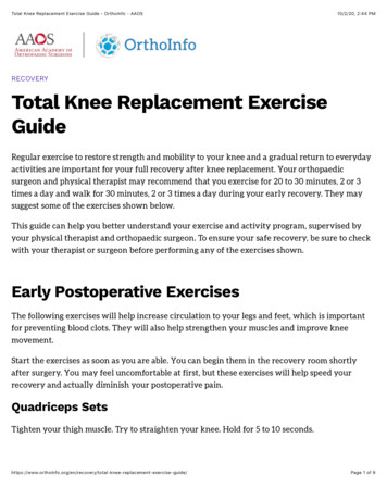
Larrainzar-Garijo et al.Fig. 1 Patient flow diagram. TKA, total knee arthroplasty.Table 1 Study population baselinePatients characteristicsValuesFemale n (%)46 (64.79)Male n (%)25 (35.21)Right knee n (%)42 (58.33)Left knee n (%)30 (41.67)Age (y)Mean SD74.42 6.49Median (Q1, Q3)75.5 (71, 78.25)BMI (kg/m2)Mean SD33.03 5.44Median (Q1, Q3)32.60 (29.29, 36.15)Follow-up (y)Mean SD8.15 2.03Median (Q1, Q3)8.77 (7.88, 9.34)Abbreviations: BMI, body mass index; SD, standard deviation.90 degrees were assessed after all bone cuts, and the finalfemoral rotation was measured by the navigation systemreferenced to the preoperative posterior condyle axis.The HKA angle was reevaluated at the end of the surgicalprocedure once the cementation process was completed, andthe tourniquet was deflated. The navigation system calculatedthe final gaps in full extension and 90 degrees of flexion. TheHKA angle was measured in the same manner as was donepreoperatively, and the range of movement from extension tofull flexion was recorded.Preoperative and Postoperative HKA Angle Data Analysisat 0, 30, 60, and 90 DegreesThe authors previously described the preoperativevarus osteoarthritic dynamic behavior through a clusteringmethod in five segments (expected, severe, varus–valgus,concave, and structured) and three patterns (Varus-Neutral,Varus-Valgus, and Varus-Varus).16 The segmentation andpatterns obtained were named empirically by the authorsat that moment.In the present study, we distinguish among three preoperative dynamic behaviors corresponding to the three abovementioned patterns: Varus-Neutral: (grouping together expected and severesegments) when the HKA angle trends to neutral as theknee flexes. Varus-Valgus: representing a starting varus alignment at0 degrees and valgus alignment as the knee flexes. Varus-Varus: (grouping together concave and structuredsegments) if the HKA angle stays approximately constantand does not reach 0 degrees at any degree of flexion.Postoperative Static Coronal AlignmentWe believed that an optimal static alignment was achievedwhen the HKA angle value measured at 0 degrees was in therange of 0 3 degrees of varus/valgus.Postoperative Dynamic Coronal AlignmentWe defined a “within-range” dynamic alignment as whenthere was no more than 3 degrees of deviation from 0 degreein any of the HKA angle values measured at 0, 30, 60, and90 degrees.Statistical AnalysisFor a coverage probability of at least 95%, a minimum samplesize of 71 knees was required to estimate the likelihood ofbeing dynamically within range after surgery with a samplingerror of 11%.Continuous data were assessed for normality using theShapiro–Wilk test. In the case of normality, analysis ofvariance (ANOVA) was used to compare the three preoperative dynamic patterns (PDPs; Varus-Neutral, Varus-Valgus,and Varus-Varus). Otherwise, the Kruskal–Wallis test wasused. The three PDPs were comparable in terms of BMI, age,and follow-up period.For each PDP and knee flexion angle (KFA), the Wilcoxontest was used to detect differences between pre- and postsurgery HKA angle values.For each phase (pre- and postsurgery) and PDP, the Friedman’s test was used to detect differences in HKA angle valuesacross the four KFAs considered. In the case of a significantThe Journal of Knee Surgery 2021. Thieme.This document was downloaded for personal use only. Unauthorized distribution is strictly prohibited.Dynamic Coronal HKA after Mechanical Alignment in TKA
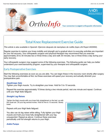
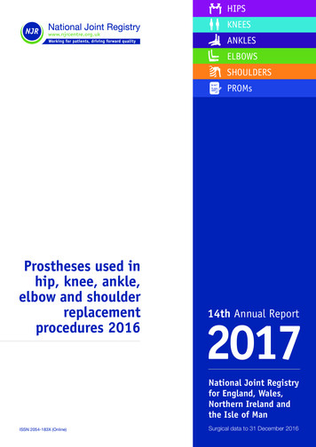
result, post hoc pairwise comparisons between KFAs wereperformed based on the Wilcoxon test (with Bonferroni’scorrection to keep the global type-I error at an α level of 5%).For each phase and PDP, the Cochran Q-test was used todetect differences in the proportions of patients whose HKAangle values deviated 3 degrees from 0 degrees across thefour KFA considered. In the case of a significant result, posthoc pairwise comparisons were performed based on theMcNemar test with Bonferroni’s correction.For each phase and KFA, an extension of Fisher’s exact test(the Freeman–Halton test) was used to detect differences inthe proportions of patients who were out of range across thethree groups defined by PDPs.Statistical analysis was performed using R software.A result of p 0.05 was considered statistically significant.ResultsPreoperative and Postoperative HKA Angle Fig. 2A graphically shows the distribution of the HKA anglevalues for each PDP in each of the four measured kneeangulations before knee replacement. The black line betweenthe boxplot represents the trend in the distribution of HKAangle values throughout the measured range of motion. Itshould be noted that between the Varus-Valgus and theVarus-Neutral, the slopes of the lines are practically similar;they differ only in the starting point.Before the surgical procedure, statistically significant differences were found between patterns at any angle of flexion(with p 0.0001 for Varus-Neutral vs. Varus-Valgus at 0, 30, 60,and 90 degrees; Varus-Neutral vs. Varus-Varus at 0 and30 degrees; and Varus-Valgus vs. Varus-Varus at 60 and90 degrees, and with p 0.05 for Varus-Valgus vs. Varus-Varusat 30 degrees). These results confirm well-differentiated preoperative dynamic behavior among the three groups. Fig. 2B shows the distribution of the HKA angle valuesfor each PDP in any of the four measured knee angulationsafter knee replacement. Again, the black line between theboxes represents the trend in the HKA angle distributionthroughout the measured range of motion. The three PDPshave very similar postoperative dynamic behavior, maintaining a constant HKA angle value, close to neutral, as theknee bends and within the 3-degree zone represented inthe graph by the dotted lines. Based on the post hoc Wilcoxontest (with Bonferroni’s correction), there were statisticallysignificant differences only when comparing the HKA anglevalues between 0 and 30 degrees in the Varus-Varus group.Graphically, it can be seen that the behavior of the knee istotally different before and after surgery and that the HKAangle shows more homogeneous and consistent values oncethe prosthesis has been implanted.The gray zone in the middle of Fig. 2 shows the statisticalanalysis using the Wilcoxon test comparing each PDP and KFAand the HKA angle distribution pre- and postsurgery. For abetter understanding, the analysis relates, for instance, theblue boxplot (representing the HKA angle measurements at0 degrees of flexion) against itself before and after surgery. Thisanalysis was done for each of the colored boxplots, representThe Journal of Knee Surgery 2021. Thieme.Larrainzar-Garijo et al.ing the four angles at which the HKA angle has been measured(blue ¼ 0 degrees, yellow ¼ 30 degrees, gray ¼ 60degrees, andred ¼ 90 degrees). Statistically significant differences aredetected in all the comparisons (except for the Varus–Valgusbehavior pre- versus postsurgery at 90 degrees of flexion), thussupporting the alternative hypothesis considered in the study. Table 2 summarizes postoperative femoral, tibial position, and flexion–extension gaps. Statistically nonsignificantdifferences were detected across the PDPs in terms of thedata collected. Table 2: femoral rotation was obtained with respect to theposterior condyle line. The joint orientation angles in thefrontal and sagittal planes were assessed according to Paley’srecommendation.26 The surgical navigator determined thecalculation of the spaces in millimeters on the medial andlateral compartment.Preoperative and Postoperative Static Coronal HKAAngle AnalysisBefore surgery, only 15.3% of the knees (11 out of 72) werepreoperatively within range at full extension (see top panelof Fig. 2).After the surgical procedure, 98.6% of the knees (71 out of72) were within 3 degrees of the HKA angle at full extension according to computer measurement. This outlier kneewas preoperatively clustered in the Varus-Neutral dynamicbehavior.Preoperative and Postoperative Dynamic Coronal HKAAngle Analysis Table 3 shows the percentage of knees that were withinrange after knee arthroplasty for any KFA assessed, considering the whole sample (fourth column) and distinguishingby PDP (the first three columns). Fifty-eight knees (80.6%)exhibited a “within-range” postoperative dynamic alignment at any KFA. Of the remaining 14 knees, 85.7% (12 outof 14) presented a similar behavior in the sense that theyremained within range up to a certain grade of flexion and,from there, remained out of range.As the knee bends, the rate of being out of range increases.According to Cochran’s Q-test, this result is statistically significant when the entire sample of knees is considered. Post hocpairwise comparison based on the McNemar test with Bonferroni’s correction finds a significant difference when comparingthe percentage of knees within range at 90 degrees (83.3%) withthe percentage at 0 (98.6%) and 30 degrees (97.6%).Despite the fact that the percentage of patients within rangeat 90 degrees and at any KFA seems to be higher in the VarusVarus group, the different PDPs do not show a statisticallysignificant difference based on the Freeman–Halton extensionof Fisher’s exact test. This absence of statistical significancemay correspond with the relatively low number of patients ineach PDP.DiscussionThe most important finding of the present study is that thereare differences in the dynamic coronal HKA angle betweenThis document was downloaded for personal use only. Unauthorized distribution is strictly prohibited.Dynamic Coronal HKA after Mechanical Alignment in TKA
Larrainzar-Garijo et al.This document was downloaded for personal use only. Unauthorized distribution is strictly prohibited.Dynamic Coronal HKA after Mechanical Alignment in TKAFig. 2 The difference in dynamic behavior between the three groups proposed by the authors can be seen graphically through colored boxplotscorresponding to each KFA (blue ¼ 0, yellow ¼ 30, gray ¼ 60, and red ¼ 90 degrees). The spot in the middle of the box represents the mean value. Thehorizontal line in the box represents the median value. The height of the box is the interquartile range, Q1–Q3, representing that central 50% of the mostrepresentative values. The vertical outbox lines represent the minimum and maximum of the nonoutlier values; when a value deviated from the top or bottomof the box more than 1.5 times the interquartile range, it was identified as an outlier and expressed as a dot. Horizontal black lines across the four coloredboxplots of each panel are regression lines that represent how HKA angle changes as the knee is bent. (A) Preoperative: in the so-called Varus-Neutral behavior,the HKA angle tends to reach a neutral value, despite having pronounced varus initial values. The Varus-Valgus pattern presents an initial HKA angle valueslightly in varus, and it reaches valgus values as it bends. The Varus-Varus cluster always maintains an HKA angle value in varus throughout the full range ofmovement. (B) Postoperative: in the three PDPs, the postoperative HKA angle is homogeneous and well established in three degrees from 0. Only the “VarusVarus” PDP shows statistically significant differences between 0 and 30 degrees. It should be noted that all patients in the Varus-Valgus group showed valgusHKA angle mean values throughout the entire range of knee motion, and only a few individual patients ranged in varus or valgus values. Gray zone: for each PDPand any KFA, there are statistically significant differences when comparing HKA angle measurements between phases (pre- versus postsurgery), except in thecase of Varus-Valgus at 90 degrees of flexion. HKA, hip–knee-ankle; KFA, knee flexion angle; ns, nonsignificant; PDP, preoperative dynamic pattern; p 0.05; p 0.01; p 0.001; p 0.0001. Statistically significant pairwise comparisons based on post hoc Wilcoxon’s tests with Bonferroni’s correction (for eachpanel, i.e., for each PDP and phase) or based on Wilcoxon tests (gray zone, i.e., for each PDP and KFA).The Journal of Knee Surgery 2021. Thieme.
Dynamic Coronal HKA after Mechanical Alignment in TKALarrainzar-Garijo et al.Table 2 Femoral rotation was obtained with respect to the posterior condyle obalPreoperative dynamic behaviorn (%)31 (43.06)26 (26.11)15 (20.83)72 (100)Femoral coronal position (degree)Mean SD90.26 0.7790.35 0.8990.20 1.2190.28 0.9195% 0.49Median (Q1, Q3)90 (90, 91)90 (90, 91)91 (89.5, 91)90 (90, 91)Mean SD90.29 1.1090.31 0.8889.93 0.5990.22 0.9495% 0.44Median (Q1, Q3)90 (90, 91)90 (90, 91)90 (90, 90)90 (90, 91)Mean SD1.55 1.461.96 1.311.80 1.151.75 1.3495% ral rotation (degree)Median (Q1, Q3)2 (1, 2.5)2 (1, 3)2 (1, 3)2 (1, 3)Femoral rotation 0–3n (%)28 (90.32)23 (88.46)15 (100)66 (91.67)Femoral rotation 3n (%)1 (3.23)3 (11.54)04 (5.56)Femoral rotation internaln (%)2 (6.45)002 (2.78)Tibial coronal position (degree)Mean SD90.42 0.6290.35 0.7490.20 0.6890.35 0.6895% 0.50Median (Q1, Q3)90 (90, 91)90 (90, 91)90 (90, 91)90 (90, 91)Mean SD92.45 1.4392.46 1.4892.53 1.4192.47 1.4295% 2.80Median (Q1, Q3)92 (92, 93.5)93 (91.25, 94)93 (92, 93)93 (92, 94)Mean SD1.55 1.911.50 1.391.40 2.061.50 1.7595% CI0.88, 2.220.96–2.040.36–2.441.10–1.90Median (Q1, Q3)1 (0.50, 2.50)1 (0.25, 2)1 (0.5, 2)1 (0, 2)Tibial slope position (degree)Medial extension gap (mm)Lateral extension gap (mm)Mean SD2 2.212.12 1.772.07 1.942.06 1.9895% an (Q1, Q3)2 (0.5, 3)2 (1, 2)2 (0.5, 3)2 (1, 3)Medial flexion gap (mm)Mean SD3.19 1.682.69 2.002.60 1.722.89 1.8095% an (Q1, Q3)3 (2, 4)3 (2, 4)3 (2, 4)3 (2, 4)3.03 1.832.65 1.622.13 2.362.71 1.89Lateral flexion gap (mm)Mean SD95% an (Q1, Q3)3 (2, 4)3 (2, 3.75)3 (1, 4)3 (1, 4)Abbreviations: CI, confidence interval; SD, standard deviation.Note: The joint orientation angles in the frontal and sagittal planes were assessed according to Paley’s recommendation.26 The surgical navigatordetermined the calculation of the spaces in millimeters on the medial and lateral compartment.The Journal of Knee Surgery 2021. Thieme.This document was downloaded for personal use only. Unauthorized distribution is strictly prohibited.Femoral slope (degree)
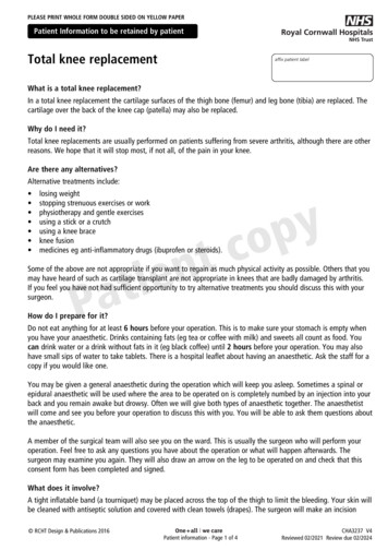
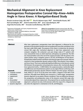
Dynamic Coronal HKA after Mechanical Alignment in TKALarrainzar-Garijo et al.VariablePreoperativeVarus-Neutraln ¼ 31PreoperativeVarus-Valgusn ¼ 15PreoperativeVarus-Varusn ¼ 26Globaln ¼ 72PO knees within range at 0 degrees% (n)95% CI96.8 (30)100 (15)100 (26)98.6 (71)88.2–99.2PO knees within range at 30 degrees% (n)95% CI96.8 (30)100 (15)96.2 (25)97.2 (70)86.9–97.9PO knees within range at 60 degrees% (n)95% CI87.1 (27)86.7 (13)88.5 (23)87.5 (63)78.2–89.2PO knees within range at 90 degrees% (n)95% CI80.6 (25)80 (12)88.5 (23)83.3 (60)a74.5–85.5PO knees within range at any KFA% (n)95% CI74.2 (23)80 (12)88.5 (23)80.6 (58)72.0–83.0Abbreviations: CI, confidence interval; KFA, knee flexion angle; PO, postoperatively.ap 0.05. Statistically significant pairwise comparisons based on the McNemar test with Bonferroni’s correction: 90 versus 0 degrees with 83.3versus 98.6% and 90 versus 30 degrees with 83.3 versus 97.2%.the preoperative and postoperative status after mechanicallyaligned knee replacement. It is remarkable that there are alsodifferent behaviors in the four measurement flexion valuesanalyzed. Except for the Varus-Varus pattern, it has beenproven that, preoperatively, the HKA angle values at 0 and30 degrees are alike but differ from those at 60 or 90 degreesof flexion. Postoperatively, these differences are stronglyattenuated.This finding suggests that, preoperatively, the geometry ofthe articular surface and the static and dynamic stabilizersplay a crucial role in the dynamic HKA angle. Once thesurgery is complete, everything relies on the prostheticdesign, position, and, to a lesser extent, the soft tissueenvelope.Among knee surgeons, the concept of KA has gainedinterest. This approach assumes that the aim of knee arthroplasty must be the natural alignment of the knee, rather thanthe restoration of alignment to neutral. This philosophy issupported by the fact that a slight varus alignment may bephysiological,27 based on the current knowledge and kinematics of the normal knee.28–32 In a certain way, it is true, aswhen standing or walking, the load transmission is close tothe medial side of the knee, and the muscles of the hip mustact as a stabilizer. It is unclear how this compensatory musclesystem is affected in a patient with osteoarthritic knees.The authors have previously described that, regardless ofvarus or valgus in extension, the preoperative HKA angle at90 degrees of flexion is close to neutral, and this situation canbe considered normal in an osteoarthritic knee.16 It is alsoknown that knees that are in varus or up to 2.5 degrees ofvalgus in the supine position tend to become more varusupon standing.33–35 This kinematic pattern suggests that thesoft tissue constraints, rather than the underlying deformity,are more influential in the control of the alignment fromlying to standing.36 This trend is also observed in a flexedposition when compared with hyperextension. Based on thisknowledge, it is easily understood that the alignment of theknee cannot be considered as static but “dynamic.”37The importance of the dynamic behavior of knee prostheses is clear from the fact that dynamic models of finiteelement models on knee arthroplasty are described.38,39The clinical relevance of our study resides in the fact that,focusing only on the full-extension HKA angle as a finalobjective, despite our philosophy of kinematic, mechanical,or functional strategy alignment, some critical data in thedynamic HKA angle that may affect the outcome of the surgicalprocedure are missed.40 It is not clearly established whatthe optimal postoperative alignment is.41 Traditionally,the gold standard has been considered a mechanical axis of0 3 degrees. Nevertheless, it has been described that thisideal mechanical axis does not ensure the optimal functionand survival of the prosthesis.10,42 This lack of certainty is dueto wide variations between individual patients6 and, as previously mentioned, because there are changes in the coronalmechanical axis from extension to flexion of the knee.4,40,43–45In our series, 98.6% of the patients were within 0 3 degrees ofthe HKA angle at full extension after surgery, with an excellentresult, minimizing the outliers. Nevertheless, we also haveproven that there is postoperative kinematic variation in thecoronal HKA angle plane between flexion and full extension,even though we achieved similar mean values in the flexionand extension gaps.18 For this reason, we consider in our dailypractice that an excellent dynamic alignment has beenachieved not only when the HKA angle value at full extensionis within the 0 3 degrees of range but also when there are notmore than 3 degrees of deviation from 0 in the HKA anglevalues measured at full extension and at 30, 60, and 90 degrees,as shown in Fig. 3.As far as we know, nobody has previously proposed thisdefinition of an optimal postoperative dynamic alignment,46The Journal of Knee Surgery 2021. Thieme.This document was downloaded for personal use only. Unauthorized distribution is strictly prohibited.Table 3 “Within range” ¼ 0 3 degrees of the hip–knee–ankle angle
Larrainzar-Garijo et al.Fig. 3 shows a suitable dynamic postoperative alignment, as there are no more than three degrees of difference between the HKA angle in thefull range of movement. (A) Full-extension, (B) midflexion, (C) 90 degrees of flexion, and (D) full flexion. HKA, hip–knee-ankle.and it may be interesting for knee surgeons to consider it asan objective while performing knee replacement that ismainly assisted by computer. We want to emphasize thatwe still recommend establishing a postoperative HKA anglevalue within 0 3 degrees of the HKA angle at full extension,33 while paying attention to the dynamic HKA angle thatprovides the surgeon with important information regardingmid-flexion stability, especially when using a cruciateretained
After total knee arthroplasty (TKA), the postoperative lower limb alignment is influenced by the geometry of the pros- . Braun Aesculap, Tuttlingen, Germany). A distal femoralcut wasaimedtobedoneata90-degreesagittaland coronal plane with respect to the hip center. The tibial cut was intended to be done at 90-degree coronal and 2-degree posterior slope sagittal to the ankle center. Gaps at 0 .