Transcription
Jiang et al. Diagnostic Pathology(2022) 17:4https://doi.org/10.1186/s13000-021-01188-yCASE REPORTOpen AccessNUT midline carcinoma as a primary lungtumor treated with anlotinib combinedwith palliative radiotherapy: a case reportJin Jiang1, Yikun Ren1, Chengping Xu2 and Xing Lin3*AbstractBackground: NUT (nuclear protein in testis) midline carcinoma (NMC) is a rapidly progressive tumor arising frommidline structures. Recent cases have reported that the poor prognosis with a median survival of 6.7 months and a2 years overall survival of 19% due to limited treatment. Based on the effect of arotinib on inhibiting tumor growthand angiogenesis. We present one patient case treated with anlotinib and radiotherapy.Case presentation: Here, we describe a 33-year old patient who complained of cough and chest pain and wasdiagnosed as a pulmonary NMC through CT scan, FISH and immunohistochemistry. In addition, we initiallydemonstrated that anlotinib combined with palliative radiotherapy could significantly prevent the tumor growth ina pulmonary NMC.Conclusion: The report indicated that anlotinib combined with palliative radiotherapy could inhibit the tumorprogression in a pulmonary NMC, which may provide a combined therapy to pulmonary NMC in the future.Keywords: NUT midline carcinoma, Primary lung tumor, Anlotinib, Palliative radiotherapy, Case reportBackgroundNUT midline carcinoma (NMC) is a rapidly progressivetumor arising from midline structures, including upperaerodigestive tract and mediastinum [1]. Less than onehundred cases have been reported worldwide until now.It is still conventional for its treatments, such as chemotherapy, radiotherapy and targeted therapy [2]. Recentcases have reported that the poor prognosis with a median survival of 6.7 months and a 2 years overall survivalof 19% [3–5].To be our knowledgement, angiogenesis plays an important role in tumor growth. Recent studies indicatedthat blocking angiogenesis has been a successfully alternative strategy in the managemenyt of cancer [4].Stikingly, anlotinib has inhibitory effects on tumor progression via preventing angiogenesis. Its mechanism isabout a novel receptor tyrosine kinases (RTK) inhibitortargeting vascular endothelial growth factor receptor(VEGFR)-2 and 3, fibroblast growth factor receptor(FGFR) 1–4, platelet-derived growth factor receptor(PDGFR) -α and -β, c-Kit and Ret [5]. Of note, antiangiogenesis drug has become an essential therapy fornon small-cell lung cancer (NSCLC) [5]. A pulmonaryNMC characterized by a highly aggressive cancer ofsquamous cell carcinoma.Therefore, in this report, we utilized radiotherapy andanlotinib to inhibit tumor growth, which may show annovel option to treat a pulmonary NMC.* Correspondence: lx15002345792@126.com3Department of Biological Immunotherapy, Chongqing University CancerHospital, Chongqing Cancer Institute, Chongqing Cancer Hospital, ShapingbaDistrict, Chongqing 400030, ChinaFull list of author information is available at the end of the articleCase presentationA 33 years old man with a smoking history of 18 packyears complained of cough and chest pain for 1 month.He went to Southwest Hospital of Third Military The Author(s). 2021 Open Access This article is licensed under a Creative Commons Attribution 4.0 International License,which permits use, sharing, adaptation, distribution and reproduction in any medium or format, as long as you giveappropriate credit to the original author(s) and the source, provide a link to the Creative Commons licence, and indicate ifchanges were made. The images or other third party material in this article are included in the article's Creative Commonslicence, unless indicated otherwise in a credit line to the material. If material is not included in the article's Creative Commonslicence and your intended use is not permitted by statutory regulation or exceeds the permitted use, you will need to obtainpermission directly from the copyright holder. To view a copy of this licence, visit http://creativecommons.org/licenses/by/4.0/.The Creative Commons Public Domain Dedication waiver ) applies to thedata made available in this article, unless otherwise stated in a credit line to the data.
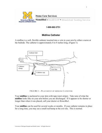
Jiang et al. Diagnostic Pathology(2022) 17:4Medical University. Transbronchial biopsy was routinelyperformed. A highly aggressive differential carcinomaconsidered as NMC was found in biopsy tissue. The immunohistochemical (IHC) markers revealed that p63( )and NUT( ). P63 was the positive one which supportedsquamous cell carcinoma (Fig. 1A). IHC staining withNUT protein showed markedly positive (Fig. 1B). Fluorescent DNA in situ hybridization (FISH) test showed aBRD4-NUT rearrangement (Fig. 2A). NMCs are difficultto morphologically different from other poorly differentiated carcinomas, and the diagnosis is usually made currently by FISH. Herbert et al. Have reported that FISHfor NUT rearrangement was used as a “gold standard”diagnostic test for NMC. C52 immunoreactivity amongcarcinomas was confined to NMCs. IHC staining had asensitivity of 87%, a specificity of 100%, a negative predictive value of 99%, and a positive predictive value of100% [6]. The diagnosis was confirmed as a pulmonaryNMC via IHC since the anti-NUT C52 monoclonal antibody had a specificity of 100%. A chemotherapy (paclitaxel liposome 300 mg day1 and cisplatin 120 mg day1).One month later, he came to our hospital because ofserious shortness of breath. Physical examinationshowed that the breathing of the left lung was low.Hematoxylin-eosin (HE) staining demonstrated a NUTmidline carcinoma with aberrant squamous differentiation (Fig. 3A). IHC staining with p63 protein showedobviously positive (Fig. 3B). Chest CT revealed that aperihilar soft tissue mass which near to hilar and mediastinal part measuring 5.6 cm*5.1 cm in the left lowerlober lung, left upper lobe obstructive inflammation,possible metastases of nodules and masses in the lowerlobe of the left lung, possible metastases of left supraclavicular, left chest wall and axillary lymph nodes andPage 2 of 6superiorvena cava and left brachiocephalic vein compression and left pleural effusion (Fig. 4A). AbdominalCT showed that neither heptic nor adrenal metastases.99mTc bone scan and brain magnetic resonance imaging(MRI) demonstrated that no evidence of bone and brainmetastases. The biopsy tissue from left supraclavicularlymph node showed that the possible metastases frompulmonary NMC. No additional treatment except a leftthoracic drainage such as chemotherapy or radiotherapywas added. Twenty-one days later, he came back to ourhospital since the shortness of breath became more serious. Physical examination showed that serious facialedema. Chest CT revealed that a perihilar soft tissuemass which became larger measuring 9.7 cm*9.0 cm inthe left lower lober lung (Fig. 4B). Emergency palliativeradiotherapy (20Gy/5F) to malignant tumor of the leftlower lober lung for alleviating superior vena caval syndrome (SVCS) and anlotinib (12 mg qd po.) for antiangiogenesis were administrated. Sixty days later, CT scanshowed that the perihilar soft tissue mass which becameabsolutely smaller measuring 5.0 cm*3.7 cm in the leftlower lober lung (Fig. 4C). However, palliative radiotherapy and targeted therapy were not subsequently administrated because of radiation esophagitis, radiationstomatitis and bone marrow suppression after ten dayslater. He passed away 4 months after diagnosis.Discussion and conclusionsNMC characterized by chromosomal rearrangement ofthe involvement of nuclear protein in testis (NUT) ofthe testis on 15q14 is a highly aggressive cancer of squamous cell carcinoma, which always arising in the midlinestructure of the body, such as head, neck, the upperaerodigestive tract and mediastinum [7]. The specificFig. 1 IHC staing showed markedly positive. A: P63 was the positive one which supported squamous cell carcinoma. B: IHC staing with NUTprotein showed obviously positive. Scale bar 100 μm
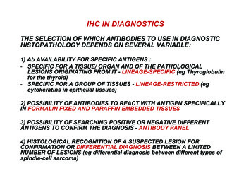
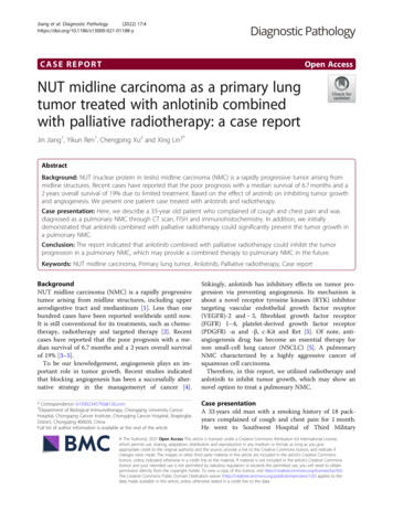
Jiang et al. Diagnostic Pathology(2022) 17:4Page 3 of 6Fig. 2 A: Fluorescent DNA in situ hybridization (FISH) demonstrated a BRD4-NUT rearrangement. A: A red probe that spans NUT splits and joinsthe green BRD4 centromeric probe. B: A negative control. Scale bar 50 μmmolecular genetics is that the fusion oncogene BRD4NUT as result from BRD4 gene translocation located at19p13.1 in the NMC [8]. Therefore, it was initially classified carcinoma with t (15;19) by WHO Classification ofTumours in 2004 [8]. Previous studies have shown it tobe associated with Epstein Barr virus (EBV) and HumanPapilloma virus (HPV) [9]. Kees and Kubonishi have reported the first two cases which were considered arisingfrom thymus in 1991 [10]. As the fusion genes discovered by FISH assay, NUT carcinoma was newly classifiedby WHO in 2015 [6].Previous studies have demonstrated that the poorprognosis in NMC cases with a median survival atless than approximate 9 months [11]. To be our bestknowledge, its incidence and underlying mechanismwere not clear. According to previous reports, patients of all age groups ranged from several monthsto 78 years old, especially children and young adults[12]. The most common clinical symptom was cough[13]. However, cough often had not been noticed during the early stage. The lung is one of the most common sites for NUT cancer, but there are few reportson lung NUT cancer. The largest centralized report isa retrospective analysis published in the Journal ofThoracic Cancer in 2015 [14], a review of the studyThe clinical features of 8 cases of lung NUT cancerwere analyzed. Unfortunately, the average survivaltime of 8 patients was only 2.2 months, which wasmuch lower than the 5.5-month average survival timeof 119 cases of NUT cancer reported by Giridha [15].It was unfortunate that most patients had lose thesurgery chance because of its distant metastases. Evenso, there is no standard treatment to NMC. We notedthat the majority of patients were administratedchemotherapy and/or radiotherapy [15]. Here, for thefirst time we revealed that the primary pulmonaryNMC, initially treated with the combination of radiotherapy and anlotinib for antiangiogenesis.
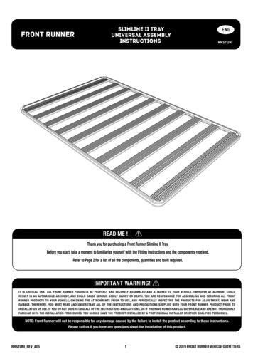
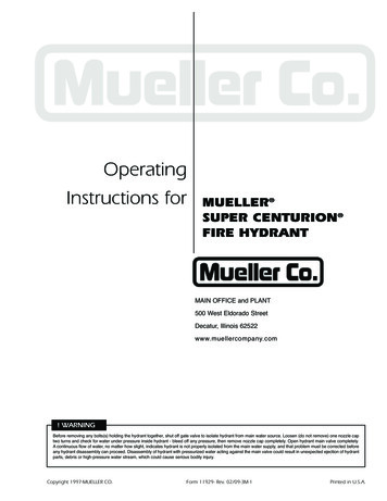
Jiang et al. Diagnostic Pathology(2022) 17:4Page 4 of 6Fig. 3 NUT midline carcinoma as a primary lung tumor. A: The NMC CT on Sept. 9th 2020. B: The NMC CT on Sept. 25th 2020. C: The NMC CT onNov. 9th 2020It has been acknowledged that anlotinib plays inhibitory effects on tumor growth and angiogenesis becauseanlotinib is a novel RTK inhibitor targeting VEGFR-2and -3, FGFR 1–4, PDGFR-α and-β, c-Kit and Ret [16].Arotinib have received its first approval as a third-linetreatment for refractory advanced NSCLC in May 2018[17]. Recent studies have demonstrated that anlotinib increases the sensitivity of pulmonary blastoma to chemotherapy in vivo [18]. As we know, activation of the FGFRsignaling pathway promotes chemotherapy resistance.Interestingly, anlotinib could inhibit the tumor growthby suppressing the activation of FGFR1–4 [18]. Thus, wespeculated that anlotinib may exert a positive role onovercoming the chemotherapy resistance. Shi et al. havesuggested that combination therapy with anlotinib andchemoradiotherapy may be an effective regimen for thetreatment of advanced esophageal squamous cell carcinoma (ESCC) [19]. A study by Wang et al. indicated thatFig. 4 The biopsy tissue from left supraclavicular lymph node showed that the possible metastases from pulmonary NMC. A: Hematoxylin-eosin(HE) staining demonstrated a NUT midline carcinoma with aberrant squamous differentiation. B: IHC staining with p63 protein showed obviouslypositive. Scale bar 50 μm
Jiang et al. Diagnostic Pathology(2022) 17:4stereotactic radiosurgery (SRS) combined with anlotiniblimited brain metastases with perilesional edema inNSCLC [20]. A recent study has reported that radiotherapy combined with anlotinib to treat with orbital NMC[21]. In this report, we for the first time used palliativeradiotherapy combined with anlotinib could significantlyalleviate primary lesion of NMC progression, which mayprovide a way to treat primary pulmonary NMC. Wespeculate that anlotinib may exert a positive role on improving the radiotherapy sensibility, which needs our advanced investigation in the future.In summary, we obtain the valuable experience torealize the characteristic of NMC in this present case.Moreover, we initially reported a case of primary pulmonary NMC, which was suppressed the progression byradiotherapy combination with anlotinib. If NMC is diagnosed at the early stage, surgery may be more effective. However, radiotherapy may play an important rolein the process of treatment during the advanced stage ofNMC. Additionally, anlotinib could be considered to bethe treatment of primary pulmonary NMC. Furtherstudies and clinic case reports are essential to exploreunderlying mechanisms in NMC.AbbreviationsNMC: nuclear protein in testis midline carcinoma; RTK: receptor tyrosinekinases; VEGFR: vascular endothelial growth factor receptor; FGFR: fibroblastgrowth factor receptor; PDGFR: platelet-derived growth factor receptor; NSCLC: non small-cell lung cancer; CT: computed tomography; FISH: fluorescentDNA in situ hybridization; MRI: magnetic resonance imaging; SVCS: superiorvena caval syndrome; EBV: Epstein Barr virus; HPV: human papilloma virus;ESCC: esophageal squamous cell carcinoma; SRS: stereotactic radiosurgeryAcknowledgementsWe thank the patient for granting permission to publish this information.Authors’ contributionsJJ designed and drafted the manuscript. XL handled the funding, gaveessential advice in the design of the report and provided the critical revisionof the manuscript. XL handled the funding and participated in the design.CX and YR collected parts of data. All authors read and approved the finalmanuscript.FundingThe study was supported by the Natural Science Foundation of Chongqing,Grand NO. cstc2019jcyj-msxmX0630, to JJ; the Natural Science Foundation ofChongqing, Grand NO. cstc2020jcyj-msxmX0325, to XL; the Chinese MedicineScience and Technology project of Chongqing Municipal Health committee,Grand NO. 2021ZY3794, to XL.Availability of data and materialsN. A.DeclarationsEthics approval and consent to participateOur manuscript report data collected from animals and this study wasapproved by the Ethics Committee for Chongqing University.Consent for publicationWritten informed consent to publish the clinical data was obtained from thepatient before the initiation of the report.Page 5 of 6Competing interestsThe authors declare that they have no competing interests.Author detailsDepartment of Neurology, The First Affiliated Hospital of ChongqingMedical University, Chongqing 400016, China. 2Department of Pathology,The Southwest Hospital, the Southwest Hospital of Army Medical University,Chongqing 400038, China. 3Department of Biological Immunotherapy,Chongqing University Cancer Hospital, Chongqing Cancer Institute,Chongqing Cancer Hospital, Shapingba District, Chongqing 400030, China.1Received: 1 February 2021 Accepted: 3 December 2021References1. Salati M, Baldessari C, Bonetti LR, Messina C, Merz V, Cerbelli B, et al. NUTmidline carcinoma: current concept sand futu reperspectives of a noveltumour entity. Crit Rev Oncol Hematol. 2019;144:102826. . French CA. Pathogenesis of NUT midline carcinoma. Ann Rev Pathol. 2012;7(1):247–65. 38.3. French CA. NUT carcinoma: Clinicopathologic features, pathogenesis, andtreatment. Pathol Int. 2018;68(11):583–95. https://doi.org/10.1111/pin.12727.4. Haefliger S, Tzankov A, Frank S, Bihl M, Vallejo A, Stebler J, et al. NUT midlinecarcinomas and their differentials by a single molecular profiling method: anew promising diagnostic strategy illustrated by a case report. VirchowsArch. 2020;478(5):1007–12. https://doi.org/10.1007/s00428-020-02869-7.5. Hellquist H, French CA, Bishop JA, Coca-Pelaz A, Propst EJ, Paiva Correia A,et al. NUT midline carcinoma of the larynx: an international series andreview of the literature. Histopathology. 2017;70(6):861–8. https://doi.org/10.1111/his.13143.6. Herbert H, Laura AJ, Christopher JF, et al. Diagnosis of NUT midlinecarcinoma using a NUT-specific monoclonal antibody. Am J Surg Pathol.2009;33(7):984–91. https://doi.org/10.1097/PAS.0b013e318198d666.7. Syed YY. Anlotinib: first global approval. Drugs. 2018;78(10):1057–62. https://doi.org/10.1007/s40265-018-0939-x.8. Gao Y, Liu P, Shi R. Anlotinib as a molecular targeted therapy for tumors(review). Oncol Lett. 2020;20(2):1001–14. https://doi.org/10.3892/ol.2020.11685.9. Benito Bernáldez C, Romero Muñoz C, Almadana PV. Carcinoma NUTpulmonar, una forma poco frecuente de cáncer de pulmón. ArchBronconeumol. 2016;52(12):619–21. https://doi.org/10.1016/j.arbres.2016.04.011.10. Wang R, Cao XJ, Kulej K, Liu W, Ma T, MacDonald M, et al. Uncovering BRD4hyperphosphorylation associated with cellular transformation in NUTmidline carcinoma. Proc Natl Acad Sci U S A. 2017;114(27):E5352–61. https://doi.org/10.1073/pnas.1703071114.11. El-Domeiri AA. Midline cancer of the floor of the mouth: role of suprahyoiddissection. Br J Surg. 1979;66(7):453–5. https://doi.org/10.1002/bjs.1800660703.12. Bauer DE, Mitchell CM, Strait KM, Lathan CS, Stelow EB, Luer SC, et al.Clinicopathologic features and long-term outcomes of NUT midlinecarcinoma. Clin Cancer Res. 2012;18(20):5773–9. https://doi.org/10.1158/1078-0432.CCR-12-1153.13. Shaikh F, Pagedar N, Awan O, McNeely P. Sinonasal NUT-midline carcinoma– a multimodality approach to diagnosis, staging and post-surgicalrestaging. Cureus. 2015;7(7):e288.14. Gökmen-Polar Y, Kesler K, Loehrer PJ Sr, Badve S. NUT midline carcinomamasquerading as a thymic carcinoma. J Clin Oncol. 013.51.1741.15. Abdelgalil AA, Al-Kahtani HM, Al-Jenoobi FI. Erlotinib. Profiles Drug SubstExcip Relat Methodol. 2020;45:93–117. https://doi.org/10.1016/bs.podrm.2019.10.004.16. Sholl LM, Nishino M, Pokharel S, Mino-Kenudson M, French CA, Janne PA,et al. Primary pulmonary NUT midline carcinoma: clinical, radiographic, andpathologic characterizations. J Thorac Oncol. 2015;10(6):951–9. https://doi.org/10.1097/JTO.0000000000000545.17. Giridhar P, Mallick S, Kashyap L, Rath GK. Patterns of care and impact ofprognostic factors in the outcome of NUT midline carcinoma: a systematicreview and individual patient data analysis of 119 cases. Eur Arch
Jiang et al. Diagnostic Pathology18.19.20.21.(2022) 17:4Otorhinolaryngol. 2018;275(3):815–21. https://doi.org/10.1007/s00405-018-4882-y.Luo Z, Cao C, Xu N, Ying K. Classic biphasic pulmonary blastoma: a casereport and review of the literature. J Int Med Res. 2020;48(10):300060520962394. https://doi.org/10.1177/0300060520962394.Shi J, Zhang Y, Wang J, Li J, Li Z. Anlotinib combined withChemoradiotherapy exhibits significant therapeutic efficacy in esophagealsquamous cell carcinoma. Front Oncol. 2020;10:995. https://doi.org/10.3389/fonc.2020.00995.Wang Y, Wang X, Guan Y, Song Y, Zhuang H, Wang E. Stereotacticradiosurgery combined with anlotinib for limited brain metastases withperilesional edema in non-small cell lung cancer: Rvision-001 studyprotocol. Thorac Cancer. 2020;11(5):1361–4. https://doi.org/10.1111/17597714.13386.Chai P, Zhou C, Jia R, Wang Y. Orbital involvement by NUT midlinecarcinoma: new presentation and encouraging outcome managed byradiotherapy combined with tyrosine kinase inhibitor: a case report. DiagnPathol. 2020;15(1):2. r’s NoteSpringer Nature remains neutral with regard to jurisdictional claims inpublished maps and institutional affiliations.Page 6 of 6
It is still conventional for its treatments, such as chemo-therapy, radiotherapy and targeted therapy [2]. Recent cases have reported that the poor prognosis with a me-dian survival of 6.7months and a 2years overall survival of 19% [3-5]. To be our knowledgement, angiogenesis plays an im-portant role in tumor growth. Recent studies indicated