Transcription
Immunohematology Review:Help, What Do I Need to Knowto Pass the ASCP Exam?Dr. Phyllis Ingham EdD. MEd. MT(ASCP)Program Director/Chair Clinical Laboratory TechnologyWest Georgia Technical CollegePhyllis.ingham@westgatech.eduAmerican Society for Clinical Laboratory Science Annual State Meeting –Georgia Chapter 2019
Take Off the Blindfold Today!
Blood Group Systems
ABO Blood Group SystemWhat We Know . Inverse reciprocal relationship between theforward and reverse type Naturally occurring antibodies ABO antibodies are predominately IgM
ABO Antibodies Activate Roomcomplementtemperature or colder Producestrong direct agglutinationreactions
ABO Blood Group Inheritance Inheritance by Mendelian genetics Codominant expression
How are ABO Red Cell Antigens Formed? ABH antigen formation results from: the interaction of genes at three separate loci (ABO, Hh,and Se) These genes produce specific glycosyltransferases thatadd sugars to a basic precursor substance.
ABO and Hh Gene Interaction Immunodominant sugars confer blood groupspecificity
H Antigen
ABO Antigens and Phenotypes
ABO Discrepancies
A SubGroups Firstdescribed in 1911 by von Dungen A1 and A2 subgroups Differences between A1 and A2 arequantitative and qualitative A subgroups generally more commonthan B subgroups
A SubGroups Forwardgrouping reagent (Anti-A) stronglyagglutinates both A1 and A2 phenotypes Anti-A1lectin reagent used in thedifferentiation of A1 and A2 phenotypes
A SubGroups Anti-A1 lectin reagent agglutinates A1 (or A1B) cells butdoes not agglutinate A2 (or A2B cells) Lectins used in blood banking Dolichosbiflorus: agglutinates A1 or A1B Bandeiraea Ulexsimplicifolia: agglutinates B cellseuropaeus: agglutinates O cells (H specificity) andother ABO blood groups depending on the amount of Hantigen available
Weak Subgroups of A Subgroups weaker than A2 occur infrequently Most often recognized through an ABOdiscrepancy (unexpected reactions in the forwardand reverse grouping ) Varying expression of four characteristics Weak A subgroups can be distinguished as A3, Ax,Aend, Am, Ay, and Ael
Weak Subgroups of B Very rare and less frequent than A subgroups Usually recognized by variations in the strength ofthe reaction using anti-B and anti-A,B Result of alternate alleles at the B locus Serologic techniques characterize B subgroups inthe following categories: B3, Bx, Bm, and Bel
Bombay Phenotype (Oh) First reported by Bhende in 1952 in Bombay, India Inheritance of a double dose of the h gene,producing the very rare genotype hh No H antigen made ABOgenes cannot be expressed ABHantigens cannot be formed
Bombay Phenotypes RBCsare devoid of normal ABH antigensand fail to react with anti-A, anti-B, andanti-H. InRBC testing using anti-A and anti-B, theBombay would phenotype as an O bloodgroup.
Bombay Phenotype Unlike the anti-H found occasionally in the serum of A1and A1B individuals, the Bombay anti-H can often bepotent and reacts strongly at 37 C . It is an IgM antibody that can bind complement and causeRBC lysis Only blood from another Bombay individual will becompatible
ABODiscrepancies
ABO Discrepancies Unexpected reactions in the forward and reverse groupingdue to Problemswith the patient’s serum (reverse grouping) Problemswith the patient’s red cells (forwardgrouping) Problemswith both the serum and cells Can appear as extra positive or weak/missing reactions Must be resolved prior to reporting a patient or donor’sABO group
ABO Discrepancy Categorization GroupI discrepancies Group II discrepancies Group III discrepancies Group IV discrepancies
Common Group Two Discrepancies Enhancingweakly reacting antigens withroom temperature incubation Pretreatment Acquiredof RBCs with enzymesB antigen
Resolving ABO Discrepancies Enhancing weakly reacting antigens with roomtemperature incubation Pretreatment of RBCs with enzymes Acquired B antigen Consideration of cold autoantibodies The patient’s RBCs can be tested with Dolichos biflorus toconfirm the presence of an ABO subgroup Unexpected alloantibodies in the patient’s serum otherthan ABO isoagglutinins may cause a discrepancy in thereverse grouping
Resolution of Group Three Discrepancies Effectsof Rouleaux Effects of Wharton’s jelly
Resolution of Common Group FourDiscrepancies Consideration of cold autoantibodies The patient’s RBCs can be tested with Dolichosbiflorus to confirm the presence of an ABOsubgroup Unexpected alloantibodies in the patient’s serumother than ABO isoagglutinins may cause adiscrepancy in the reverse grouping
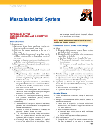
Chemistry/Antigens (Methodologies –Traditional Tube/Microwell Testing)Fig. 2-5 Summary of ABO reagents. Bloodbanks are using monoclonal antibodies forABO reagents in routine testing.Fig. 9-4 Results and interpretation of anABO/Rh phenotype. In the hemagglutinationtest, agglutination is a positive result, and noagglutination is a negative result.
Chemistry/Antigens (Methodologies – SolidPhase Red Cell Adherence Assay - SPRCA)Fig. 9-6SPRCA.Reactions and interpretation of
Chemistry/Antigens (Methodologies –Gel Test)Fig. 9-14 ID-MTS Gel Test procedure for thedetection of A, B, and D antigens.
Now You Give It a Try GroupActivity
Rh Blood Group Rh specific antigens reside on proteins versus thecarbohydrate antigens ABO and Hh. Rh antigens are very immunogenic. Rh antibodies are produced after exposure toforeign red blood cells.
Rh Blood Group Rh is the second most important blood groupsystem after ABO. Itis a complex blood group system composed ofover 50 different RBC antigens. Individualswho lack RhD are “Rh negative.” Individualswho possess RhD are “Rh positive.” Determiningthe presence or absence of RhD iscritical in pretransfusion testing.
Fisher Race-DCE Terminology Fisherand Race postulated that theantigens of the system were produced bythree closely linked sets of alleles. Eachgene was responsible for producing aproduct (or antigen) on the RBC surface.
Eachantigen and corresponding gene weregiven the same letter designation (whenreferring to the gene, the letter isitalicized). The phenotype (antigens expressed onthe RBC detected by typing) of a givenRBC is defined by the presence of D, C, c,E, and e expression.
Wiener: Rh-Hr Terminology Wienerbelieved there was one generesponsible for defining Rh that producedan agglutinogen containing a series ofblood factors. ThisRh gene produced at least threefactors within an agglutinogen.
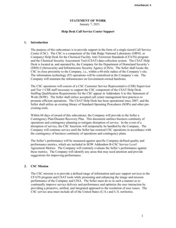
Comparison of Rh Genetic Theories
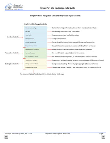
Rh Phenotypes and Conversions
Weak D: Variations of D expression Someindividual’s RBCs possess weakerexpression of D antigen that requires anindirect antiglobulin test to detect the Dantigen. Individuals with altered D antigen are categorized into different phenotypesdefined as weakened D. Cin trans to RHD Weak D Partial D
Weak D
Detection of Rh Antibodies and Antigens Most Rh antibodies are IgG and react optimally at37 C or after AHG testing. Rhantibodies are usually produced followingexposure to foreign RBCs. Rh Rhantibodies may show dosage.antibodies are enhanced when testing withenzyme-treated RBCs.
Weak D Procedure
Other Blood GroupSystems
The Lewis System The Lewis blood group system is unique because the Lewisantigens are not intrinsic to RBCs but are on type 1glycosphingolipids that are passively adsorbed onto theRBC membrane from the plasma. Thereare several Lewis antigens, but the two ofprimary concern are Lea and Leb. Twoalleles at the Lewis locus, Le and the amorph le,and two alleles at the Secretor locus, Se and theamorph se.
Lewis Antigens
Lewis Antibodies Frequently naturally occurring antibodies made byLe(a b ) persons; that is, they occur without any knownRBC stimulus. Generally NotIgM and do not cross the placenta.well developed on fetal RBCs. Anti-Leais the most commonly encountered. Anti-Lebis not as common or generally as strong asanti-Lea.
Synthesis of Lewis Antigens Lewisantigens produced in saliva andother secretions are glycoproteins, butLewis cell-bound antigens absorbedfrom plasma onto the RBC membranesare glycolipids
Development of Lewis Antigens Dependingupon the genes inherited,Lea and Leb glycoproteins will bepresent in the saliva of newborns, butLewis glycolipids are not detectable inthe plasma until about 10 days afterbirth.
All Other Blood Group SystemsMust Know for each Group: Antigen frequency (high or low) Phase most likely to be detected at (IS: Coldreacting, AHG: 37 degrees) Reactivity with enzyme treated cells (are theyenhanced or destroyed) Clinically significant – capable of causinghemolytic transfusion reactions and/or HDFN
Antibody Specificities Anti-D, E, and K are most common in the US Anti-C, -c, -e, -Jka, -Jkb–Fya, -S, -s sometimesseen Anti-Fya and anti-Fyb rarely exist as singlealloantibodies
Antibody Specificities Temperature(IgM cold reactive,usually not clinically significant) ”Lemon Pie is best served cold” (Lewis M, N, P1)
Knowledge of Antibody Specificities Antibodies to high incidence antigens are rarely seen(few people lack the antigen and can therefore formthe antibody): k (Cellano), Kpb, Jsb, P, Pk, U, Lub,Vel, etc. Antibodies to low incidence antigens are rarely seen(although most people can form the antibody, theantigens are rarely found on donor blood—antibodiesusually formed through HDFN) Cw, Kpa, Jsa, Lua, etc.
Knowledge of Antibody Specificities Usually clinically significant: ABO, Rh, Kidd, Duffy, S, s, U, P Rarely (if ever) cause clinically obvious symptoms:Bg(HLA), Ch/Rg(C4), Leb, JMH, Xga Sometimes: Cartwright (Yt), Lutheran (Lu),Gerbich(Ge), Dombrock(Do), M,N, Lea, Vel, LW, Ii,H, Ata, Inb, Mia, Csa
Blood Group Disease Associations Hemolytic anemias (Rhnull, McLeod phenotype) McLeod Syndrome (Chronic Granulomatous Disease) Malaria (Duffy) Cold Hemagglutinin Disease (I, IH) Paroxysmal Cold Hemoglobinuria (P – IgG biphasichemolysin; Donath Landsteiner antibody test)
Antibody Screening andIdentification
Immune Response what reallyhappens?
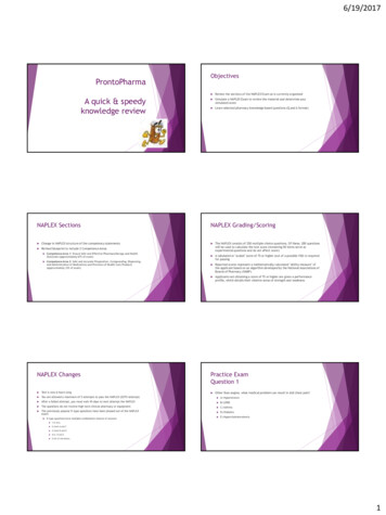
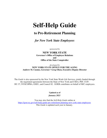
Immune Response(Primary vsSecondary)Fig. 1-6 Primary and secondaryimmune responses. The initialexposure to an antigen elicits theformation of IgM, followed by IgGantibodies and memory B cells.The second response to the sameantigen causes much greaterproduction of IgG antibodies andless IgM antibody secretion.
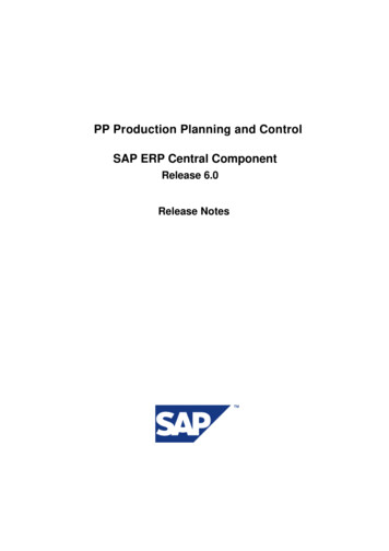
Immune Response (Macrophages)Fig. 1-4 Antibody attaches to the Fc receptor on a macrophage to signalclearance. The variable portion of the immunoglobulin attaches to the antigen onthe red cell, while the macrophage attaches to the Fc portion. The red cell istransported to the spleen and liver for clearance.
ImmunoglobulinsTABLE 1-2Comparisonof IgM andIgG
Immunoglobulin Structure
ComplementSerum proteins that assistwith the clearance ofantibody-coated red cells.Biologic functions include: Opsonization – enhancingphagocytosis of antigens Chemotaxis – attractingmacrophages and neutrophils Cell Lysis – rupturing membranesof foreign cells Agglutination – clustering andbinding of pathogens together(sticking)
Stages- Agglutination ReactionsTABLE 1-5 Factors AffectingAgglutination
PotentiatorsTABLE 2-14 Summary of AntibodyPotentiators
Direct and Indirect Antiglobulin TestComparison
Sources of Error in Antiglobulin Testing
Clinical Examples Causing a PositiveDirect Antiglobulin Test
Applications of Indirect Antiglobulin Testin the Immunohematology Laboratory
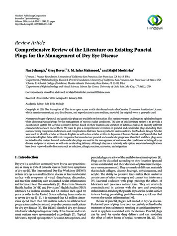
Antibody ScreeningFig. 7-1 Screening cell antigram.Methodology (Tube, Gel,Solid Phase)
AntibodyScreeningFig. 7-2 Screen interpretations.Tentative interpretations thatcan be made after testing ofthe antibody screen and directantiglobulin test. IS,Immediate-spin; 37 C, 37 Cincubation; AHG, antihumanglobulin; CC, check cells; ,check cells agglutinate; NT,not tested; Poly, polyspecificantiglobulin reagent; C3,anticomplement reagent.
Guidelines for Interpretation of a Panel
Antibody Panel
Multiple Antibodies
Select Cell Panel
Ficin Treated Panel
Strong Cold Autoantibody
Mini Cold Panel
Warm Autoantibody
Elution Technique
Special Procedures for Antibody IDNeutralization – good for Lewis (Lewis Substance); P1 antibodies (hydatid cystfluid), anti-Sda (fresh urine) Ch and Rg (plasma).Adsorptions:Autologous – can only be performed if patient has not been recentlytransfused or unless cell separation procedure is performed. If DAT is positive,antibody must be removed before procedure can be performed.Allogeneic – use R1R1, R2R2 and rr cells. Must be careful during interpretationas clinically significant antibodies can be excluded.Commercially available W.A.R.M and RESt kits are available.RBC Phenotyping – DAT must be negative. If DAT is positive and patient has notbeen recently transfused, Chlorquin Diphosphate can be used to remove IgG frompatient’s red cells. Recent transfusion requires cell separation procedure to beperformed prior to phenotyping.
Special Reagents to help in Antibody ID 2-ME (2-Mercaptoethanol) and DTT (Dithiothreitol) –cleaves disulfide bonds of IgM antibody molecules. Helpsdistinguish between IgM and IgG.AET (2-aminoethylisothiouronium bromide) – creates redcells that lack Kell antigensZZAP (DTT papain) –used to remove immunoglobulinsand complement from the surface of red blood cells,commonly when evaluating a potential autoantibody. ZZAPalso deactivates a multitude of red cell antigens on thered cell surface.
Prewarm Technique
Strategies for the weak antibody or one thatdoes not fit a pattern
Saline Replacement for Rouleaux
Antibody ID You Give it a Try!Group Activity
Crossmatching, Here we come!
Compatability ProcessPre-Analytical: Label theTube Correctly!
Comparison of immediate-spin, computer, andantiglobulin crossmatch re
Immunohematology Review: Help, What Do I Need to Know to Pass the ASCP Exam? Dr. Phyllis Ingham EdD. MEd. MT(ASCP) Program Director/Chair Clinical Laboratory Technology West Georgia Technical College Phyllis.ingham@westgatech.edu American Society for Clinical Laboratory Science Annual State Meeting – Georgia Chapter 2019. Take Off the Blindfold Today! Blood Group Systems.