Transcription
Hindawi Publishing CorporationJournal of OphthalmologyVolume 2016, Article ID 9312340, 22 pageshttp://dx.doi.org/10.1155/2016/9312340Review ArticleComprehensive Review of the Literature on Existing PunctalPlugs for the Management of Dry Eye DiseaseNaz Jehangir,1 Greg Bever,2 S. M. Jafar Mahmood,3 and Majid Moshirfar41Francis I. Proctor Foundation, University of California San Francisco, San Francisco, CA 94143, USADepartment of Ophthalmology, Francis I. Proctor Foundation, University of California San Francisco, San Francisco, CA 94143, USA3Charles E. Schmidt College of Medicine, Florida Atlantic University, Boca Raton, FL 33431, USA4Department of Ophthalmology and Visual Sciences, Moran Eye Center, University of Utah, Salt Lake City, UT 84132, USA2Correspondence should be addressed to Majid Moshirfar; cornea2020@me.comReceived 12 November 2015; Accepted 11 January 2016Academic Editor: Edit Tóth-MolnárCopyright 2016 Naz Jehangir et al. This is an open access article distributed under the Creative Commons Attribution License,which permits unrestricted use, distribution, and reproduction in any medium, provided the original work is properly cited.Numerous designs of punctal and canalicular plugs are available on the market. This variety presents challenges to ophthalmologistswhen choosing punctal plugs for the management of various ocular conditions. The aim of this literature review is to provide aclassification system for lacrimal occlusive devices based on their location and duration of action as well as to identify differentcharacteristics of each one of them. We want to give a comprehensive overview on punctal and canalicular plugs including theirmanufacturing companies, indications, and complications that have been reported in various articles. PubMed and Google Scholarwere used to identify articles written in English as well as few articles written in Japanese, Chinese, Slovak, and Spanish that hadabstracts in English. Nine different companies that manufacture punctal and canalicular plugs were identified and their plugs wereincluded in this review. Punctal and canalicular plugs are used in the management of various ocular conditions including dry eyedisease and punctal stenosis as well as in ocular drug delivery. Although they are a relatively safe option, associated complicationshave been reported in the literature such as infection, allergic reaction, extrusion, and migration.1. IntroductionDry eye is a condition commonly seen by eye care practitioners; as many as 25% of patients seen in clinic have symptomsof dry eye [1]. The International Dry Eye Workshop (DEWS)defines dry eye as a multifactorial disease of tears and ocularsurface with symptoms of visual disturbance, discomfort,and tear film instability with associated ocular inflammationand increased tear film osmolarity [2]. Data from Women’sHealth Studies (WHS) and Physicians’ Health Studies (PHS)estimates 3.2 million women and 1.6 million men aged 50years or older in the United States suffering from moderateto severe dry eye [3–5]. It is estimated that 8.5 million Americans spend more than 300 million dollars on artificial tearpreparations and other related over-the-counter medicationsfor dry eye disease [6]. The DEWS classified dry eye diseaseinto four levels depending on severity of the disease and treatment options were recommended accordingly [7]. Topicallubricants, topical cyclosporine (Restasis), tetracyclines, andpunctal plugs are a few of the available treatment options [8].Plugs can be classified according to their location (punctalversus canalicular) and their duration of placement (temporary versus permanent). They are made of different materialsthat include collagen, silicone, hydrogel, polydioxanone, andacrylic. The ability to preserve tears makes them useful incertain cases of refractive surgery and contact lens intolerance[9]. Lacrimal occlusion with plugs prolongs the effects oflubricants and preserves natural tears. They are relativelycontraindicated in patients with dry eyes and coexistinginflammation. Blocking the puncta exposes the ocular surfaceto tears having preexisting proinflammatory cytokines thatworsen the ocular inflammation [10].The use of punctal plugs is not limited to dry eye disease.Perforated punctal plugs have been successfully utilized in thetreatment of punctal stenosis resulting in significant improvement in epiphora associated with the stenosis. Punctal plugscan be used for ocular drug delivery and can modulatethe effect of other forms of topical treatment [11, 12]. This
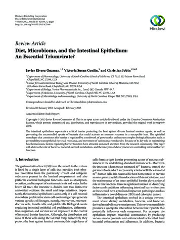
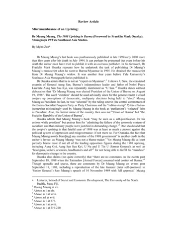
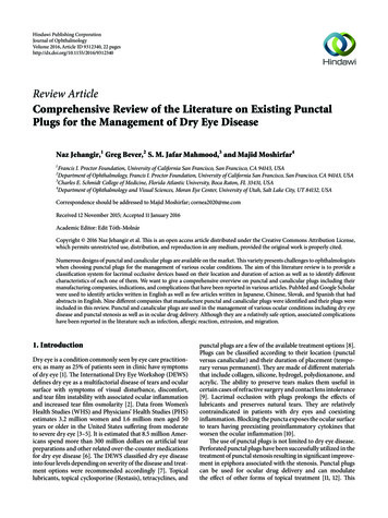
2can be utilized in the treatment of glaucoma by increasingthe drug retention time [9]. Both punctal and canalicularplugs have been associated with complications that havebeen reported in the literature. They can result in infectionssuch as canaliculitis, biofilm formation, extrusion, migration,epiphora, and chronic irritation [9].The purpose of this literature review is to give cliniciansan update on different types of punctal and canalicular plugs,with recent advancements in designs and techniques. Choosing the best suitable punctal/canalicular plug for treatmentof various ocular surface disorders (dry eye disease, punctalstenosis, epithelial erosions, and ocular drug delivery) maybe difficult for clinicians as a large variety of punctal andcanalicular plugs in different shapes, designs, and materialsare available. It is important for the clinicians to be familiarwith the complications that have been reported with differentlacrimal plugs and to evaluate patients for any preexistingocular or lid abnormalities. This paper provides a comprehensive overview of all the available punctal and canalicular plugsand can serve as a guide for clinicians to choose the mostsuitable lacrimal plug when treating the above-mentionedconditions.2. Materials and MethodsPubMed and Google Scholar were searched for studiespublished up to October 2015. Eligibility criteria includedstudies evaluating indications, contraindications, adverseeffects, shapes, designs, and characteristics of different punctal and canalicular plugs. Using Google, we searched fordifferent manufacturers of punctal and canalicular plugsand used pictures (after getting permission from respectivemanufacturers) and characteristic features of the plugs tocompile classification tables. We also evaluated differenttypes of punctal and canalicular plugs microscopically toevaluate their characteristics. Keywords included punctalplugs, dry eyes, punctal stenosis, silicone plugs, perforatedplugs, drug delivery, collagen plugs, SmartPlug, EagleFlex,Lacrimal Gland Occlusion, and intracanalicular plugs. Avariety of articles related to punctal plugs were included inthis review.2.1. Classification of Punctal and Canalicular Plugs. Lacrimalocclusive devices can be classified into punctal and canalicular plugs (Figure 1). Freeman, in 1975, developed the dumbbell shaped punctal plug made of silicone and this conceptof punctal plug is still in use [13]. Punctal plugs rest at thepunctal opening making them easily visible and, hence, areremovable without much difficulty. In contrast, canalicularplugs are not visible as they are placed inside the canaliculus(either the vertical or the horizontal canaliculus), makingextrusion unlikely but increasing the risk of migration anddifficulty in localizing their position without ultrasound [14].Occlusion of the lacrimal drainage system with temporaryor permanent plugs is a widely used nonpharmacologicaltherapy for conserving tears. A wide variety of lacrimal plugswith specific indications are in use. Both horizontal andvertical canalicular plugs can be further classified into temporary and permanent. Temporary short duration canalicularJournal of Ophthalmologyplugs (Figure 2(a)) are used before attempting extendedduration or permanent occlusion to assess risk of epiphoraand the probability of symptomatic relief [15]. Temporaryshort duration ones plugs are usually made of animal collagenand last for 4–14 days. Temporary extended duration plugs(Figure 2(b)) are used following refractive surgery, for dryeye disease and for ocular retention of medications [16].Temporary extended duration plugs can last from 2 to 6months [17]. They are made of different materials such asglycolic acid with trimethylene carbonate, E-CaprolactoneL-Lactide copolymer (PCL), and polydioxanone (PDS).2.2. Characteristics of Punctal Plugs. Different designs andshapes of plugs have been developed to increase their effectiveness and to minimize complications. Generally punctalplugs have a head on the top and are shaped like anumbrella. The head facilitates removal of the plug if necessary[26]. They usually have a slender neck and a cone-shapedthicker base. The majority of the punctal plugs are madeof silicone, but teflon, hydroxyethylmethacrylate (HEMA),and polymethylmethacrylate (PMMA) have been tested [9].We evaluated different punctal plugs under the microscopeand classified them based on their shapes (Table 1). Punctalplugs have different shaft designs (e.g., tapered shafts andstraight shafts) with pros and cons of different styles. The headportion can have reservoirs in some designs for increasedtrapping of tears. There are variations in the collarette suchas a slanted collarette, which improves the fit. Some designshave tractional ribs for greater flexibility while some havecollapsible noses that spring open once inside the puncta(Table 1). Perforated punctal plugs have a central lumen; theyare used in treating punctal stenosis and partial occlusionby allowing some tear flow through the plug [27]. Punctalplug manufacturers, sizes, and characteristics are discussedin Table 2.2.3. Characteristics of Canalicular Plugs. Canalicular plugsfor temporary use are usually rod-shaped and available indifferent sizes and colors depending on the punctal size.They are inserted into the canaliculus making it difficult tovisualize them or monitor their position. To achieve completeocclusion of the lacrimal drainage system, the diameter of theplug is more important than its length [9]. Special designsfor permanent use have been developed. The Form Fit plug(Figure 2(c)) is a vertical canalicular plug made of hydrogelthat expands into a soft gelatinous material after contact withthe tear film, filling and conforming to the shape of the vertical canaliculus [17]. Thermosensitive acrylic canalicular plugs(Medennium SmartPlug) have been in use since 2002. Theybecome shorter and thicker at body temperature. The plug hasa diameter of 0.4 mm and length of 9 mm before insertionthat change after insertion to a diameter of 1 mm and alength of 2 mm [28]. Newer materials are thought to reducebacterial adhesion and chances of infections [9]. Horizontalcanalicular plugs can be temporary or permanent (Figure 3).Herrick canalicular plugs, made of silicone, are placed in thehorizontal canaliculus and shaped like a golf tee. They do notrequire punctal dilation prior to insertion. Removal of thesehorizontal canalicular plugs may prove challenging, in some
Journal of Ophthalmology3Lacrimal occlusive devicesPunctalComplete occludersCanalicularPartial occludersHorizontal canaliculusVertical canaliculusSoft PlugTemporaryPlug 1SuperEagle PlugeFlex PlugEagleFlexPermanentTemporaryPermanentForm FitSoft Plug FlowControlSmartPlugHerrick plugEaglePlu TearFlowwEaglePlugePlugEaglePlugEagle FloFlow ControlLacriProiProParasol PunctalOccluderShort durationMicrofloMicroflowPerforated punctalplugSoft Oasis collagenOasis Extended DurationEagle collagenBeaver-VisitecExtended DurationFCI collagenUltraPlugextended durationBeaver-VisiteccollagenDuraPlugVisiPlugVera C7FCI prolongMedennium XsorbrbUltraPlug collagenVera90Snugg PlugReady-Setdy-SetShort durationTearSaverSaverExtended durationUltraPlugPlVeraPlugQuintess PunctaltOccluderLacrimedics collagenExtended durationFigure 1: Classification of lacrimal occlusive devices based on shape, location, and duration of action.(a)(b)(c)Figure 2: Vertical canalicular plugs. (a) Schematic of the temporary short duration plug made of collagen and effective for 4–14 days.(b) Representation of the temporary extended duration plug made of different materials (polydioxanone, glycolic acid and trimethylenecarbonate, and E-Caprolactone-L-Lactide copolymer) and effective for about 2–6 months depending on the manufacturer. (c) Schematicof the hydrogel (Form Fit) plug that expands with hydration to mold into canaliculus and be permanently effective (image C courtesy ofhttp://www.oasismedical.com).
4Journal of OphthalmologyTable 1: Shapes of punctal plugs. Several designs are made by different companies to increase the effectiveness of punctal plugs with differentshapes of the shafts, collapsible noses, reservoirs, and traction ribs.DesignAdvantagesNameEaglePlugTapered shaftDesigns extra force horizontally to keep plug inplace.TearSaverSuperEagle PlugCollapsible noseCollapsible hollow nose adheres the plug to theshape of ampulla and springs open once inside thepuncta.Parasol PunctalOccluderLacriProReservoired headIndentations trap tears and minimize foreignbody sensation.Quintess PunctalOccluderRibbed shaftGreater flexibility.EagleFlex PlugModel
Journal of Ophthalmology5Table 1: Continued.DesignAdvantagesNameSuperFlexSoft Plug FlowControlEagle Flow ControlPerforated shaftSlantedcollaretteperforated lumen.EaglePlugTearFlowMicroflowPerforated punctalplugSlanted lipResists migration and prevents rubout. Conformsto the natural anatomy of eyelid.Ready-SetModel
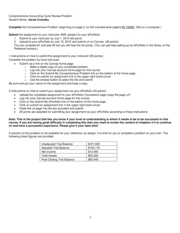
6Journal of OphthalmologyTable 1: Continued.DesignAdvantagesNameDual lobe tipFits a range of punctal sizes.Plug 1Stretched shaftReturns to natural shape after insertion.ModelSnug PlugVeraPlugStraight shaftParasol PunctalOccluderLow profile dome and easyinsertion.Soft PlugUltraPlug(a)(b)(c)Figure 3: Horizontal canalicular plugs. (a) Collagen plug (Lacrimedics) that is meant to last for about two weeks. (b) Temporary extendedduration plug (VisiPlug by Lacrimedics) made of polydioxanone does not swell with moisture and lasts about 6 months. (c) Permanentcanalicular plug (Herrick plug by Lacrimedics) is shaped like a golf tee (photos courtesy of http://www.lacrimedics.com).
SuperFlexPlugSuperEaglePlugPlug 1Soft PlugName11 sizes (combinationswith 0.3–1.3 mmdiameter, 1.1–2.0 mmlength)Small (0.4–0.6 mm)Medium (0.6–0.8 mm)Large ( 0.8 mm)Values puncta sizeSiliconeLow durometer siliconeSiliconeMedical grade silicone5 sizes (0.4–0.8 mm)(micro, mini, petite,small, and medium)1 size(fits 0.5–0.8 mm puncta)MaterialSizeBetter fit with good retention. Easyinsertion.Low durometer silicone with lowprofile rim for more comfort.Tapered shape and wide flexed nosedesign with a goal of good retention.Dual lobed design to fit a range ofpunctal sizes.Pointed nose simplifies insertion;large anchor secures plug.CharacteristicsTable 2: Punctal plugs [18–25] (types of punctal plugs, manufacturers, and characteristics discussed in facturerJournal of Ophthalmology7
Snug PlugParasolPunctal OccluderLacriPro PunctumEagleFlex PlugEaglePlugName1 size fits allX-small (0.25–0.35 mm)Small (0.35–0.65 mm)Medium (0.6–0.85 mm)Large ( 0.9 mm)Values puncta size4 sizes (0.3, 0.5, 0.7, and0.9 mm)(X-small, small,medium, and large)SiliconeSiliconeMedical grade siliconeSiliconeSilicone5 sizes (0.4–0.8 mm)6 sizes (0.4–0.9 mm)MaterialTable 2: Continued.SizePreloaded in a stretched positionand returns to natural shape afterinsertion (Figure 4).Hollow nose. Umbrella-like headwhich squeezes as it is inserted andsprings open once in place. No needfor punctal dilation.Reservoir indentations trap tearsand decrease foreign bodysensation. The number ofindentations corresponds to the sizeof the sionTapered shaft exerts horizontalforce to keep plug in place. Easyinsertion and removal.Tapered shaft with traction ribs forbetter retention and flexibility.External ribs give 30% more surfacearea. Thin rim decreases cornealcontact.ManufacturerCharacteristics8Journal of Ophthalmology
Quintess Ready-SetNameSiliconeSiliconeSmall (0.4–0.6 mm)Medium (0.6–0.7 mm)Large (0.7–0.8 mm)X-Large (0.8–1.0 mm)Values puncta size4 sizes (0.3, 0.5, 0.7,0.9 mm)(X-small, small,medium, and large)Silicone5 sizes (0.4–0.8 mm)SiliconeSilicone7 sizes (0.4–1.0 mm)(slim mini, slim petite,micro, mini, small,medium, and large)5 sizes (0.4–0.8 mm)MaterialSizeTable 2: Continued.Reservoir indentations are designedto decrease foreign body sensation.Indentations correspond to plugsize (Figure 5).Low profile dome for more comfort.Proprietary shaft design for easyinsertion and good fit (Figure 5).Straight shaft. Low profile capdesign minimizes foreign bodysensation.Tapered shaft exerts extra horizontalforce to keep plug in place.Slanted lip in sizes 0.6 mm andlarger makes it a better fit.Conforms to natural anatomy of theeyelid. Sizes 0.6 mm and larger alsohave slightly larger bulbs to resistmigration and prevent cialtiesCorporationFCIFCIManufacturerJournal of Ophthalmology9
MicroflowEaglePlugTearFlowEagle FlowControllerSoft Plug FlowControlNameSmall (0.4–0.55 mm)Medium (0.55–0.7 mm)Large (0.7–0.85 mm)Values punctal size6 sizes:0.5 mm plug, 0.2 mmlumen0.6 mm plug, 0.3 mmlumen0.7 mm plug, 0.3 mmlumen0.8 mm plug, 0.3 mmlumen0.9 mm plug, 0.4 mmlumen1.0 mm plug, 0.5 mmlumenSiliconeSiliconeSiliconeSoft silicone3 sizes (0.6–0.8 mm)4 sizes (0.5–0.8 mm)MaterialTable 2: Continued.SizePartial occluder. Patent lumenallows for use in punctal stenosis.Partial occluder. Lumen size is 150%larger than Eagle Flow Control plugallowing for increased tear flow.Tapered shaft gives more controlledretention. Patent lumen allows foruse in punctal stenosis.Tapered shaft. Partial occluder.Patent lumen allows for use inpunctal stenosis. Tapered shaftincreases vector force to keep theplug in position.Partial occluder. Opening throughnose portion for partial occlusion.Low profile dome head. Softer,flexible silicone makes it morecomfortable. Pointed nose secures itfirmly. Patent lumen allows for usein punctal gleVisionOasisManufacturer10Journal of Ophthalmology
Perforatedpunctal plugNameMaterialSilicone coated with PVP(polyvinylpyrrolidone)2 sizes (0.7, 0.9 mm)(mini, medium)Table 2: Continued.SizePartial occluder. Slanted collarette.PVP (polyvinylpyrrolidone) ishydrophobic and allows tear to flowsmoothly through the perforation.Used in treating punctal stenosis.CharacteristicsFCIManufacturerJournal of Ophthalmology11
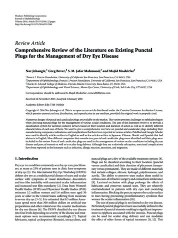
12Journal of Ophthalmology(a)(b)Figure 4: Snug Plug (FCI) is a punctal plug that is preloaded in a stretched position (a) compared to its natural shape (b) after release frominserter. The plug is on stretch for insertion with the goal of eliminating the step of punctal dilation prior to insertion. The widened bulb atthe plug’s base in the natural position acts to prevent the plug from falling out spontaneously. Each dash in scale represents 1 mm.(a)(b)Figure 5: Punctal plug without reservoirs (VeraPlug) on (a) compared with punctal plug with reservoirs (Quintess Punctal Occluder) on (b).The Quintess Punctal Occluder was designed to have reservoir indentations in the collar to trap tears.cases, due to their intracanalicular location [29]. The Herrickplug is partially radiopaque and dyed blue in color to makelocalization possible with transillumination [9]. Horizontaland vertical canalicular plugs are discussed in Tables 3 and 4.2.4. Indications of Punctal and Canalicular Plugs(i) Dry Eye Disease. Occlusion of the lacrimal drainage systemwith plugs is considered an option in patients with moderatedry eye syndrome. An article by the American Academyof Ophthalmology reviewed literature to assess efficacy andsafety of punctal and canalicular plugs for treatment ofdry eye disease. The use of lacr
Review Article Comprehensive Review of the Literature on Existing Punctal Plugs for the Management of Dry Eye Disease NazJehangir, 1 GregBever, 2 S.M.JafarMahmood, 3 andMajidMoshirfar 4 Francis I. Proctor Foundation, University of California San Francisco, San Francisco, CA , USACited by: 20Publish Year: 2016Author: Naz Jehangir, Greg Bever, S.