Transcription
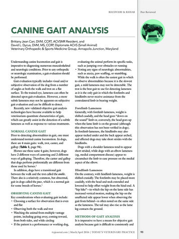
RECOVERY & REHABPeer ReviewedCANINE GAIT ANALYSISBrittany Jean Carr, DVM, CCRT, ACVSMR Resident, andDavid L. Dycus, DVM, MS, CCRP, Diplomate ACVS (Small Animal)Veterinary Orthopedic & Sports Medicine Group, Annapolis Junction, MarylandUnderstanding canine locomotion and gait isimperative to diagnosing numerous musculoskeletaland neurologic conditions. Prior to any orthopedicor neurologic examination, a gait evaluation shouldbe performed.Gait evaluation typically includes visual and/orsubjective observation of the dog from a numberof angles at both the walk and trot on a flatsurface. To the trained eye, lameness can often bedetected upon gait evaluation. However, a moresubtle lameness may not be apparent on subjectivegait evaluation and can be difficult to detect.Recently, new validated objective gait analysistechnologies have become available to helpveterinarians quantitate characteristics of gait,which can greatly assist in the detection of a subtlelameness as well as response to various treatments.NORMAL CANINE GAITPrior to detecting abnormalities in gait, one mustunderstand normal canine locomotion. In dogs,there are 4 main gaits: walk, trot, canter, andgallop (Table 1, page 94).Horses use these same 4 gaits; however, dogshave 2 different ways of cantering and 2 differentways of galloping. Therefore, the canter and gallopthat dogs perform preferentially are different fromthose used by horses.1In addition, dogs have a transitional gaitbetween the walk and the trot called the amble.There also is a relatively common, but abnormal,gait in dogs called the pace, which is a normal gaitfor some breeds of horses.1OBSERVING CANINE GAITKey considerations when evaluating gait include: Choosing a surface for observation that is evenand flat Observing both the walk and trot Watching the animal from multiple vantagepoints, including going away, coming toward,from both sides, and while circling If the patient is a performance or working dog,evaluating the animal perform its specific tasks,such as jumping over obstacles or running Noting any signs of neurologic abnormalities,such as ataxia, paw scuffing, or stumbling.While the walk is often the easiest gait in whichto observe abnormalities because it is the slowestgait, a mild lameness may not be detectable. Thetrot is the best gait to use for detecting lamenessas it is the only gait in which the forelimbs andhindlimbs never receive assistance from thecontralateral limb in bearing weight.Forelimb LamenessGenerally, with forelimb lameness, weight isshifted caudally, and the head goes “down onthe sound” limb or, conversely, the head goes upwhen the lame limb is on the ground (althoughthis observation has not been verified in dogs).In forelimb lameness, the hindlimbs may alsoappear tucked under and the back appear arched,and affected dogs may take short strides with thehindlimbs.Dogs with a shoulder lameness tend to appearshort strided, while dogs with an elbow lameness(eg, medial compartment disease) appear tocircumduct the limb to ease pressure on the medialaspect of the elbow.Hindlimb LamenessOn the contrary, with hindlimb lameness, weight isshifted cranially. The forelimbs may be placed morecaudally, with the head and neck extended andlowered to help offset weight from the hind end. A“hip hike”—in which the hip on the lame side hasincreased vertical motion, making the hip on theunaffected side appear lower when observing thegait from behind—is often noted on the same sideas the lameness. The tail may also rise as the lameleg contacts the ground.METHODS OF GAIT ANALYSISIt is imperative to have a means for objective gaitanalysis because gait is difficult to consistently andtvpjournal.com March/April 2016 TODAY’S VETERINARY PRACTICE93
Peer ReviewedRECOVERY & REHABTABLE 1.Summary of Canine GaitsGAITFORWARD MOVEMENTEXAMPLECOMMENTSWalk1. One rear foot2. Ipsilateral front foot3. Other rear foot4. Front foot on that side1. RR2. RF3. LR4. LF 2 or 3 feet on the ground at any given time Only canine gait in which there are ever 3 feet on1. One rear foot2. Quickly followed by ipsilateral front foot3. Other rear foot4. Quickly followed by front foot on that side1. RR2. RF3. LR4. LF A faster form of the walk but not preferred gait Moments when 3 feet are on the ground ob-AmblePaceRotaryCanterto side; rear feet are not lifted very high, oftenscuffling along the ground; wasted horizontalenergy and less pleasing to the eye Should only be used for short periods: transitioning from walk to trot or resting trotting muscles1. Ipsilateral legs move forward together1. RF–RRwhile other 2 legs bear weight2. No ground2. Moment when body is suspended in the aircontact3. Other 2 legs move forward together3. LF–LR Abnormal gait for all dog breeds Very inefficient gait: Center of gravity keepsshifting from side to side; energy expended tokeep recentering weight Cannot respond quickly when a change in speedis required Wide range of speeds for movement unavailableunless dog slows to amble or speeds up to trot1. Two diagonal front and rear feet1. RF–LR2. Moment when body is suspended in the air 2. No ground3. Other diagonal front and rear feetcontact3. LF–RR Most efficient gait Often gait of choice for gait evaluation: The front1. One rear leg2. Both legs on other side of body together3. Other front leg Predominant canter in dogs (90%) Allows very sharp turns with greater drive from1. RR2. LR–LF3. RF1. One rear foot2. Other rear foot and diagonal front foottogether3. Other front footand rear leg must support the body without helpfrom opposite legthe rear Referred to as cross cantering in horsesDifferent lead legs in front and rearTransverseCanterserved on close inspection or slow motion video Very inefficient gait: Rear end sways from sideBegins to appear as if ipsilateral feet aremoving forward togetherOnly 2 feet on ground at any given timeTrotthe ground1. RR2. LR–RF3. LF Less efficient for dogs; only used in about 10% ofcantering Used predominantly by horsesSame side leads in the front and rearRotaryGallop1. Spine flexes, with both rear feet on ground; 1. RR–LR2. LF–RFlead foot slightly ahead of the other2. Spine extends, stretching front feet3. RR–LRforward, which hit ground at same time,one slightly ahead of the other, with leadfront on opposite side of lead rear3. Spine flexes to bring rear feet forward Used by dogs preferentiallyDifferent lead legs in front and rearTransverseGallop1. Spine flexes, with both rear feet on ground; 1. RR–LRlead foot slightly ahead of the other2. RF–LF2. Spine extends, stretching front feet3. RR–LRforward, which hit ground at same time,one slightly ahead of the other, with leadfront on same side as lead rear3. Spine flexes to bring rear feet forwardSame side leads in the front and rear Very uncommon dog gait Gallop used by horsesLF left front; LR left rear; RF right front; RR right rear94TODAY’S VETERINARY PRACTICE March/April 2016 tvpjournal.com
RECOVERY & REHABPeer ReviewedTABLE 2.Methods for Canine Gait AnalysisSubjective: Visual observation of gait (eg, numericalrating scale, visual analog scale)Objective: Kinematic gait analysis Kinetic gait analysis (force plate analysis) Temporospatial gait analysis (pressuresensing walkways)reliably assess subjectively. Objective analysis isespecially important when developing treatmentplans and monitoring patient progress. Numerousmethods for gait analysis have been developed(Table 2).Visual Observation of GaitA systematic and disciplined approach must beused to clinically evaluate a patient’s gait. Todocument this clinical evaluation in the medicalrecord, findings are often semiquantified using anumerical rating scale (Table 3) or visual analogscale (Figure 1).Both types of scales were developed to providea systematic approach to visual observation ofgait. However, it is important to realize that,while visual or subjective gait analysis is oftenhelpful in identifying lameness, the gold standardfor quantifying lameness is quantification ofgait characteristics with a form of objective gaitanalysis, such as force plate analysis.Evans and colleagues compared visual observationof gait to force plate analysis.2 This study evaluated148 Labrador retrievers—131 that were 6 monthspost surgery for unilateral cranial cruciate ligamentinjury and 17 that were free of orthopedic disease.FIGURE 1. Example of the visual analog scale (VAS): The animal is gradedon a 10-cm line, with one end of the line representing “sound” and theother end representing “non–weight-bearing.” An “X” is placed alongthe scale, noting the degree of lameness, and then the VAS can beplaced into the patient’s record. Either the veterinarian or a trained staffmember typically completes the VAS. Previous VASs can be compared todetermine if there is improvement, decline, or no change.The observer only identified 11% of the 131 dogsthat were 6 months post surgery as being abnormalcompared with force plate analysis, which revealedthat 75% of the 131 dogs failed to achieve groundreaction forces consistent with sound Labradorretrievers.While force plate analysis has been shown to besuperior to visual observation, visual observationis still a practical tool in clinical practice, and itsimportance should not be discounted.Kinematic Gait AnalysisKinematic gait analysis quantifies the positions,velocities, acceleration/deceleration, and anglesTABLE 3.Example of a Numerical Rating Scale for Visual Assessment of GaitLAMENESS GRADEDESCRIPTIONGrade 1Sound at the walk, but weight shifting and mild lameness noted at trotGrade 2Mild weight-bearing lameness noted with the trained eyeGrade 3Weight-bearing lameness, typically with distinct “head bob”Grade 4Significant weight-bearing lamenessGrade 5Toe-touching lamenessGrade 6Non–weight-bearing lamenessFor moreinformation,visit tvpjournal.comand select ClinicalResources todownload thenumerical ratingscale and visualanalog scaledescribed inthis article. Thescales have beenkindly providedby VeterinaryOrthopedic &Sports MedicineGroup.Note: Grades 2 through 6 lameness can be observed at the walk or trot.tvpjournal.com March/April 2016 TODAY’S VETERINARY PRACTICE95
Peer ReviewedRECOVERY & REHABFIGURE 2. Example of how reflective markers would be placed for apatient undergoing kinematic gait analysis.FIGURE 4. Diagram showing the forcesmeasured during force plate analysis: Theforces in the Z direction are generated by avertical compression, which results in largerforces than those in the X or Y direction.Because of this, most veterinary publicationsderive and use the peak vertical force from theZ direction. Courtesy en.wiktionary.orgTABLE 4.Types of Ground Reaction Forces FIGURE 3. A force plate walkway; note the graysquare in the center of the mat—this is theforce plate that the dog must step on duringthe analysis.of various anatomic structures in space. Mostkinematic gait analysis systems use colored,retroreflective, or light-emitting diode (LED)markers that identify specific anatomic landmarks(Figure 2).A few of the most common locations formarkers include the dorsal scapular spine,acromion/greater tubercle, lateral humeralepicondyle, ulnar styloid process, iliac crest,femoral greater trochanter, femorotibial joint,lateral malleolus of the distal tibia, and spinousprocess at T13. Typically, markers are attached byshaving and cleaning the skin with alcohol; thenpressing the marker’s adhesive back directly to96Peak vertical forceVertical impulseRising and falling slopeBraking forceBraking impulsePropulsive forcePropulsive impulseMediolateral forcethe skin. The marker can be further secured withtape, if needed.When the dog is in gait, the movement of themarkers is tracked by a series of cameras. Thelocations of the markers over time are then usedto create a 2- or 3-D model of the dog’s gait withcalculations of bone and joint excursions.Kinematic parameters include displacements,angular velocities, and range of motion3: Displacement is the distance recorded when amarker changes position. Angular velocity is the speed at which this changeoccurs. Range of motion is calculated from thedisplacement at a specific joint.While a multitude of information can begathered from this form of gait analysis, onemajor limitation is the variation of structuresbetween breeds, as well as within breeds.3Further limitations include the potential for skinTODAY’S VETERINARY PRACTICE March/April 2016 tvpjournal.com
RECOVERY & REHABmovement, and accuracy and repeatability ofmarker placement.Kinetic Gait AnalysisKinetic gait analysis measures the ground reactionforces that are the result of an individual’s step.The most commonly used method for kineticgait analysis is force plate analysis, in which metalplates are mounted on the floor or walkway(Figure 3) to measure ground reaction forces(Table 4 and Figure 4). The forces are measuredin 3 dimensions: vertical, craniocaudal, andmediolateral.These forces are often presented graphically, withthe peak forces as the maximum forces generatedin the described phase of gait, represented by theforce–time curve. The impulse is then representedas the area under the force–time curve. Peak vertical force (PVF) is the single largestforce during the stance phase and represents onlya single data point on the force–time curve. Vertical impulse (VI) can be derived bycalculating the area under the vertical force curveusing time. PVF and VI are the two most commonly usedindices to detect lameness3-5 and, in general, adog with lameness has a lower PVF and VI inthat limb.While braking, propulsion, and mediolateralforces may be useful in evaluating mechanismsof locomotion, they are not commonly used fordiagnostic purposes or to assess outcome.4Force plate measurements have been themost widely used and validated quantitative gaitapplication in veterinary medicine to date.3 Thus,force plate analysis is considered the optimumapproach to quantification of gait characteristics byobjective gait analysis.However, there are disadvantages to force plateanalysis. Limitations include:Peer ReviewedTips for Obtaining an Acceptable Recording on a PressureSensing Walkway Walk or trot the dog down the mat at a consistent velocity or steadystate gait. Keep dogs on a loose leash, their heads looking forward in thedirection of travel, and moving down the center of the mat. One recent study has shown that the side the handler is on may affectgait analysis, particularly when evaluating the forelimbs; thus, it isrecommended to perform multiple passes with the handler switchingsides between passes.12 Place the mat in a quiet location where dogs won’t become easilydistracted. Start the pass 2 meters before the mat to encourage the dog to bestraight and maintain a steady gait prior to stepping on the mat. If the dog has been trained for competitions, such as agility orobedience, it is most helpful to have the owner run the dog on themat; otherwise, have the owner leave the room and have technical staffrun the dog over the mat. Practice with the dog before recording to acclimate the dog to the matand surroundings. Do not look at the dog while it is gaiting; this almost invariably makesthe dog look at the owner or handler, although it might take three tofour passes for this to work. If there is uncertainty regarding whether or not the pass down themat was acceptable, it is often possible to review the video footagecaptured by the software’s camera. Inability to measure stride or step length Need for consistent velocity, long dedicatedwalkway, and multiple trials Difficulty in setting up, breaking down, andmoving Complexity of software and data analysis Cost and impracticality for clinical practice.Temporospatial Gait Analysis with PressureSensing WalkwaysPressure sensing walkways have been validated toanalyze normal and abnormal gaits in dogs.6-13Having this information aids in diagnosingorthopedic, muscular, and neurologic disordersTABLE 5.Description of Measurements Calculated in Temporospatial Gait AnalysisTERMDEFINITIONStance timeStance phase of gait cycle and time paw is in contact with groundSwing timeSwing phase of gait cycle and time paw is in airStride lengthDistance from one footfall to the next footfall of same limbStep lengthDistance between heel point of one foot to heel point of contralateral footTotal pressure index Sum of peak pressure values recorded from each activated sensor by a pawduring mat contact; related but not equal to peak vertical forcetvpjournal.com March/April 2016 TODAY’S VETERINARY PRACTICE97
Peer ReviewedRECOVERY & REHABFIGURE 5. Results from temporospatial gait analysis using a pressure sensing walkway in a patientwith a left cranial cruciate ligament rupture. Dogs carry about 60% of body weight on theirforelimbs and 40% of body weight on their hindlimbs. Thus, the total pressure index percentageshould be 30% in each forelimb and 20% in each hindlimb. This patient is placing 17% on the lefthindlimb and 21% on the right hindlimb.that affect gait. These measures provide novelinformation about temporal and spatial gaitcharacteristics (Figure 5).The portable pressure walkway system wasoriginally developed for use in the human medicalfield9,14,15 and has since been adapted and validatedfor use in veterinary medicine. Previous studieshave established a protocol for temporal–spatialanalysis and determined reference values andsymmetry ratios for various breeds.6-11Temporospatial gait analysis uses a pressuresensing mat (Figure 6, page 100) and computersoftware system to calculate velocity, stance time,swing time, stride length, step length, and totalpressure index (Table 5, page 97).Forces exerted to change speed, changedirection, or maintain balance can interfere andcomplicate measurement interpretation.12,16 Thepressure sensing walkway does not measure forcedirectly but does measure the influence of theseforces. Therefore, by limiting excess externalinfluences, the measurements may be more98representative of the dog’s true gait.As with any other gait analysis system, there areadvantages and disadvantages to using a pressuresensing walkway. Advantages include: No size restrictions Multiple readings from a single pass Determination of stride and step length Information on limb placement User-friendly software Portability.However, just as with every other system, thereare disadvantages, including: Ability to only measure total ground reactionforces Inability to separate the 3 dimensions as withforce plate analysis Cost.IN SUMMARYWith more dogs participating in activities withtheir owners, canine sports, or a working role,it is essential for owners and veterinarians toTODAY’S VETERINARY PRACTICE March/April 2016 tvpjournal.com
Peer ReviewedRECOVERY & REHABReferencesFIGURE 6. A temporospatial gait analysissystem; note the series of sensors on the rightside of the mat that are used to record eachfoot strike to calculate velocity, stance time,swing time, stride length, step length, and totalpressure index. The sensors are connected to acomputer (not pictured) with software to allowthe gait to be analyzed.understand canine gait. Early signs of lamenessmay be as subtle as a shortened stride or shorterstance time on the injured leg. Both subjective andobjective gait analyses are important to not onlyestablish a diagnosis but also monitor progressionof treatment.LED light-emitting diode; PVF peak verticalforce; VAS visual analog scale; VI verticalimpulseBRITTANY JEAN CARRBrittany Jean Carr, DVM, CCRT, is currently a rehabilitation therapist andAmerican College of Veterinary SportsMedicine and Rehabilitation resident atVeterinary Orthopedic & Sports Medicine Group (VOSM) in Annapolis Junction, Maryland. Dr. Carr earned her DVMfrom Virginia–Maryland College of Veterinary Medicine (Virginia Institute of Technology). She completed a small animalrotating internship at the Animal Specialty Group in Los Angeles, California, anda surgical internship at VOSM.1001. Zink MC. Locomotion and athletic performance. InZink MC, Van Dyke JB (eds): Canine Sports Medicine andRehabilitation. Ames, IA: Wiley-Blackwell, 2013, pp 19-31.2. Evans R, Horstman C, Conzemius M. Accuracy andoptimization of force platform gait analysis in Labradors withcranial cruciate disease evaluated at a walking gait. Vet Surg2005; 34(5):445-449.3. Gordon-Evans WJ. Gait analysis. In Tobias KM, Johnston SA(eds): Veterinary Surgery: Small Animal. St. Louis: Elsevier,2012, pp 1190-1196.4. Gillette RL, Angle TC. Recent developments in caninelocomotor analysis: A review. Vet J 2008; 178:165-176.5. Nunamaker DM, Blauner PD. Normal and abnormal gait. InNewton CD, Nunamaker DM (eds): Textbook of Small AnimalOrthopaedics. Philadelphia: JB Lippincott, 1985, pp 1084-1085.6. Light VA, Steiss JE, Montgomery RD, et al. Temporalspatial gait analysis by the use of a portable walkway systemin healthy Labrador retrievers at a walk. Am J Vet Res 2010;71(9):997-1002.7. Besancon MF, Conzemius MG, Derrick TR, Ritter MJ.Comparison of vertical forces in normal greyhounds betweenforce platform and pressure walkway measurement systems.Vet Comp Orthop Traumatol 2003; 16(3):153-157.8. Lascelles BD, Roe SC, Smith E, et al. Evaluation of a pressurewalkway system for measurement of vertical limb forces inclinically normal dogs. Am J Vet Res 2006; 67:277-282.9. Webster KE, Wittwer JE, Feller JA. Validity of the GAITRitewalkway system for the measurement of averaged and individual step parameters of gait. Gait Posture 2005; 22:317-321.10. Gordon-Evans WJ, Evans RB, Conzemius MG. Accuracy ofspatiotemporal variables in gait analysis of neurologic dogs. JNeurotrauma 2009; 26:1055-1060.11. LeQuang T, Maitre P, Roger T, Viguier E. Is a pressurewalkway system able to highlight a lameness in dog? J AnimVet Adv 2009; 8:1936-1944.12. Keebaugh AE, Redman-Bentley D, Griffon DJ. Influenceof leash side and handlers on pressure mat analysis ofgait characteristics in small breed dogs. JAVMA 2015;246(11):1215-1221.13. Carr BJ, Canapp SO Jr, Zink MC. Quantitative comparisonof the walk and trot of border collies and Labrador retrievers,breeds with different performance requirements. PLoSONE 2015; 10(12): e0145396. doi:10.1371/journal.pone.0145396.14. McDonough AL, Batavia M, Chen FC, et al. The validity andreliability of the GAITRite system’s measurements: A preliminary evaluation. Arch Phys Med Rehabil 2001; 82:419-425.15. Bilney B, Morris M, Webster K. Concurrent related validity ofthe GAITRite walkway system for quantification of the spatialand temporal parameters of gait. Gait Posture 2003; 17:68-74.16. Voss K, Galeandro L, Wiestner T, et al. Relationships of bodyweight, body size, subject velocity, and vertical ground reaction forces in trotting dogs. Vet Surg 2010; 39(7):863-869.DAVID L. DYCUSDavid L. Dycus, DVM, MS, CCRP, DiplomateACVS (Small Animal), is a staff orthopedicsurgeon at Veterinary Orthopedic & SportsMedicine Group (VOSM) in Annapolis Junction, Maryland. He has presented at nationalmeetings, lectured second- through fourthyear veterinary students, and published anarray of research articles and a book chapter.Dr. Dycus received his DVM from MississippiState University, completed a rotating internship at Auburn University, and earned his MSand completed a small animal surgical residency at Mississippi State University.TODAY’S VETERINARY PRACTICE March/April 2016 tvpjournal.com
In dogs, there are 4 main gaits: walk, trot, canter, and gallop (Table 1, page 94). Horses use these same 4 gaits; however, dogs have 2 different ways of cantering and 2 different ways of galloping. Therefore, the canter and gallop that dogs perform preferentially are different from those used by horses.1 In addition, dogs have a transitional gait