Transcription
Ekere et al. BMC Infectious Diseases(2020) RCH ARTICLEOpen AccessCysteine-cysteine chemokine receptor 5(CCR5) profile of HIV-infected subjectsattending University of Calabar TeachingHospital, Calabar, Southern NigeriaEkerette Friday Ekere1, Monday F. Useh2, Henshaw Uchechi Okoroiwu3*and Tatfeng Youtchou Mirabeau4AbstractBackground: Cysteine-cysteine chemokine receptor 5 is the main HIV co-receptor involved in the virus and cell-tocell spread. A variant of the CCR5 gene known as CCR5-Δ32 which is a product of 32 base pair deletion in thegene plays critical role in the infection and progression to AIDS. The study was carried out to determine the CCR5genotype of HIV-infected subjects attending University of Calabar Teaching Hospital, Calabar.Methods: A total of 100 subjects attending HIV clinic, University of Calabar Teaching Hospital were purposivelyrecruited for this study. DNA was extracted from each sample using the Quick gDNA miniprep DNA extraction kit,Zymo Research. Polymerase chain reaction (PCR) was used in the amplification of CCR5 gene in each DNA in a9700 ABI Thermo cycler and then resolved on 4% agarose gel electrophoresis.Result: Out of the 100 samples assessed, 100 (100%) were homozygous for the CCR5 wild type gene (CCR5-wt),while none (0%) was homozygous for the CCR5-Δ32 (mutant type), and heterozygosity was not observed.Conclusion: This study observed absence of CCR5-Δ32 deletion gene among the studied subjects in Calabar,implying lack of genetic advantage in HIV infection and possible rapid progression towards AIDS if otherprecautions are not checked.Keywords: CCR5, HIV, CCR5-Δ32, CCR5 genotype, Mutant CCR5, Wild type CCR5BackgroundHuman immunodeficiency virus (HIV)/ Acquired immunodeficiency syndrome (AIDS) despite the campaigns has remained a public health concern [1] withapproximately 36.7 million people living with HIVglobally at the end of 2016. The burden of the epidemic varies between regions and countries with subSaharan Africa being the most affected having nearly1 in every 25 adult living with HIV accounting fortwo-third of people living with HIV globally [2–4].According to the 2014 Gap report, 9% of those livingwith HIV reside in Nigeria [5]. With 3.2 million (9%)people living with HIV in Nigeria as of 2016, she has* Correspondence: okoroiwuhenshaw@gmail.com3Hematology Unit, Department of Medical Laboratory Science, University ofCalabar, Calabar, NigeriaFull list of author information is available at the end of the articleranked the second largest HIV global disease burdenbehind South Africa that housed 7.1 million (19%)people living with HIV [6, 7]. Infection with HIV isassociated with progressive loss of cellular immunitywhich results to life threatening opportunistic infections and progressive development of acquired immunodeficiency syndrome [8].The major event in HIV infection is the continuousdecrease in CD4 T cells that leads to deficient immune system that is unable to withstand the actionsof opportunistic pathogens. The more interesting partis the significant disparity of individuals in the susceptibility to the infection, the time lag for depletionof CD4 T – lymphocytes, as well as time lag to progress to AIDS-defining state [9–11]. Host genes collectively known as AIDS restriction genes (ARGs); CC type chemokine receptor 5 (CCR5) and C-X-C The Author(s). 2020 Open Access This article is distributed under the terms of the Creative Commons Attribution 4.0International License (http://creativecommons.org/licenses/by/4.0/), which permits unrestricted use, distribution, andreproduction in any medium, provided you give appropriate credit to the original author(s) and the source, provide a link tothe Creative Commons license, and indicate if changes were made. The Creative Commons Public Domain Dedication o/1.0/) applies to the data made available in this article, unless otherwise stated.
Ekere et al. BMC Infectious Diseases(2020) 20:5Page 2 of 9Fig. 1 Map of study areachemokine receptor type 4 (CXCR4) have beenknown to modify individual response to HIV-1 exposure, infection and pathogenesis markedly [12–14].The human C-C type chemokine receptor (CCR5)belongs to the G-protein-coupled receptor family andis found mostly on the cell surface of some whiteblood cells such as T4 cells, monocytes and macrophages [15, 16]. The CCR5 wild type protein (CCR5wt) is made up of 352 amino acids folded into 7membrane-spanning domains linked by 3 intracellularloops. Aside the wild type, there is CCR5 allele witha 32 base pair deletion (CCR5-Δ32) in the coding region. This mutation results in a truncated proteinthat cannot be detected on the cell surface [14, 15].The CCR5-Δ32 mutation has been shown to significantly affect the entry of the HIV and also theprogression to AIDS [14]. Individuals who are homozygous for this defect (polymorphism) appear to beresistant to HIV-1 infection [12, 14] and same retardsTable 1 Virological and clinical characteristics of the studiedsubjectsParameterCD4 categoryFrequency (%)TotalABCD4 500 cells/μL45 (45)43 (95.6)2 (4.4)0 (0.0)CD4 200–499 cells/μL41 (41)29 (70.7)9 (22.0)3 (7.3)CD4 200 cells/μL14 (14)4 (28.6)7 (50.0)3 (21.4)ART intakeOn ART100 (100)ART Naïve0 (100)GenderMale35 (35)Female65 (65)Mode of acquisitionVertical0 (0)0 (0)Blood transfusion0 (0)Sexual0 (0)Not sure100 (100)C
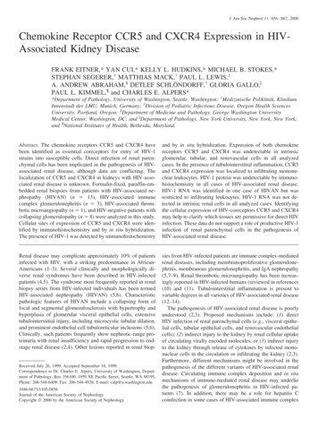
Ekere et al. BMC Infectious Diseases(2020) 20:5Page 3 of 9disease progression in heterozygotes [17]. HIV gainsentry into the host’s cell through its glycoprotein 120receptor (gp120) which binds to the hosts cell receptor cluster of differentiation four (CD4 ) on T lymphocytes. For the virus to enter the host cell, itrequires membrane fusion. This occurs through thevirus glycoprotein 41 (gp41) which catalyzes the fusion of the virus to a host co -receptor being eitherCCR5 or CXCR4, and anchors the virus to the hostmembrance [17]. In the case of the mutated protein(in this case CCR5-Δ32) prevents the fusion of thevirus to the host’s cell surface, thereby denying ifentry into the cell, hence, rendering the cell resistantto HIV [12, 17]. The R5-tropic virus strain of HIV-1prevail using CCR5 as co-receptor for fusion with thecell in the early stage of infection [18–20], while theX4-tropic strain of HIV-1 utilizes CXCR4 as coreceptor [21–23]. Probably due to the emergence ofthe X4-tropic HIV strains, the protective effect of thehomozygous deletion in the CCR5 gene is not entire [24].The CCR5- Δ 32 mutatiion is predominantly found inEuropean population and rare or no occurrence in theAsians and African population [12, 14, 25].Considering the lack of information of CCR5 profile inCalabar, Cross River State, this study set off to bridgethe gap in literature.Table 2 Sequence profiling of CCR5 receptor proteinMethodStudy locationThis cross sectional study was carried out in University of Calabar Teaching Hospital, Cross River State,Nigeria. Cross River State is located in SouthernNigeria [26] in the Niger Delta Region. It is boundedin the north by Benue State, the west by Ebonyi andAbia State, the east by Cameroon Republic and thesouth by Akwa Ibom State and the Atlantic Ocean[27]. Cross River State has an area of 21,787km2 anda population of 2,892,988 (2006 census) [28]. University of Calabar is sited in Calabar Metropolis which isa fusion of Calabar south local government andCalabar municipality. Calabar has a geographical coordinates of 8019′37.02E with an estimated populationof 375,196 (2006 census) [28, 29]. The map showingthe study area is in indicated in Fig. 1.Study populationOne hundred HIV-infected clients attending antiretroviral therapy (ART) clinic of University of CalabarTeaching Hospital were purposively recruited for thisstudy. Positive subjects who have been confirmed andwere on ART treatment who gave their consent were recruited. HIV negative and HIV positive subject who declined were excluded.SEQ NameProtein sequence1 R5F H10 22FWKNFQTLKI VILGLVLPLL VMVICYSGILKTLLRCRNEK KRHR2 R5F A11 02FWKNFQTLKI VILGLVLPLL VMVICYSGILKTLLRCRNEK KRHR3 R5F B11 05FWKNFQTLKI VILGLVLPLL VMVICYSGILKTLLRCRNEK KRHR5 R5-F D05 11FWKNFQTLKI VILGLVLPLL VMVICYSGILKTLLRCRNEK KRHR7 R5-F F05 17FWKNFQTLKI VILGLVLPLL VMVICYSGILKTLLRCRNEK KRHR8 R5-F G05 20FWKNFQTLKI VILGLVLPLL VMVICYSGILKTLLRCRNEK KRHR9 R5-F H05 23FWKNFQTLKI VILGLVLPLL VMVICYSGILKTLLRCRNEK KRHR10 R5-F A06 03FWKNFQTLKI VILGLVLPLL VMVICYSGILKTLLRCRNEK KRHR11 R5-F B06 06FWKNFQTLKI VILGLVLPLL VMVICYSGILKTLLRCRNEK KRHR12 R5-F C06 09FWKNFQTLKI VILGLVLPLL VMVICYSGILKTLLRCRNEK KRHR13 R5-F D06 12FWKNFQTLKI VILGLVLPLL VMVICYSGILKTLLRCRNEK KRHR14 R5-F E06 15FWKNFQTLKIVILGLVLPLL VMVICYSGIL KTLLRCRNEK KRHR15 R5-F F06 18FWKNFQTLKI VILGLVLPLL VMVICYSGILKTLLRCRNEK KRHR16 R5-F G06 21FWKNFQTLKI VILGLVLPLL VMVICYSGILKTLLRCRNEK KRHR17 R5-F H06 24FWKNFQTLKI VILGLVLPLL VMVICYSGILKTLLRCRNEK KRHR18 R5-F A07 01FWKNFQTLKI VILGLVLPLL VMVICYSGILKTLLRCRNEK KRHR19 R5-F B07 04FWKNFQTLKI VILGLVLPLL VMVICYSGILKTLLRCRNEK KRHR20 R5-F C07 07FWKNFQTLKI VILGLVLPLL VMVICYSGILKTLLRCRNEK KRHRCode NameRatioCodeNameRatioPhenylalanine 2 (4.5%)IIsoleucine4 (9.1%)WTryptophan1 (2.3%)VValine3 (6.4%)KLysine5 (11.4%)CCysteine2 (4.5%)NAsparagine2 (4.5%)SSerine1 (2.3%)QGlutamine1 (2.3%)RArginine4 (9.1%)TThreonine1 (2.3%)HHistidine1 (2.3%)LLeucine7 (15.9%)PProline1 (2.3%)MMethionine1 (2.3%)EGlutamate1 (2.3%)GGlycine1 (2.3%)YTyrosine1 (2.3%)F
Ekere et al. BMC Infectious Diseases(2020) 20:5Page 4 of 9Fig. 2 Profile of CCR5 gene among HIV-infected subjects using PCRDNA extractionDNA was extracted using gDNA mini prep DNA extraction kit, Zymo Research supplied by Inqaba Biotechnological South Africa. One hundred microliter(100 μL) of whole blood was pipetted into a 1.5 ml.Four hundred microliters (400 μL) of the genomiclysis buffer were added. The samples were mixed byvotexing for 5 s and were allowed to stand at roomtemperature for 10 min. The mixtures were transferred to a zymo-spin column in a collection tube. Itwas then centrifuged at 12,000 rpm for 1 min. Theflow through and collection tubes were discarded.The zymo-spin columns were transferred to a newcollection tube and 200 μL of DNA pre-wash bufferwas added and centrifuged at 12,000 rpm for 1 min.Five hundred microliter (500 μL) of gDNA was buffered and was added to the spin column and centrifuged at 12,000 rpm for 1 min. The spin column wasthen transferred into another micro-centrifuge tubeand 50 μL of DNA elution buffer added to the spincolumn and incubated at room temperature for 5 minfollowed by centrifugation for 30 s to elute the DNA.The eluted DNA was then used for PCR.CCR5 gene amplificationThe CCR5 gene was amplified by polymerase chainreaction (PCR) utilizing primers that flank the 32base pair deletion; primary: P1: P1:5’CAAAAGGTCTTCATTACACC3′ and secondary: P2: P2:5’CCTGTGCCTCTTCTCATTTC-3’ on a 9700 ABI thermalcycler. The PCR mixture included 1X master mix(which contains Tq polymerase, DNTPS, MgCl2), forward and reverse primers at concentration of 0.4 Mand 3 μL of the extracted DNA. DNAse free waterwas used to make up the PCR to a final volume of20 μL. The PCR conditions used for the amplificationof the gene was as follows: 5 cycles of 94 C for 3 min(initial denaturation), 94 C for 45 s (denaturation),55 C for 45 s (annealing), 72 C for 1.5 s (extension)and and a final extension of 72 C for 5 min and 30cycles at 94 C for 30 s (denaturation), 60 C for 30 s(annealing) and 72 C for 30 s (extension) and a finalextension of 72 C for 3 min. A 5 μl aliquot of eachamplicon were resolved on 4% agarose gel at 120 Vfor 20 min and visualized in an ultraviolet transilluminator. The presence of a single fragment of 185bp was considered to represent homozygote for CCR5gene.HIV-V3 region amplificationA two-step PCR amplification, first with outerprimers and then with nested or inner primers, wasperformed to detect the presence of HIV-1 in infectedTable 3 Frequency of CCR5 genotype of the studied HIVinfected populationGene variantFrequencyPercentage (%)Wild type (CCR5-wt)100100Mutant type (CCR5-Δ32)00
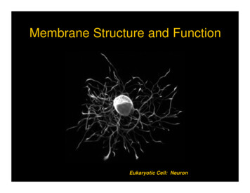
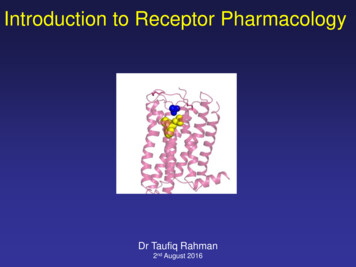
Ekere et al. BMC Infectious Diseases(2020) 20:5Page 5 of 9Fig. 3 PCR product showing amplified V3 genepatients’ PBMC. The DNA oligonucleotide primersHIV-19: 5′-AATGTCAGCACAGTACAATGTACA-3′and HIV-20: 5′-CAGTAGAAAAAATTCCCCTCCACAATT-3′ and HIV-21: 5′-CTGCTGTTAAATGGCAGTCTAGC-3′, and HIV-22: 5′-TCTGGGTCCCCTCCTGAGGA-3′ were used The PCR componentscomprised of 5 PCR buffer (100 mM Tris-HCl [pH8.3], 500 mM KCl, 15 mM MgCl2, 0.01% gelatin), 200mM each dATP, dCTP, dGTP, and TTP, 0.2 to 1.0mM each HIV-1 outer primer pair. The reactionswere carried out at 94 C for 1.5 min, 45 C for 2 min,and 72 C for 3 min for 35 cycles. The amplified DNAproducts were analyzed by electrophoresis on a 1.2%agarose gel. Negative controls consisting of DNAfrom PBMC of seronegative individuals were includedin each set of reactions, which were negative in all assays. After the first round PCR, 1 ml of the productwas amplified for 25 cycles with the correspondinginner primers at 94 C for 1.5 min, 50 C for 2 min,and 72 C for 3 min. The PCR products were analyzedby electrophoresis on a 1.2% agarose gel.Sequencing of CCR5 and HIV-V3 geneThe sequencing of the V3 gene and CCR5 was doneusing the Big Dye Terminator kit on ABI 3500 sequenceby Inquaba, South Africa.Phylogenic analysisObtained sequences were edited using the Bioinformatic software Bioedit. Similar sequences were downloaded from the National Centre for BioinformaticsInformation (NCBI) using Blast N. Downloaded sequence were aligned using the alignment softwareMAFT and the evolutionary relationship was eliminated at 500 bootstrap using MEGA.ResultsTable 1 shows the virological and clinical details of thestudied population. Majority (65%) of the studied subjects were females. Subjects with CD4 200 cells/μL,CD4 200–499 cells/μL and CD4 500 cells/μL consisted14, 41 and 45% of the studied population. Majority ofthe studied subjects fell into the CDC category A. Thestudied participants weren’t sure of their mode of acquisition of HIV (Table 1).Table 2 shows the sequencing of CCR5 receptor protein among the HIV-infected subjects and reveals a similar protein sequence without any mutation amongamino acid sequence with high concentration of Leucine7 (15.99%) followed by Lysine 5 (11.6%).Figure 2 shows the profiling of CCR5 gene among theHIV-infected participants using PCR. The amplifiedCCR5 gene resolved on agarose at 120 V for 20 minshowed that CCR5 DNA bands were slightly ahead of
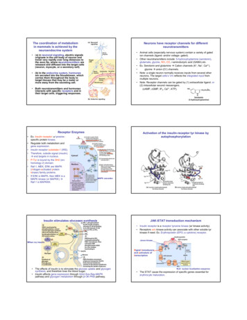
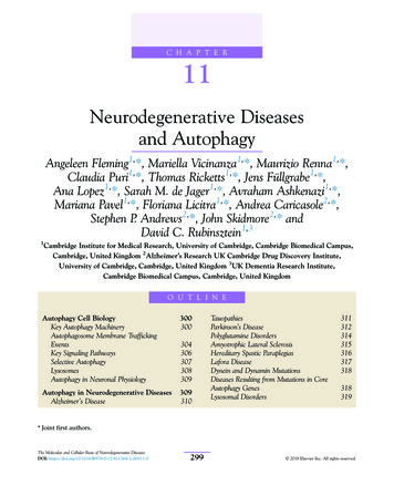
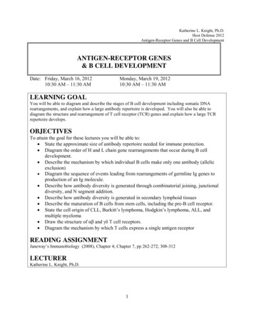
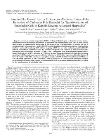
Ekere et al. BMC Infectious Diseases(2020) 20:5200 bp of the ladder and the sizes were estimated at 189bp. The gel revealed that all he subjects (100%) werehomozygote for CCR5-wt gene, as no band was detectedat 157 bp indicating a 32 bp deletion.Table 3 shows the frequency of CCR5 gene genotypein the studied population. All the assessed subjects(100%) were homozygous for the wild type CCR5(CCR5-wt). No homozygous mutant type (CCR5-Δ32)nor heterozygote was observed.The amplified V3 gene was resolved on agarose at 120V for 20 min and visualized on UV trans-illuminator.The subjects amplicons were ran along a 100 bp Biolabsmolecular ladder. The V3 DNA bands aligned with the300 bp of the ladder and the sized were estimated to be300 bp as expected (Fig. 3).Figure 4 shows sequence alignment of the V3 geneshowing the various nucleotide polymorphisms, whileFig. 5 shows the phylogenic tree showing the geneticrelationship between the HIV strains. The obtainedV3 sequences from the patients produced an exactmatch during the megablast search for highly similarsequences from the NCBI non-redundant nucleotidePage 6 of 9(nr/nt) database. The V3 sequence showed a percentage similarity to other V3 region ranging from 98 to100%. The evolutionary distances computed using theJukes-Cantor method were in agreement with thephylogenetic placement of the V3 sequences withinthe HIV and revealed a closely relatedness to HIV-1isolate 97CIRMF09 (gi\24,061,974) from Senegal,HIV-1 HC 19 (gi/5932488) from Gabon and HIV-1(gi/918567001) from Cameroon. Furthermore, thegene2pheno software revealed that all the strains wereHIV1 subtypes C (Table 4) though the sequencealignment showed some variations in the nucleotidessequence of the V3 gene of various strains (Fig. 4).DiscussionThe CCR5 gene profiling among the HIV-infectedsubjects in Calabar showed that all (100%) subjectsthat participated in the study were homozygotes forthe normal allele/wild type (CCR5-wt). There was nodetection of CCR5 Δ 32 mutation. The absence ofCCR5 Δ 32 mutation observed in this study is consistent with 1% earlier report by Martinson andFig. 4 Sequence alignment of the V3 gene showing the various nucleotide polymorphisms
Ekere et al. BMC Infectious Diseases(2020) 20:5Page 7 of 9Fig. 5 Phylogenic tree showing the genetic relationship between the HIV strainsTable 4 HIV-1 strains usedStrainHIV-1 eagues [25] for Nigeria and a more recent reportof Nil value by Kenneth et al., 2016 [30] in BayelsaState, Nigeria. However, higher frequencies of CCR5Δ 32 mutation has been reported in northern Europe,particularly the Baltic region as represented by databy Roy and Chakrabarti [9] in Sweden, Belarus,Finland, Lithuania and Estonia. Other areas with reported high frequency of CCR5- Δ 32 polymorphismincluded northern coast of France, the Russian citiesof Moscow and Ryazan, portions of Volga-Ural regionof Russia [31]. Population based studies reported thephenotype of the wild type gene as 75.56% in Rusia[32], 87.5% in Turkey [25], 98.21% in Cyprus Greek[32], 91.22% in Jordan [32], 97.16% in Syria [32],97.96% in Kuwait [31], 100% in Yemen [25] beingstudies in Euro - Asia while 100% has been reportedin Kenya [25] and Sudan [33]. The relatively higherfrequency of the wild type gene of the CCR5 inAfrican region is suggestive that the mutant variant(CCR5- Δ 32) is fairly recent in terms of human evolution [34, 35].
Ekere et al. BMC Infectious Diseases(2020) 20:5The implication of the absence of the CCR5- Δ 32gene in the studied population revealed a poor resistance to HIV and above all, underlined the factthat our HIV infected subjects are likely rapid progressors. The V3 sequencing showed genetic relationship between HIV strains isolated in this studyand those isolated from Senegal P3764 (V3e), Garbon (V3d and Veg), V3b from Cameroon (V3b) and(V3e) from Senegal, this relatedness among strainscould be due to trans-border movement of the population which may facilitate the transfer of one stainfrom a region to the other,ConclusionThis study has revealed the absence of the mutant genetype of CCR5 (CCR5-Δ32) in the studied subjects implying that the studied population lack one of the prominent genetic advantage against HIV-infection as well aspossible rapid progression to AIDS, hence the need tostrengthen management and diagnostic strategies.AbbreviationsAIDS: Acquired immunodeficiency syndrome; ART: Antiretroviral therapy;CCR5: C-C- type chemokine receptor; CD: Cluster of differentiation; CXCR4: CX-C chemokine receptor 4; DNA: Deoxyrebonucleic acid; HIV: HumanImmunodeficiency virus; PCR: Polymerase chain reactionAcknowledgementsNot applicable.Authors’ contributionsEFE conceived the study, designed the study, performed analyticalprocedures, analysed data and edited the initial manuscript; MFU designedthe study, analysed data, supervised the study. HUO analysed data,performed statistical analysis, performed literature search, wrote the initialmanuscript draft. TYM participated in the analytical procedure, analysed data.All authors approved the final manuscript.FundingThis study was funded by the authors.Availability of data and materialsDatasets generated and analysed in this study are available from thecorresponding author on request.Ethics approval and consent to participateThis study was approved by Health Research Ethical Committee (HREC) ofthe University of Calabar Teaching Hospital. Informed consent (written) wasobtained from the study participants before commencing the study.Consent for publicationNot applicable.Competing interestsThe authors declare that they have no competing interests.Author details1Hematology Laboratory, University of Calabar Teaching Hospital, Calabar,Nigeria. 2Microbiology Unit, Department of Medical Laboratory Science,University of Calabar, Calabar, Nigeria. 3Hematology Unit, Department ofMedical Laboratory Science, University of Calabar, Calabar, Nigeria.4Department of Medical Laboratory Science, College of Health Sciences,Niger Delta University, Amassama, Nigeria.Page 8 of 9Received: 23 July 2019 Accepted: 24 December 2019References1. Asemota EA, Okafor IM, Okoroiwu HU, Ekong ER, Anyanwu SO, Effiong EE,et al. Zinc, copper, CD4 T – cell count and some hematological parametersof HIV – infected subjects in southern Nigeria. Integr Med Res. 2018;7:53–60.2. World Health Organization (WHO). Global Health Observation (GHO) data:HIV/AIDS. Available at: www.who.gho/hiv/en. Accessed 26 May 2019.3. Okoroiwu HU, Okafor IM, Asemota EA, Okpokam DC. Seroprevalence oftransfusion-transmissible infections (HBV, HCV, syphilis and HIV) amongprospective blood donors in a tertiary health care facility in Calabar, Nigeria;an eleven years evaluation. BMC Public Health. 2018;18:645.4. Coco-Bassey SB, Asemota EA, Okoroiwu HU, Etura JE, Efiong EE, Inyang IJ,et al. Glutathione, glutathione peroxidase and some hematologicalparameters of HIV-seropositive subjects attending clinic in University ofCalabar Teaching Hospital, Calabar, Nigeria. BMC Infect Dis. 2019;19:944.5. Joint United Nations Programme on HIV and AIDs (UNAIDS). The Gapreport: children and pregnant women living with HIV. Geneva: UNAIDS;2014.6. Joint United Nations programme on HIV and AIDs (UNAIDS). Country factsheets; Nigeria. 2017. Available at: www.unaids.org/en/regionscountries/nigeria. Accessed 26 May 2019.7. Joint United Nations Programme on HIV and AIDs (UNAIDS). Country fastsheets; South Africa. 2017. Available at: www.unaids.org/en/regionscountries/southafrica. Accessed 26 May 2019.8. Nsonwu-Anyanwu AC, Ighodalo EV, King D, Agu CE, Jeremiah S, SolomonOT, et al. Biomarken of oxidative stress in HIV seropositive individuals onhighly active antiretroviral therapy. Reactive Oxygen Species. 2017;3(9):1–11.9. Roy CP, Chakrabarti S. Mutation in AIDS restriction gene affecting the HIVinfection and disease progression in a high risk group from northeasternIndia. Med J Armed Forces India. 2016;722:111–5.10. Buchbinder SP, Katz MH, Hessol NA, O’Malley PM, Holmberg SD. Long-termHIV-1 infection without immunologic progression. AIDS. 1994;8:1123–8.11. Winkler C, Ping AN, Stephen J, Brien O. Patterns of ethnic diversity amongthe genes that influence AIDS. Hum Mol Gent. 2004;13(1):R1–R19.12. Samson M, Libert F, Doranz BJ, et al. Resistance to HIV-1 infection inCaucasian individuals bearing mutant alleles of the CCR-5 chemokinereceptor gene. Nature. 1996;382:722–5.13. O’Brien SJ, Nelson GW. Human genes that limit AIDS. Nat Genet. 2004;36:565–74.14. Liu R, Paxton WA, Choe S, et al. Homozygous defect in HIV-1 coreceptoraccounts for resistance of some multiply – exposed individuals to HIV-1infection. Cell. 1996;86:367–717.15. Solloch UV, Lang K, Lang V, Bohne I, Shmidt AH. Frequencies of genevariant CCR5-Δ32 in 87 countries based on next generation sequencing of1.3 million individuals sampled from 3 national DKMS donor centres. HumImmunol. 2017;78:710–7.16. Wu L, Paxton WA, Kassam N, Ruffin N, Rottman JB, Sullivan N, et al. CCR5levels and expression pattern correlate with infectability by macrophagetropic HIV-1, in vitro. J Exp Med. 1997;185:1681–91.17. Barmania F, Potgiefer M, Pepper MS. Mutation in C-C chemoreceptor type 5(CCR5) in south African individuals. Int J Infect Dis. 2013;17:e1148–53.18. Connor RI, Sheridan KE, Ceradini S, Choe S, Landau NR. Change incoreceptor use correlates with disease progression in HIV-1 infectedindividuals. J Exp Med. 1997;185:621–8.19. Dragic T, Litwin V, Allaway G, Martin SR, Huang Y, Nagashima KA, et al. HIV-1entry into CD4 cells in mediated by the chemokine receptor CCR5. Nature.1996;381:667–73.20. Deng H, Lin R, Ellmeier W, Choe S, Unutrnaz D, Burkhart M, et al.Identification of a major co-receptor for primary isolates of HIV-1. Nature.1996;381:661–6.21. Feng Y, Broder CC, Kennedy PI, Berger EA. HIV-1 entry co-factor: functionalcDNA cloning of a seven-transmembrane, G protein-coupled receptor.Science. 1996;872:877.22. Khorramdelazad H, Bagheri V, Hassanshahi G, Zeinali M, Vakilian A. Newinsight into the role of stromal cell-derived factor 1 (SDF-1/CXCL12) in thepathophysiology of multiple schlerosis.23. Karimbadad MN, Falahati-Pour SK, Hassanshabi G. Significant role(s) ofCXCL12 and SDF-1 31A genetic variant in the pathophysiology of multiplesclerosis. Neuroimmunomodulation. 2016;23:197–208.
Ekere et al. BMC Infectious Diseases(2020) 20:524. Michael NI, Nelson JA, Kewal Ramani VN, Chang G, O’Brien SJ, Mascola JR,et al. Exclusive and persistent use of the entry co-rector CxCR4 by humanimmunodeficiency virus type 1 form a subject homozygous for CCR5 delta32. J Virol. 1998;72:6040–7.25. Martinson JJ, Chapman NH, Rees DC, Liu YT, Clegg JB. Global distribution ofthe CCR5 gene 32 base pair deletion. Nat Genet. 1997;16:100–3.26. Nwabueze BO. A constitutional history of Nigeria. United Kingdom: C. Hurstand Co. LTD.; 1982.27. Wikipedia, the free encyclopedia. Geography of Cross River State. Availableat: https//en.wikipedia.org/wiki/Cross River State . Accessed 27 May 2019.28. National Bureau of Statistics. Annual abstract of statistics 2011. NBS FederalRepublic of Nigeria 2011.29. Okoroiwu HU, Asemota EA. Blood donors deferral prevalence and causes ina tertiary health care hospital, southern Nigeria. BMC Health Serv Res. 2019;19:510.30. Kenneth OZ, Opuata S, Tatfeng YM TCA, Zaccheaus AJ. Null singlenucleotide polymorphism in Cysteine – cysteine chemokine receptor 5genes among Ijaw Etnic Population of Nigeria. Open J Blood Dis. 2016;2(3):1–6.31. Novembre J, Galvani AP, Slatkin M. The geographic spread of the CCR5Δ32HIV-resistance allele. Plos Biol. 2005;3(11):e339.32. Salem AH, Batzer MA. Distribution of the HIV resistance CCR5-delta 32 alleleamong Egytians and Syrians. Mutat Res. 2007;616:175–80.33. Su B, Sun G, Lu D, Xiao J, Hu F, Chakraborty R, Deka R, Jin L. Distribution ofthree HIV-1 resistance – conferring Polymorphisms (SDF1–3’A, CCR2–641and CCR5 -Δ32) in global populations. Eur J Hum Genet. 2000;8:975–9.34. Galvani AP, November J. The evolutionary history of the CCR5-Delta 32 HIVresistance mutation. Microbes Infect. 2005;7:302–9.35. Picton ACP, Shalekoff S, Paximadis M, Tiemessen CT. Marked differences inCCR5 expression and activation levels in two South African Populations.Immunology. 2012;136:397–407.Publisher’s NoteSpringer Nature remains neutral with regard to jurisdictional claims inpublished maps and institutional affiliations.Page 9 of 9
Nigeria. Cross River State is located in Southern Nigeria [26] in the Niger Delta Region. It is bounded in the north by Benue State, the west by Ebonyi and Abia State, the east by Cameroon Republic and the south by Akwa Ibom State and the Atlantic Ocean [27]. Cross River State has an area of 21,787km2 and a population of 2,892,988 (2006 census .