Transcription
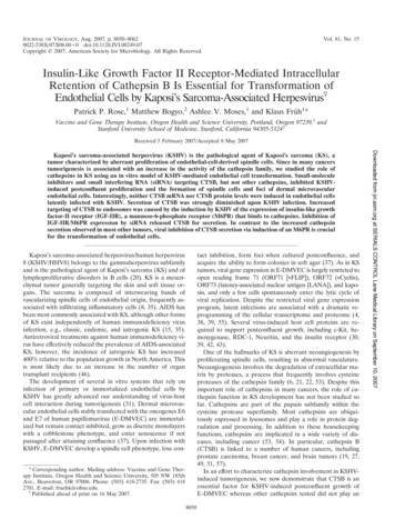
JOURNAL OF VIROLOGY, Aug. 2007, p. 8050–80620022-538X/07/ 08.00!0 doi:10.1128/JVI.00249-07Copyright 2007, American Society for Microbiology. All Rights Reserved.Vol. 81, No. 15Insulin-Like Growth Factor II Receptor-Mediated IntracellularRetention of Cathepsin B Is Essential for Transformation ofEndothelial Cells by Kaposi’s Sarcoma-Associated Herpesvirus!Patrick P. Rose,1 Matthew Bogyo,2 Ashlee V. Moses,1 and Klaus Früh1*Vaccine and Gene Therapy Institute, Oregon Health and Science University, Portland, Oregon 97239,1 andStanford University School of Medicine, Stanford, California 94305-53242Received 5 February 2007/Accepted 8 May 2007tact inhibition, form foci when cultured postconfluence, andacquire the ability to form colonies in soft agar (37). As in KStumors, viral gene expression in E-DMVEC is largely restricted toopen reading frame 71 (ORF71 [vFLIP]), ORF72 (vCyclin),ORF73 (latency-associated nuclear antigen [LANA]), and kaposin, and only a few cells spontaneously enter the lytic cycle ofviral replication. Despite the restricted viral gene expressionprogram, latent infections are associated with a dramatic reprogramming of the cellular transcriptome and proteome (4,38, 39, 55). Several virus-induced host cell proteins are required to support postconfluent growth, including c-Kit, hemoxygenase, RDC-1, Neuritin, and the insulin receptor (30,39, 42, 43).One of the hallmarks of KS is aberrant neoangiogenesis byproliferating spindle cells, resulting in abnormal vasculature.Neoangiogenesis involves the degradation of extracellular matrix by proteases, a process that frequently involves cysteineproteases of the cathepsin family (6, 21, 22, 53). Despite thisimportant role of cathepsins in many cancers, the role of cathepsin function in KS development has not been studied sofar. Cathepsins are part of the papain subfamily within thecysteine protease superfamily. Most cathepsins are ubiquitously expressed in lysosomes and play a role in protein degradation and processing. In addition to these housekeepingfunctions, cathepsins are implicated in a wide variety of diseases, including cancer (53, 54). In particular, cathepsin B(CTSB) is linked to a number of human cancers, includingprostate carcinoma, breast cancer, and brain tumors (19, 27,49, 51, 57).In an effort to characterize cathepsin involvement in KSHVinduced tumorigenesis, we now demonstrate that CTSB is anessential factor for KSHV-induced postconfluent growth ofE-DMVEC whereas other cathepsins tested did not play anKaposi’s sarcoma-associated herpesvirus/human herpesvirus8 (KSHV/HHV8) belongs to the gammaherpesvirus subfamilyand is the pathological agent of Kaposi’s sarcoma (KS) and oflymphoproliferative disorders in B cells (20). KS is a mesenchymal tumor generally targeting the skin and soft tissue organs. The sarcoma is composed of interweaving bands ofvascularizing spindle cells of endothelial origin, frequently associated with infiltrating inflammatory cells (8, 35). AIDS hasbeen most commonly associated with KS, although other formsof KS exist independently of human immunodeficiency virusinfection, e.g., classic, endemic, and iatrogenic KS (15, 35).Antiretroviral treatments against human immunodeficiency virus have effectively reduced the prevalence of AIDS-associatedKS; however, the incidence of iatrogenic KS has increased400% relative to the population growth in North America. Thisis most likely due to an increase in the number of organtransplant recipients (46).The development of several in vitro systems that rely oninfection of primary or immortalized endothelial cells byKSHV has greatly advanced our understanding of virus-hostcell interaction during tumorigenesis (31). Dermal microvascular endothelial cells stably transfected with the oncogenes E6and E7 of human papillomavirus (E-DMVEC) are immortalized but remain contact inhibited, grow as discrete monolayerswith a cobblestone phenotype, and enter senescence if notpassaged after attaining confluence (37). Upon infection withKSHV, E-DMVEC develop a spindle cell phenotype, lose con* Corresponding author. Mailing address: Vaccine and Gene Therapy Institute, Oregon Health and Science University, 505 NW 185thAve., Beaverton, OR 97006. Phone: (503) 418-2735. Fax: (503) 4182701. E-mail: fruehk@ohsu.edu.!Published ahead of print on 16 May 2007.8050Downloaded from jvi.asm.org at SERIALS CONTROL Lane Medical Library on September 10, 2007Kaposi’s sarcoma-associated herpesvirus (KSHV) is the pathological agent of Kaposi’s sarcoma (KS), atumor characterized by aberrant proliferation of endothelial-cell-derived spindle cells. Since in many cancerstumorigenesis is associated with an increase in the activity of the cathepsin family, we studied the role ofcathepsins in KS using an in vitro model of KSHV-mediated endothelial cell transformation. Small-moleculeinhibitors and small interfering RNA (siRNA) targeting CTSB, but not other cathepsins, inhibited KSHVinduced postconfluent proliferation and the formation of spindle cells and foci of dermal microvascularendothelial cells. Interestingly, neither CTSB mRNA nor CTSB protein levels were induced in endothelial cellslatently infected with KSHV. Secretion of CTSB was strongly diminished upon KSHV infection. Increasedtargeting of CTSB to endosomes was caused by the induction by KSHV of the expression of insulin-like growthfactor-II receptor (IGF-IIR), a mannose-6-phosphate receptor (M6PR) that binds to cathepsins. Inhibition ofIGF-IIR/M6PR expression by siRNA released CTSB for secretion. In contrast to the increased cathepsinsecretion observed in most other tumors, viral inhibition of CTSB secretion via induction of an M6PR is crucialfor the transformation of endothelial cells.
VOL. 81, 2007CATHEPSIN B ACTIVITY IN KS TUMORIGENESISessential role. Unexpectedly, secretion of CTSB was inhibitedin latently infected endothelial cells. CTSB is retained by theinsulin-like growth factor-II receptor/mannose-6-phosphatereceptor (IGF-IIR/M6PR), which we previously reported to betranscriptionally induced during KSHV-mediated transformation of E-DMVEC (43). Therefore, in contrast to the increasedsecretion of CTSB observed in several other tumor models,CTSB is retained in the endosomal/lysosomal compartment inKS, where it likely regulates the processing of growth factors orgrowth factor binding proteins. Given the recent progress indeveloping CTSB inhibitors for cancer treatment (10), ourdata suggest that such inhibitors might be a novel treatmentfor KS.Viruses, cell culture, and reagents. KSHV-infected E-DMVEC were established and maintained as previously described (37). KSHV-infected DMVECwere used in experiments when "90% of the cells expressed LANA-1. CA-074(catalog no. 205530), CA-074ME (catalog no. 205531), and hydroxy-2-naphthalenyl-methyl phosphonic acid trisacetoxymethyl ester [HNMPA-(AM3)] (catalogno. 397100) were obtained from EMD Biosciences (San Diego, CA). Antibodiesagainst cathepsin S (catalog no. IM1003), cathepsin L (catalog no. 219387), andCTSB (catalog no. IM27L) were purchased from EMD Biosciences (San Diego,CA). A mouse monoclonal antibody against calreticulin (SPA-601) was purchased from Stressgen (Victoria, British Columbia, Canada). Purified humannative CTSB (catalog no. JA7741) was obtained from EMD Biosciences (SanDiego, CA). A polyclonal antibody against IGF-IIR/M6PR (H-20) was obtainedfrom Santa Cruz Biotechnology (Santa Cruz, CA).Microarray experiments and analysis. cDNA synthesis, hybridization toHG U133A and B arrays (Affymetrix), and signal intensity normalization wereperformed at the Oregon Health and Science University (OHSU) microarraycore facility (www.ohsu.edu/gmsr/amc). GeneChip data were analyzed with Arrayassist (Stratagene). Comparisons were made between passage-matchedKSHV-infected E-DMVEC and mock-infected E-DMVEC in four separate experiments. A master data table consisting of expression values extracted from allCEL files was created by the probe logarithmic error intensity estimate (PLIER)method. To define the baseline from which changes in gene expression data weredetermined for KSHV-infected samples, a virtual chip comprising all mockinfected control expression data was used. Significant changes with a P value of0.01 were determined by analysis of variance with variance stabilization and noP value correction.Western blotting and immunoprecipitations. For immunoprecipitation, cellswere washed twice with ice-cold phosphate-buffered saline (PBS) and lysed inNP-40 buffer (1% NP-40, 150 mM NaCl, 10% glycerol, 20 mM Tris-HCl [pH 8.0],1 mM EDTA [pH 8.0], 0.2% sodium dodecyl sulfate [SDS]) containing proteaseinhibitors (Roche Diagnostics; Indianapolis, IN), 1 mM NaVO4, and 1 mg/mlpepstatin. Lysates were cleared, and the protein concentration was determined.For IGF-IIR/M6PR, 500 #g of whole-cell lysate protein was immunoprecipitatedwith 10 #g of an anti-IGF-IIR/M6PR antibody and incubated overnight at 4 Cwith rocking. A 50-#l volume of a protein A/G-agarose bead slurry (Upstate) wasadded for 45 min with rocking at 4 C. Three washes were performed, and thepellet was boiled in 2 SDS sample buffer. The beads were spun down, and thesupernatant was separated by 10% SDS-polyacrylamide gel electrophoresis. ForWestern blotting, cells were directly lysed in 1 SDS sample buffer (100 mMTris-Cl [pH 6.8], 2% SDS, 10% glycerol) without dyes or with dithiothreitol–2mercaptoethanol and were subsequently boiled for 5 min. Protein content wasnormalized and determined using a bicinchoninic acid protein assay kit (catalogno. 23227; Pierce, Rockford, IL). Before resolution of samples by 10% SDSpolyacrylamide gel electrophoresis, 100 mM dithiothreitol and 0.01% bromophenol blue were added to the lysates and samples were boiled for 5 min. Gelswere transferred to nylon membranes, blocked for 20 min with 10% milk in 1 PBS supplemented with 0.1% Tween 20, and probed with the respective antibodies. Bound antibodies were detected by enhanced chemiluminescence(Pierce) and exposed to film.Reverse transcriptase PCR (RT-PCR). Quantitative PCR was performed onan ABI7700 sequence detection system (Applied Biosystems, Foster City, CA).Target gene expression was compared to expression of the glyceraldehyde-3phosphate dehydrogenase housekeeping gene. Total RNA was purified fromprimary DMVEC using RNeasy spin columns (QIAGEN Inc, Valencia, CA).Human tissue was a generous gift from H. Koon and B. J. Dezube and wasprepared as previously described (42). Total RNA was treated with DNase I(DNase free; Ambion, Austin, TX) before synthesis of cDNA with randomhexamers and Superscript III (Invitrogen, Carlsbad, CA). The following primerswere selected by using Primer Express software (Applied Biosystems): IGF-IIR/M6PR 6333F (GCAGAAGCTGGGTGTCATAGG), 6420R (5%-CACGGAGGATGCGGTCTTAT), IGF-IR 2184F (GGAGGAGGCTGAATACCGC), and2252R (5%-TCAGGTCTGGGCACGAAGAT). Reactions were performed usingSYBR green PCR core reagents. Relative expression values for uninfected versus KSHV-infected cells and for normal skin versus KS tissue were calculated bythe comparative CT method (28). Dissociation curves were performed on humanreference RNA after each amplification run to control for primer dimers. Foranalysis of CTSB splicing, the following primers were used as previously published for PCR: CTSB full length fwd (5%-CAGCGCTGGGCTGGTGTG),CTSB(&2) fwd (5%-CAGCGCTGGGTGGATCTA), CB(&2/3) fwd (5%-CAGCGCTGGGCCGGGCAC), and CB EXON 11 rev (5%-CCAGGACTGGCACGACAGGC). PCR to determine CTSB isoforms was performed by standard approaches (95 C for 5 min; 30 cycles of 95 C for 1 min, 55 C for 1 min, and 72 Cfor 2 min; and 72 C for 10 min).siRNA treatments. KSHV-infected E-DMVEC were seeded the day prior tosmall interfering RNA (siRNA) treatment at 30% confluence in 35-mm-diameter polystyrene dishes (Corning). On the day of treatment, for each sample, 3 #lOligofectamine (Invitrogen) was mixed with 12 #l OptiMEM (GIBCO-Invitrogen) for 5 min at room temperature. A mixture of 200 nM SMARTpool siRNA(Dharmacon) and 180 #l OptiMEM was added to the Oligofectamine mixtureand incubated for 20 min at room temperature. For IGF-IIR/M6PR siRNAtreatment, single sense strand siRNAs were employed: AGACCAGGCUUGCUCUAUAUU (On-Targetplus set 6), CAACUUUCCUCCAUCACAAUU(On-Targetplus set 7), and CGAUACCUCUCAAGUCAAAUU (On-Targetplus set 8) (catalog no. J-010601-06; Dharmacon). Cells were washed once inOptiMEM, and 800 #l OptiMEM was added to the dish prior to addition of 200#l siRNA mixture. Cells were incubated for 6 h before receiving a secondtreatment of siRNA as before. Cells were incubated overnight, and the siRNAmixture was removed and replenished with 2 ml of complete medium. siRNAtreated cells were incubated for 4 to 6 days to determine the peak of downmodulation, at which point protein levels were assayed by Western blotting. Forlong-term observation of phenotypic changes, siRNA-treated cells were treatedagain with siRNA 1 week after the first treatment to ensure efficient knockdown.One day after the second treatment series, CTSB activity was measured by azymogen assay.Focus inhibition assay. KSHV-infected E-DMVEC were seeded at 30 to 50%confluence on 35-mm-diameter dishes (Corning) and treated either with siRNAor with the CTSB small-molecule inhibitor CA-074 or CA-074ME for a 2-weekperiod. Inhibitors (10 #M) were added during the change of medium (every 2days), whereas a second round of siRNA treatment was performed 7 days afterthe first treatment to maintain the knockdown of specified genes. For siRNArescue, 10 ng/ml of native human CTSB was added every 2 days after initialsiRNA treatment and continued for 2 weeks total. Cells were examined forspindle cell formation, loss of contact inhibition (a result of adherent foci in themonolayer), or virus-induced cytopathic effect by microscopy. Cells were washedtwice in PBS and fixed in 3.7% paraformaldehyde for 20 min before anotherwash and addition of a coverslip over 80 #l glycerol.Immunofluorescence microscopy. Cells were washed twice with 1 PBS andfixed with 3.7% formaldehyde (in PBS) for 20 min at room temperature. Cellswere then washed twice with 1 PBS and permeabilized with 0.1% Triton X-100(in PBS) for 5 min. Subsequently, cells were washed three times with 2% PBA(bovine serum albumin in PBS). To measure KSHV infection after inhibitortreatment, cells were probed with a primary rabbit polyclonal antibody againstLANA-1 (ORF73) (a gift from Bala Chandran, Rosalind Franklin University,Chicago, IL) at 1:500 for 2 h at 37 C. Cells were washed three times with 2%PBA and then incubated with a goat anti-rabbit secondary antibody conjugatedwith Alexa Fluor 594 (Molecular Probes, Eugene, OR) at 1:500 for 45 min at37 C. Images were analyzed with a Nikon fluorescence microscope. For livestaining, KSHV-infected E-DMVEC were seeded at confluence on Lab-Tek II1.5 borosilicate chambered coverglass slides (catalog no. 155382; Electron Microscopy Sciences, Hatfield, PA). The next day, cells were either left untreated ortreated with either CA-074 (10 #M), CA-074ME (10 #M), or HNMPA-(AM3)(50 #M) overnight before being probed with GB-111 FL (1 #M) for 60 min.Images were acquired by Aurelie Snyder of the OHSU Molecular Microbiologyand Immunology Research Core Facility (http://www.ohsu.edu/research/core)with an Applied Precision DeltaVision RT image restoration system. This includes the API chassis with a precision-motorized XYZ stage, an Olympus IX-71inverted fluorescent microscope with standard filter sets, mercury illuminationDownloaded from jvi.asm.org at SERIALS CONTROL Lane Medical Library on September 10, 2007MATERIALS AND METHODS8051
8052ROSE ET AL.J. VIROL.RESULTSCathepsins expressed in KSHV-infected E-DMVEC are required for postconfluent growth. Since cathepsins have beenimplicated in a variety of human cancers, we wanted to assessthe role of cathepsins in KSHV-mediated tumorigenesis byusing the E-DMVEC in vitro model. We initially monitoredpostconfluent growth of E-DMVEC latently infected withKSHV in the presence or absence of the broad-spectrum cysteine protease inhibitor butane ethyl ester loxistatin (EST). KSHV-infected E-DMVEC were seeded at 30% confluence and treatedwith EST (58 #M) for 2 weeks; the inhibitor was refreshedevery third day. While untreated cells develop a spindle cellphenotype and form foci, EST prevented postconfluent growthand caused KSHV-infected E-DMVEC to remain cobblestone-like as a single monolayer (Fig. 1A). Similar observationswere made with a series of other broad-spectrum cysteineprotease inhibitors (data not shown). These studies suggestedFIG. 1. Cysteine protease inhibitors effectively prevent KSHV-induced formation of spindle cell-containing foci. (A) Phase-contrastimages of KSHV-infected E-DMVEC treated with cysteine proteaseinhibitor EST or LHVS. Both compounds effectively inhibit spindlecell and focus formation. (B) Western blot analysis comparing levels ofprotein expression for cathepsins S, L, and B in KSHV-infected anduninfected E-DMVEC. To control for equal loading, antibodies to thecellular chaperone calreticulin were used.that cysteine proteases, presumably cathepsins, are importantfor the continuous growth of KSHV-infected E-DMVEC whenthe cells reach confluence.To determine which cathepsins are expressed in KSHVinfected DMVEC, the gene expression profiles for all knowncathepsin transcripts were extracted from complete genomeexpression arrays (Affymetrix U133A and U133B). Resultsfrom four independent experiments with KSHV-infected EDMVEC cultures were compared with results for their passage- and age-matched cultures of uninfected E-DMVEC.Cathepsin transcripts were scored with the Stratagene algorithm and the PLIER method. Whereas a number of cathepsins were expressed in both uninfected and KSHV-infectedE-DMVEC, only cathepsin S, represented by two Affymetrixprobe sets, was induced upon KSHV infection (Fig. 2). Thisexpression profile was confirmed at the protein level by usingspecific antibodies in immunoblotting (Fig. 1B).siRNA screen against cathepsin S, L, and B suggests CTSBinvolvement in KSHV-mediated transformation of DMVEC.Using a series of more selective cathepsin inhibitors, we narrowed down the list of potential candidates responsible for theinhibitory effects observed with broad-spectrum inhibitors. InDownloaded from jvi.asm.org at SERIALS CONTROL Lane Medical Library on September 10, 2007with an API light homogenizer, a Nikon CoolSnap camera, and DeltaVisionsoftware. Deconvolution using the iterative constrained algorithm of Sedat andAgard and additional image processing were performed on a Linux OS platformwith the SoftWoRx program. Further technical specifications are available fromApplied Precision.Cell viability assay. Cell viability by ATP determination was measured according to the manufacturer’s protocol (Molecular Probes; catalog no. A-22066).KSHV-infected E-DMVEC were treated with either CA-074 (10 #M), CA074ME (10 #M), or HNMPA-(AM3) (100 #M) for 4 days in 35-mm-diameterdishes and were lysed in 1% Triton X-100 for 10 min. A D-luciferin–luciferasemixture (5 #l per sample) was added, and the level of ATP was immediately readin a luminometer.Annexin V-FITC apoptosis detection assay. Apoptosis in uninfected andKSHV-infected E-DMVEC was analyzed with the R&D Systems annexin Vfluorescein isothiocyanate (FITC) apoptosis detection kit (catalog no. TA4638).Cells were washed once in PBS, plated in serum-free medium, and either treatedovernight with staurosporine (1 #M), CA-074 (10 #M), or CA-074ME (10 #M)or left untreated. Cells were trypsinized and washed in 2% PBA before eachsample was resuspended in 100 #l annexin reagent (10 #l 10 binding buffer, 10#l propidium iodide, 1 #l annexin V-FITC, and 79 #l double-distilled H2O) for15 min at room temperature. A 400-#l volume of 1 binding buffer was addedbefore samples were analyzed by flow cytometry.Proliferation assay. Uninfected or KSHV-infected E-DMVEC were seeded at3 105 cells (for preconfluence) or 5 105 cells (for postconfluence) per35-mm-diameter dish. Cells were grown in the presence or absence of CA-074(10 #M), CA-074ME (10 #M), or HNMPA-(AM3) (50 #M) for 5 days beforebeing trypsinized, stained with trypan blue, and counted. For the postconfluenceassay, cell numbers were normalized to cell numbers in a confluent monolayer ofuninfected E-DMVEC (set at 100%) and are presented as percentages of thenumber of uninfected E-DMVEC.Soft agar assay. Forty thousand KSHV-infected E-DMVEC, either treatedwith CA-074 or CA-074ME or left untreated for 14 days, were plated in 1.5 mlendothelial-SFM growth medium (11111-044; Invitrogen) supplemented with10% human AB serum, 25 #g/ml endothelial cell growth supplement, and 0.4%melted agarose onto a 3-ml bottom layer of 0.5% agarose medium per well of asix-well dish. The cells were fed several drops of medium every 3 days, andcolonies were photographed after 3 weeks.Zymogen assay. CTSB activity was measured with the InnoZyme CTSB activity assay kit (catalog no. CBA001; EMD Biosciences, San Diego, CA). To testinhibition by CA-074 and CA-074ME on native CTSB, 10 #M of either inhibitorwas combined with 50 ng of native CTSB enzyme in vitro. The fluorogenicsubstrate Z-Arg-Arg 7-amino-4-methylcoumarin (AMC) was added, and therelease of AMC was assayed as recommended by the manufacturer. To measureCTSB enzymatic activity in uninfected and KSHV-infected E-DMVEC lysatesand supernatants, cells were seeded in 35-mm-diameter dishes (Corning) in 200#l medium. Dishes were rocked constantly to cover all cells with medium. Themedium was collected, and cells were lysed as suggested in the protocol. Fluorescence was measured using the Flexstation plate reader and analyzed usingSoftworx Pro (version 4.0.1) software (Molecular Devices, Sunnyvale, CA).
VOL. 81, 2007particular, the vinyl sulfone LHVS fone-phenyl) has specificity forcathepsins S, L, and B (9, 41, 50). KSHV-infected E-DMVECwere treated with LHVS (25 #M) as described above. As with8053broad-spectrum inhibitors, inhibition of postconfluent growthwas observed (Fig. 1A), suggesting that at least one of theseproteases is important for KSHV-mediated transformation ofE-DMVEC.To distinguish which of these three cathepsins is necessary forKSHV-induced tumorigenesis, SMARTpool siRNAs were employed against each candidate protease. KSHV-infected EDMVEC either were treated separately with SMARTpoolsiRNAs against cathepsin S, L, or B or were mock treated with aCy3-labeled siRNA against luciferase. siRNA-treated KSHV-infected cultures were monitored over 2 weeks for spindle cell andfocus formation. Transfection efficiency was visualized with theCy3 label conjugated to the luciferase siRNA and was estimatedto be consistently around 95%. Western blot analysis of siRNAtreated cultures revealed that all three SMARTpool siRNAs efficiently reduced target protein levels (Fig. 3A). Furthermore,cathepsin L siRNA inhibited its target cathepsin but did not affectprotein expression of CTSB, and vice versa. Despite efficient geneknockdown, cathepsin S and L SMARTpool siRNAs did notimpede KSHV-induced tumorigenesis. Compared to that for untreated cells, there was a slight reduction in the level of focusformation in cathepsin L and cathepsin S siRNA-treated cells,probably due to the multiple rounds of transfection, since a reduction was also observed with Cy3-labeled control siRNA (notshown). In contrast, CTSB siRNA dramatically inhibited spindlecell and focus development in KSHV-infected E-DMVEC (Fig.3B). These data suggested that CTSB was responsible for theinhibition of KSHV-mediated E-DMVEC transformation by cathepsin inhibitors.Intracellularly active CTSB is essential for transformationof DMVEC by KSHV. We initially hypothesized that secretedor extracellular matrix-associated CTSB was involved in tumorigenesis by degrading extracellular matrix components (25,33, 49). To test this hypothesis, we examined whether postconfluent growth of CTSB siRNA-treated cells could be rescuedby exogenous addition of native human CTSB to the mediumFIG. 3. Downregulation of CTSB mRNA inhibits KSHV-mediated focus formation. (A) Western blot (WB) analysis of cathepsin S, L, and Bprotein levels after siRNA treatment. An antibody to calreticulin ('-CRT) was used to control for equal loading. siRNAs to cathepsins L and Bwere target specific as determined by Western blotting. (B) Representative phase-contrast microscopy comparing untreated samples to cathepsinS, L, or B siRNA-treated, KSHV-infected E-DMVEC. Control siRNA-treated samples were indistinguishable from untreated samples and are notrepresented here. Only CTSB siRNA reduced focus formation. Immunofluorescence analysis of Cy3-labeled siRNA transfectants indicated atransfection efficiency of "95% (data not shown).Downloaded from jvi.asm.org at SERIALS CONTROL Lane Medical Library on September 10, 2007FIG. 2. Gene expression profiling of cathepsin genes in KSHV-infectedand uninfected E-DMVEC. (A) Affymetrix HG U133A GeneChip data forcathepsins were obtained from four infected samples and compared to theirrespective controls. Asterisks indicate significant changes (P " 0.01 by analysis of variance). Genes with multiple probe sets are reflected in the table.NC, no change in gene expression; A, the probe set scored absent on themicroarray; I, changes where gene expression was induced.CATHEPSIN B ACTIVITY IN KS TUMORIGENESIS
8054ROSE ET AL.J. VIROL.of KSHV-infected E-DMVEC. Even after prolonged treatment with 10 ng/ml native human CTSB, the siRNA treatmentstill inhibited postconfluent growth of spindle cells (Fig. 4A),suggesting that endogenous rather than secreted CTSB is essential for KSHV-mediated transformation.The role of intracellular versus extracellular CTSB was further examined by using two different versions of the activitybased CTSB suicide inhibitor CA-074. CA-074 is unable tocross the cell membrane and consequently can inhibit onlyextracellular CTSB activity. A methyl ester-modified version ofCA-074, proinhibitor CA-074ME, can cross the membrane andinhibit intracellular CTSB, although the modification reducesselectivity for CTSB (34). To ensure that both versions ofCA-074 inhibited CTSB activity, they were tested against native human CTSB in vitro. At a concentration of 10 #M, theinhibitors were combined with native human CTSB in thepresence of the synthetic fluorogenic CTSB substrate AMC.The release of AMC was measured by fluorescence. Both versions of CA-074 successfully reduced CTSB activity comparedto that with no treatment (Fig. 4B). To examine whether CA074 or CA-074ME inhibited KSHV-mediated transformationof E-DMVEC, uninfected or KSHV-infected E-DMVEC cultures were treated with 10 #M of the respective CTSB inhibitors over a period of 2 weeks. Untreated KSHV-infected EDMVEC developed a spindle cell morphology and formed foci(Fig. 4C1). The impermeant version of the inhibitor, CA-074,Downloaded from jvi.asm.org at SERIALS CONTROL Lane Medical Library on September 10, 2007FIG. 4. Intracellular CTSB is essential for KSHV-induced spindle cell and focus formation. (A) Phase-contrast images of CTSB siRNA-treatedcultures incubated with purified human native CTSB. Treatment of KSHV-infected E-DMVEC with 10 ng/ml native CTSB (rescue) did not reversethe effect of CTSB siRNA knockdown. The upper left panel shows KSHV-infected E-DMVEC without treatment. (B) Zymogen assay to determinethe effectiveness of the two inhibitors of CTSB enzymatic activity. Analysis by cleavage of the CTSB fluorogenic substrate AMC showed that bothinhibitors effectively reduced CTSB activity. (C) Representative phase-contrast images of KSHV-infected E-DMVEC that were either leftuntreated (panel 1) or treated with CA-074ME (panel 3) or CA-074 (panel 2). Only the membrane-permeant version of the CTSB inhibitor,CA-074ME, was able to inhibit spindle cell and focus formation in KSHV-infected E-DMVEC (panel 2). (D) Microscopy images of thefluorescently tagged cathepsin L and B activity probe GB-111 FL after treatment with a competitive CTSB inhibitor or a noncompetitive insulinreceptor inhibitor [HNMPA-(AM3)]. KSHV-infected E-DMVEC were treated with the various inhibitors overnight before being probed with theactivity-based small-molecule inhibitor GB-111 FL. Only CA-074ME treatment dramatically reduced the staining pattern of GB-111 FL (panel 4).Untreated (panel 1), HNMPA-(AM3)-treated (panel 2), and CA-074-treated (panel 3) cells showed no change in their staining. Some cells treatedwith CA-074 did show a reduction in vesicular staining similar to that with CA-074ME, and it has been documented that CA-074 can permeatethe cells; however, at this concentration the inhibitor had no effect on KSHV-induced transformation of E-DMVEC.
VOL. 81, 2007CATHEPSIN B ACTIVITY IN KS TUMORIGENESIS8055was unable to inhibit spindle cell and focus formation (Fig.4C3). In contrast, the development of spindle cell morphologywas inhibited by CA-074ME (Fig. 4C2). This result supportsthe conclusion that intracellular rather than secreted CTSB isrequired for DMVEC transformation by KSHV.To locate enzymatically active CTSB in live cells, a fluorescein-conjugated activity-based probe, GB-111 FL, which bindsto both active cathepsins L and B, was employed. Prior toprobing with GB-111 FL, KSHV-infected E-DMVEC weretreated with CA-074 (10 #M) or CA-074ME (10 #M). As acontrol, a noncompetitive inhibitor of the insulin receptor,HNMPA-(AM3), was used. As shown in Fig. 4D1, untreatedcells were probed with GB-111 FL, indicating that active cathepsins L and B are localized to small vesicles. In Fig. 4D2, EDMVEC treated with the noncompetitive inhibitor HNMPA(AM3) showed no difference in staining pattern from untreatedcells. Probe staining of most cells treated with the impermeantinhibitor CA-074 was similar to that for untreated cells. Somecells did
program, latent infections are associated with a dramatic re-programming of the cellular transcriptome and proteome (4, 38, 39, 55). Several virus-induced host cell proteins are re-quired to support postconßuent growth, including c-Kit, he-moxygenase, RDC-1, Neuritin, and the insulin receptor (30, 39, 42, 43).