Transcription
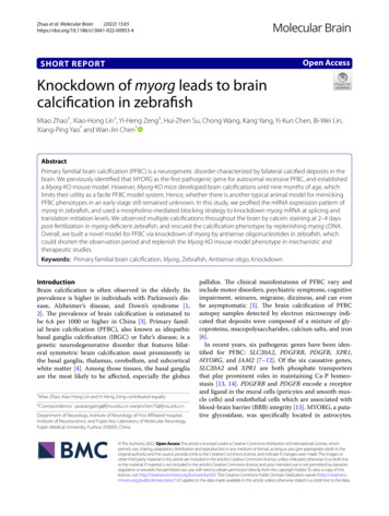
(2022) 15:65Zhao et al. Molecular n AccessSHORT REPORTKnockdown of myorg leads to braincalcification in zebrafishMiao Zhao†, Xiao‑Hong Lin†, Yi‑Heng Zeng†, Hui‑Zhen Su, Chong Wang, Kang Yang, Yi‑Kun Chen, Bi‑Wei Lin,Xiang‑Ping Yao* and Wan‑Jin Chen*AbstractPrimary familial brain calcification (PFBC) is a neurogenetic disorder characterized by bilateral calcified deposits in thebrain. We previously identified that MYORG as the first pathogenic gene for autosomal recessive PFBC, and establisheda Myorg-KO mouse model. However, Myorg-KO mice developed brain calcifications until nine months of age, whichlimits their utility as a facile PFBC model system. Hence, whether there is another typical animal model for mimickingPFBC phenotypes in an early stage still remained unknown. In this study, we profiled the mRNA expression pattern ofmyorg in zebrafish, and used a morpholino-mediated blocking strategy to knockdown myorg mRNA at splicing andtranslation initiation levels. We observed multiple calcifications throughout the brain by calcein staining at 2–4 dayspost-fertilization in myorg-deficient zebrafish, and rescued the calcification phenotype by replenishing myorg cDNA.Overall, we built a novel model for PFBC via knockdown of myorg by antisense oligonucleotides in zebrafish, whichcould shorten the observation period and replenish the Myorg-KO mouse model phenotype in mechanistic andtherapeutic studies.Keywords: Primary familial brain calcification, Myorg, Zebrafish, Antisense oligo, KnockdownIntroductionBrain calcification is often observed in the elderly. Itsprevalence is higher in individuals with Parkinson’s disease, Alzheimer’s disease, and Down’s syndrome [1,2]. The prevalence of brain calcification is estimated tobe 6.6 per 1000 or higher in China [3]. Primary familial brain calcification (PFBC), also known as idiopathicbasal ganglia calcification (IBGC) or Fahr’s disease, is agenetic neurodegenerative disorder that features bilateral symmetric brain calcification most prominently inthe basal ganglia, thalamus, cerebellum, and subcorticalwhite matter [4]. Among those tissues, the basal gangliaare the most likely to be affected, especially the globus†Miao Zhao, Xiao-Hong Lin and Yi-Heng Zeng contributed equally*Correspondence: yaoxiangping@fjmu.edu.cn; wanjinchen75@fjmu.edu.cnDepartment of Neurology, Institute of Neurology of First Affiliated Hospital,Institute of Neuroscience, and Fujian Key Laboratory of Molecular Neurology,Fujian Medical University, Fuzhou 350005, Chinapallidus. The clinical manifestations of PFBC vary andinclude motor disorders, psychiatric symptoms, cognitiveimpairment, seizures, migraine, dizziness, and can evenbe asymptomatic [5]. The brain calcification of PFBCautopsy samples detected by electron microscopy indicated that deposits were composed of a mixture of glycoproteins, mucopolysaccharides, calcium salts, and iron[6].In recent years, six pathogenic genes have been identified for PFBC: SLC20A2, PDGFRB, PDGFB, XPR1,MYORG, and JAM2 [7–12]. Of the six causative genes,SLC20A2 and XPR1 are both phosphate transportersthat play prominent roles in maintaining Ca-P homeostasis [13, 14]. PDGFRB and PDGFB encode a receptorand ligand in the mural cells (pericytes and smooth muscle cells) and endothelial cells which are associated withblood-brain barrier (BBB) integrity [15]. MYORG, a putative glycosidase, was specifically located in astrocytes. The Author(s) 2022. Open Access This article is licensed under a Creative Commons Attribution 4.0 International License, whichpermits use, sharing, adaptation, distribution and reproduction in any medium or format, as long as you give appropriate credit to theoriginal author(s) and the source, provide a link to the Creative Commons licence, and indicate if changes were made. The images orother third party material in this article are included in the article’s Creative Commons licence, unless indicated otherwise al significance was performed using a Student’s t-test, where appropriate. Statistical significance is indicated by *P 0.05, **P 0.01,***P 0.001, and ****P 0.0001.Resultsmyorg expression analysis in zebrafishThe myorg protein sequence shows about 67% 68%identity between the zebrafish and human or mice usingPage 3 of 9BLAST, which could be regarded as average to high conservation between those species. (Fig. 1A). To analyze themyorg expression pattern in zebrafish, we first performedthe whole-mount in situ hybridization and quantitativemRNA analysis of the zebrafish brain. myorg mRNA washighly expressed in the cerebellum in the central nervoussystem (Fig. 1B, C) in zebrafish, while myorg was mosthighly expressed in the muscle in the peripheral tissues(Fig. 1C). Also, myorg mRNA exhibited relative abundant expression in the hypothalamus, medulla oblongata,eyes, kidney, and intestine, but had low expression levels in the heart (Fig. 1C; Additional file 1: Table S1). Themyorg expression pattern in zebrafish was similar to thatof mice, especially in the brain [11].As the Expression Atlas database (https:// www. ebi. ac. uk/ gxa/ home) displayed, myorg is highly expressed in thedevelopment periods from blastula to larval day 5 exceptfor few stages (like segmentation 14–17 somites andpharyngula prim-25) (Additional file 1: Fig. S1). A singlecell transcriptome atlas for zebrafish development suggested that the distribution of myorg RNA ranged fromdifferent cell types, including neuroblasts, neuron, cranial neural crest, and glial cells [25]. Collectively, myorgexpressed broadly in the brain of zebrafish, including thecritical regions, stages, and cell types.Calcification deposits in myorg knockdown zebrafishWe previously found that Myorg-KO mice could developbrain calcifications in the thalamus when they were ninemonths old. To examine the loss-of-function effect ofmyorg in zebrafish, we designed and injected zebrafishwith an antisense oligo targeting Exon 2 and Intron 2(E2I2-MO) of myorg to block its splicing sites (Fig. 2Aa).The effectiveness of myorg knockdown was confirmed byPCR of cDNA after morpholino injection of zebrafish larvae at 2 dpf (Fig. 2B). As a comparison, e1fα showed similar transcript abundance under both the myorg-E2I2-MOand Control-MO intervention (Fig. 2B). Sanger sequencing also validated that E2I2-MO resulted in a completeskipping of Exon 2 in the transcript (Fig. 2C), leadingto a knockdown of myorg in zebrafish. We also utilizedanother morpholino targeting the start codon of myorg(ATG-MO), in order to prevent the protein expression byblocking its translation initiation (Fig. 2Ab).We observed the calcifications in the head of myorgknockdown zebrafish before skeletal formation around5 dpf using calcein, a calcium-binding fluorescentchromophore. The fluorescent chromophore, calcein (C30H26N2O13), specifically binds to calcium, fluorescently staining the calcified structures and allowingfor highly sensitive analysis of brain calcifications inlive zebrafish larvae [24]. After microinjection of themyorg-E2I2-MO and myorg-ATG-MO into fertilized
Zhao et al. Molecular Brain(2022) 15:65Page 4 of 9Fig. 1 Expression pattern of myorg in zebrafish. A Sequence analysis of homology of amino acids of MYORG among different species (https:// www. ncbi. nlm. nih. gov/ gene/ 57462/ ortho log/? scope 7776& term MYORG). The color code legends could be found at the NCBI database (https:// www. ncbi. nlm. nih. gov/ tools/ msavi ewer/ tutor ial1/# conse r vati on). B Whole-mount in situ hybridization analysis of myorg in the zebrafish brain. Whitearrows indicate the regions of positive signals. C The quantitative real-time PCR analysis of myorg in different tissues in zebrafish. Means SEM,n 3, Student’s t-test. Scale bar, 50 μm. DV dorsal view, LV lateral viewone-cell stage embryos, the calcification number in themyorg-knockdown zebrafish brain was 35.6 (11–80) and10.6 (9 odeling thePFBC phenotype. We also rescued the phenotype bysupplementing the myorg cDNA, which served as a keycontrol to validate the fidelity of brain calcification inmyorg-knockdown zebrafish. However, the knockdownstrategy also has limitations, for example, ASO knockdown effects cannot be steadily passed on to its offspring,and the myorg-knockdown zebrafish could not survivemore than one week because of the inability to obtainfeed. This hinders us from continuously tracking thepathogenic characteristics.Conventional calcification detection methods haveused histochemical stainings including Alcian blue, Alizarin red, and Von Kossa. The widespread calcification invasculature could be observed in the α-klotho knockoutzebrafish at five months old [46]. However, it is also confirmed that Alcian blue and Alizarin red are not sensitive enough to recognize the calcified bone structure inzebrafish embryos [24]. Our myorg knockdown larvalzebrafish is more fragile with PFA fixing when preparedfor immunohistochemical staining, which hinders usfrom recognizing their physical structures. Accordingly,the results were not stable. Compared to the above twobone markers, calcein staining is more convenient, inclusive, and accurate without toxicity in a live state.In summary, we established zebrafish as a novelmodel to simulate brain calcifications in animals otherthan mice. Our results demonstrate that knockdown ofmyorg by ASO could lead to calcification in the brainof zebrafish embryos. This could promote further studyof the molecular mechanisms and precise therapies forPFBC.AbbreviationsPFBC: Primary familial brain calcification; dpf: Days post-fertilization; ASO: Anti‑sense oligonucleotides; BBB: Blood–brain barrier; CNS: Central nervous system;GCR : Genetic compensation response.Supplementary InformationThe online version contains supplementary material available at https:// doi. org/ 10. 1186/ s13041- 022- 00953-4.Additional file 1: Fig. S1. myorg expression of zebrafish in different devel‑oping stages (https:// www. ebi. ac. uk/ gxa/ home). Table S1. The mean Ct
Zhao et al. Molecular Brain(2022) 15:65values for myorg and ef1α mRNA expression in different regions of adultzebrafish.Page 8 of 97.8.AcknowledgementsNot applicable.Author contributionsWJC, XPY, and MZ designed and supervised the study, and drafted themanuscript. WJC critically revised the important intellectual content of themanuscript. MZ, XHL, and YHZ generated and collected data, designed andmade diagrams, and performed analyses and interpretation. HZS, CW, KY, YKC,and BWL collected data and provided technical support. The final version ofthe manuscript was approved by all authors. All authors read and approvedthe final manuscript.FundingThis work was supported by the grants 82025012 (W.-J.C.), U1905210 (W.-J.C.)82171841 (X.-P.Y.), and 81901158 (C.W.) from the National Natural ScienceFoundation of China, the Natural Science Foundation of Fujian Province2019J02010 (W.-J.C.) and 2021J01221 (M.Z.), the Joint Funds for the Innovationof Science and Technology, Fujian Province 2020Y9118 (X.-P.Y.), China Postdoc‑toral Science Foundation 2022M710704 (M.Z.), the Post-doctoral Startup Fundfor Scientific Research of the First Affiliated Hospital of Fujian Medical Univer‑sity BSH3606 (M.Z.), and the Scientific Research Foundation for the Introduc‑tion of Talent of the First Affiliated Hospital of Fujian Medical University (M.Z.).Availability of data and materialsAll experimental protocols are described in the “Materials and methods”section or in the references therein, and resources are available upon requestfrom the corresponding authors XPY and WJC.DeclarationsEthics approval and consent to participateThis study was approved by the Institutional Ethnics Committee of theFirst Affiliated Hospital of Fujian Medical University (MRCTA, ECFAH of FMU[2019]198).Consent for publicationNot applicable.Competing interestsThe authors declare no competing or financial interests.Received: 14 October 2021 Accepted: 9 July .References1. Mann DMA. Calcification of the basal ganglia in Down’s syndrome andAlzheimer’s disease. Acta Neuropathol. 1988;76:595–8.2. Vermersch P, Leys D, Pruvo JP, Clarisse J, Petit H. Parkinson’s disease andbasal ganglia calcifications: prevalence and clinico-radiological correla‑tions. Clin Neurol Neurosurg. 1992;94:213–7.3. Chen S, Cen Z, Fu F, Chen Y, Chen X, Yang D, et al. Underestimateddisease prevalence and severe phenotypes in patients with biallelicvariants: a cohort study of primary familial brain calcification fromChina. Park Relat Disord. 2019;64:211–9.4. Quintáns B, Oliveira J, Sobrido MJ. Primary familial brain calcifications.Handb Clin Neurol. 2018;147:307–17.5. Nicolas G, Pottier C, Charbonnier C, Guyant-Maréchal L, Le Ber I,Pariente J, et al. Phenotypic spectrum of probable and genetically-con‑firmed idiopathic basal ganglia calcification. Brain. 2013;136:3395–407.6. Kobayashi S, Yamadori I, Miki H, Ohmori M. Idiopathic nonarterioscleroticcerebral calcification (Fahr’s disease): an electron microscopic study. ActaNeuropathol. 1987;73:62–6.25.26.27.28.29.30.31.Wang C, Li Y, Shi L, Ren J, Patti M, Wang T, et al. Mutations in SLC20A2 linkfamilial idiopathic basal ganglia calcification with phosphate homeosta‑sis. Nat Genet. 2012;44:254–6.Nicolas G, Pottier C, Maltête D, Coutant S, Rovelet-Lecrux A, Legallic S,et al. Mutation of the PDGFRB gene as a cause of idiopathic basal gangliacalcification. Neurology. 2013;80:181–7.Keller A, Westenberger A, Sobrido MJ, García-Murias M, Domingo A, SearsRL, et al. Mutations in the gene encoding PDGF-B cause brai
Knockdown of myorg leads to brain calcication in zebrash Miao Zhao†, Xiao‑Hong Lin†, Yi‑Heng Zeng †, Hui‑Zhen Su, Chong Wang, Kang Yang, Yi‑Kun Chen, Bi‑Wei Lin, Xiang‑Ping Yao * and Wan‑Jin Chen* Abstract Primary familial brain calcication (PFBC) is a neurogenetic disorder characterized by bilateral calcied deposits in the .