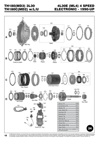
Transcription
Arcturus HistoGene Immunofluorescence Staining KitUser Guide
For Research Use Only. Not intended for any animal or human therapeutic or diagnostic use.Information in this document is subject to change without notice.APPLIED BIOSYSTEMS DISCLAIMS ALL WARRANTIES WITH RESPECT TO THIS DOCUMENT, EXPRESSED OR IMPLIED, INCLUDING BUT NOTLIMITED TO THOSE OF MERCHANTABILITY OR FITNESS FOR A PARTICULAR PURPOSE. TO THE FULLEST EXTENT ALLOWED BY LAW, IN NOEVENT SHALL APPLIED BIOSYSTEMS BE LIABLE, WHETHER IN CONTRACT, TORT, WARRANTY, OR UNDER ANY STATUTE OR ON ANY OTHERBASIS FOR SPECIAL, INCIDENTAL, INDIRECT, PUNITIVE, MULTIPLE OR CONSEQUENTIAL DAMAGES IN CONNECTION WITH OR ARISING FROMTHIS DOCUMENT, INCLUDING BUT NOT LIMITED TO THE USE THEREOF, WHETHER OR NOT FORESEEABLE AND WHETHER OR NOT APPLIEDBIOSYSTEMS IS ADVISED OF THE POSSIBILITY OF SUCH DAMAGES.TRADEMARKSThe trademarks mentioned herein are the property of Life Technologies Corporation or their respective owners.GeneChip is a trademark of Affymetrix, Inc. Cy is a trademark of GE Healthcare. Tissue-Tek is a trademark of Sakura Finetek. RNase AWAYis a trademark of Molecular Bio-Products, Inc. 2010 Life Technologies Corporation. All rights reserved.12653-00 Rev. C07/2010
Table of ContentsI. IntroductionA.B.C.D.BackgroundStorage and Stability34Safety Data Sheets4Related Products5II. Kit ComponentsA. Reagents and Supplies in KitB. Additional Lab Equipment and Materials Required66III. Preliminary StepsA. Material and Protocol ReviewB. Recommendations for RNase-free TechniqueC. Checking RNA Quality in Starting Material888IV. ProtocolA.B.C.D.E.Specimen FreezingSlide PreparationPrimary Biotinylated Antibody Solution PreparationCy3 Streptavidin Conjugate PreparationHistoGene LCM Immunofluorescence Staining Protocol99111212V. TroubleshootingA.B.C.D.Targeted Cells Do Not Lift From the Slide During LCMNo StainingBackground StainingRNA Cannot be Recovered from the Sample14141515VI. AppendicesA. Tissue Scrape Protocol for Verifying RNA Quality Usingthe PicoPure RNA Isolation Kit16
IntroductionI. IntroductionA. BackgroundThe HistoGene LCM Immunofluorescence Staining Kit isthe only kit designed to enable retrieval of high-quality RNAfrom immunofluorescently stained frozen tissue. It enablesconvenient and reliable staining, dehydration and Laser CaptureMicrodissection (LCM) of tissue sections with protocolsstreamlined and optimized both for optimal LCM capturesand maintaining RNA quality for downstream applicationsthat require intact RNA, like microarray analysis and RT-PCR.The HistoGene Immunofluorescence Staining Kit uses a twostep immunostaining technique that works with any biotinylatedmonoclonal antibody. It is based on use of biotin-avidin system:a primary biotinylated antibody and Cy 3-conjugatedStreptavidin. Avidin is an egg-white derived glycoprotein withan extraordinarily high affinity for biotin. Many biotin moleculescan be coupled to a protein, enabling the biotinylated protein tobind more than one molecule of avidin. Cy3 dye is used because itoffers the best staining intensity and works well with the biotinavidin system.The advantages of using the biotin-avidin system are that itimproves sensitivity and reduces background fluorescence.Furthermore, the high affinity between biotin and avidin assuresthe user of a rapidly formed and stable complex between primaryantibody and the avidin-fluorophore conjugate. Thus, thebiotin-avidin system provides highly specific staining with verylittle background and enables short staining times necessary tomaintain RNA integrity.Liver, kidney and some lymphoidtissues contain endogenous biotin andare therefore not recommended forstaining with Streptavidin based IHCprocedures.This kit should not be used withantibodies requiring enzyme digestionor target retrieval.The unique process is performed at 4 C using speciallyformulated Staining Buffer. This dramatically increases RNAyield from captured cells. The HistoGene Cold Block (HIS0101)makes the 4 C processing very convenient. It holds up to fourslides and has places for reagent tubes. It is designed for use withthe CoolSafe triple density polystyrene cooler (Cat. # CSF-BOX)3
and –10 C cool brick (Cat. # BRIK-1520) from DiversifiedBiotech (www.divbio.com). The proprietary Staining Bufferperserves the quality of RNA in the tissue sections and preservesgood, specific staining intensity.The HistoGene Immunofluorescence Kit requires starting withunfixed, frozen tissues known to contain intact RNA. Thus, werecommend that you verify RNA quality before you begin. SeeAppendix A. All Kit components used are premium grade,LCM certified and nuclease free to ensure RNA quality ismaintained during the process.B. Storage and Stability1.HistoGene LCM Immunofluorescence Staining KitStore the staining reagents as follows:Buffer A (–70 C)Buffer B (4 C)Cy3 Streptavidin (4 C)Store slides at room temperature and the Cold Block at4 C. Do not freeze and thaw Buffer A repeatedly.2.HistoGene Cold BlockStore at 4 C.C. Safety Data SheetsSafety Data Sheets (SDS) for kit chemicalcomponents are available from Technical Services.Call: 1 800 831-6844 option 5.You can also obtain these sheets from:www.appliedbiosystems.com.4Do not use on fixed frozen orformalin-fixed, paraffin-embeddedsections.
IntroductionD. Related ProductsPicoPure RNA Isolation KitFor extraction and isolation of total RNA from small samplesparticularly Laser Capture Microdissected (LCM) cells. ThePicoPure RNA Kit comes with optimized buffers, MiraCol Purification Columns and an easy-to-use protocol to maximizerecovery of high-quality total cellular RNA ready for amplificationwith the RiboAmp OA 1 Round RNA Amplification Kit.RiboAmp PLUS RNA Amplification KitThe RiboAmp PLUS RNA Amplification Kit enables the production of microgram quantities of antisense RNA (aRNA)from nanogram quantities of total cellular RNA. AmplifiedRNA produced using the kit is suitable for labeling and use forprobing expression microarrays. The kit achieves amplificationsof up to 1000-fold in one round of amplification, and amplifications of up to 1,000,000-fold in two rounds. The RiboAmpPLUS Kit comes with all necessary enzymes, reagents, andMiraCol purification columns needed to complete the includedamplification protocol.RiboAmp HS RNA Amplification KitThe RiboAmp HS RNA Amplification Kit starts with picogramtotal cellular RNA input and enables the production of microgramquantities of antisense RNA (aRNA). The Kit provides the greatestlevel of sensitivity in starting RNA quantities to produce enoughRNA suitable for labeling and hybridizing onto expressionmicroarrays. The RiboAmp HS Kit comes with all necessaryenzymes, reagents, and MiraCol Purification Columns neededto complete the included amplification protocol.5
PicoPure DNA Extraction KitThe PicoPure DNA Extraction Kit is optimized to maximize therecovery of genomic DNA from 10 or more cells captured byLCM. The kit comes with reagents and protocol tested to ensurecomplete extraction of DNA from LCM samples prepared withany standard tissue preparation procedure. DNA prepared usingthe kit is PCR-ready and needs no additional purification to performamplification.HistoGene LCM Frozen Section Staining KitThe HistoGene LCM Frozen Section Staining Kit is used toprocess tissue sections for LCM that maximizes the quality andyield of RNA from LCM cells. The Kit comes with all dehydrationand staining reagents, disposable staining jars, specially treatedslides, and detailed protocol and troubleshooting guide.Paradise PLUS Reagent SystemThe Paradise PLUS Reagent System is the only reagent system designed to enable gene expression studies using formalin-fixed paraffin-embedded (FFPE) tissue samples. Components include samplepreparation and staining reagents, RNA extraction and isolationreagents, RNA amplification reagents and a comprehensive userguide.6
Kit ComponentsII. Kit ComponentsA. Reagents and Supplies in KitsThe HistoGene Immunofluorescence Staining Kit includesreagents to stain 32 tissue sections. Fixation and dehydrationreagents are not supplied with the Kit. See additional materialsrequired below. Buffer A (8 x 500 µl)Buffer B (60 ml)Cy3 Streptavidin (60 µl)Slides (72)User GuideB. Additional Equipment and MaterialsRequired1. Materials PAP pen or ImmEdge pen (Vector Laboratories,Cat. # H-4000) RNase-free pipet tips 15 ml nuclease-free Falcon tubes Microcentrifuge tubes, nuclease-free RNaseAway (Life Technologies, Cat. # 10328-011) Kimwipes Tissue-Tek OCT Compound (VWR Cat. # 25608-930) Staining jars (Evergreen Scientific # 222-5450-G8S) Dry ice Disposable gloves Tissue-Tek Cryomold (VWR Cat. # 25608-916)2. Equipment -70ºC freezer Fume hood Cryostat with disposable microtome blades Pipettors: 1000 µl, 200 µl, 100 µl, 10 µl Cover glass forceps HistoGene Cold Block (Applied Biosystems Cat. # HIS0101) Thermometer, celsius Microslide box, plastic (VWR Cat. # 48444-004) Refrigerator7
2. Reagents Acetone(VWR Cat. # AX-125-4; Fisher Cat. # HC-300-GAL) Xylene(VWR Cat. # EM-XX0060-4; Fisher Cat. # HC-700-GAL) Ethanol 100%(VWR Cat. # 34172-020; Fisher Cat. # HC-800-GAL) Ethanol 95%(VWR Cat. # 34172-022; Fisher Cat. # HC-1100-GAL) Ethanol 70%(VWR Cat. # 34172-000; Fisher Cat. # HC-1000-GAL) Isopentane or 2-Methylbutane (VWR Cat. #JTQ223-7)8
Preliminary StepsIII.Preliminary StepsA. Material and Protocol ReviewThis protocol is only for immunofluorescent staining of acetonefixed frozen tissue sections.To get the most from your HistoGene LCM ImmunofluorescenceStaining Kit, take a few moments to examine the components ofthe kit and read through the information in this Section andSection IV.B. Recommendations for RNase-free TechniqueRNase contamination will cause experimental failure. MinimizeRNase contamination by adhering to the followingrecommendations throughout your experiment: Wear disposable gloves and change them frequently. Use RNase-free solutions, glassware and plasticware. Do not re-purify HistoGene LCM ImmunofluorescenceStaining Kit components. They are certified Nuclease Free. Wash scalpels, tweezers and forceps with detergent and bakeat 210 C for four hours before use. Use RNase AWAY solution (Life Technologies) accordingto the manufacturer’s instructions on any surfaces that may comein contact with the sample.C. Checking RNA Quality in Starting MaterialBefore you begin preparing specimens, it is critical that you firstcheck the quality of the RNA in the tissue to ensure that yoursamples contain intact, high-quality RNA. See Appendix A.9
IV. ProtocolA. Specimen FreezingNote that isopentane has a very low flash point and should bekept away from open flames. Perform procedure in a fumehood or a well-ventilated space.1.Place dry ice in an appropriate container.2.Slowly pour isopentane into container with dry ice, fillinguntil the isopentane level is just above the layer of dry ice.3.Bubbling of the isopentane will occur upon its addition tothe dry ice, once this has subsided the isopentane is readyfor use.4.If necessary, identify specimen on cryomold using a sharpiepen.5.Take cryomold and place a thin layer of OCT on the bottomof it.6.Collect dissected tissue specimen and place tissue in desiredorientation onto the layer of OCT in the cryomold.7.Carefully add more OCT until specimen is completelycovered and the cryomold is filled.8.Carefully place prepared cryomold into the cooledisopentane.9.Wait for OCT to completely solidify. If freezing downadditional specimens, the processed specimens can be heldin a separate container with dry ice only.For best RNA preservation, freezetissue specimens immediately afterdissection.Wear clean disposable glovesthroughout the Specimen Freezingprocedure. Use clean RNase-freeinstruments.10. Store frozen specimen in the cryomold in a –70 C freezeror proceed to slide preparation.It is okay to stop at this point in the protocol.B. Slide Preparation1. Pre-cool the cryostat to the temperature recommended bythe manufacturer for the specimen you are preparing.10Wear clean disposable glovesthroughout the Slide Preparationprocedure.
Protocol2.Remove and discard old microtome blade. Wipe down theknife holder and antiroll plate in the cryostat with 100%ethanol to avoid sample cross-contamination. Dry wetsurfaces with another dry Kimwipe.3.Install a new disposable microtome blade in the cryostat.4.Set cutting thickness to 8-10 µm.5.Place a microslide box on dry ice near the cryostat.6.Transfer the cryomold containing your specimen fromthe –70 C freezer to the cryostat, transporting on dry ice ifnecessary.7.Wait a minimum of 10 minutes for your specimen toequilibrate with the temperature of the cryostat.8.Mount specimen to specimen holder with OCT. Cut 810µm sections.9.Mount sections toward the center of a room temperatureLCM microslide. Place slide immediately into microslidebox on dry ice. Do not allow slide to dry at room temperature.Frequent cycling of the tissue blockfrom –70oC to –20oC for cryosectioningmay accelerate RNA degradation. Forbest results, cut and mount a sufficientnumber of sections for two months’use during one cryosectioning session.Store the mounted sections at –70oCuntil needed.10. Discard slides with folded or wrinkled sections.11. If you are cutting more than one specimen, use a new unusedsection of the disposable microtome blade or a new blade foreach one. In addition, wipe down the knife holder and antiroll plate with 100% ethanol in between each specimen toavoid cross-contamination.12. Upon completion of sectioning, the surface of the specimenmust be protected with a thin layer of OCT prior to storingat –70C. This can be accomplished by applying a smallamount of OCT on the surface of the block and immediatelyapplying a metal block that has been pre-cooled to thecryostat’s temperature. To separate the two objects once theOCT has solidified, take a razor blade and place it betweenthe metal block and the OCT. Once separated, carefullyremove the specimen block from the specimen holder usingthe razor blade. Remove any excess OCT surrounding theblock with the razor blade and replace the specimen into itscryomold.13. Proceed immediately to the “Staining and Dehydration”segment of the protocol or store slides in a slide box at –70 Cfor up to two months.11
C. Primary Biotinylated Antibody Solution PreparationWhen using the HistoGene LCM Immunofluorescence StainingKit, it is important to use primary antibodies proven to be ofhigh quality. They should be tested in a conventionalimmunostaining procedure and shown to produce good stainingintensity with low background before proceeding with theHistoGene LCM Immunofluorescence Staining protocol.The recommended concentration of the primary biotinylatedantibody should be between 30–100 µg/ml. The finalconcentration of the primary antibody should be optimizedby the user by performing a serial dilution staining experimentusing the HistoGene Immunofluorescence Kit’s Staining Buffer(Buffers A & B combined).Biotinylated antibodies against many different antigens arenow commercially available. If a certain antibody cannot beobtained from a commercial source the biotinylation can easilybe performed using a biotinylation kit (Pierce cat # 21338,Sigma Cat. # B-TAG).It should be noted, that some commercially availablebiotinylated antibodies have low IgG concentrations and needto be used undiluted.To perform a negative control staining, use a biotinylated controlantibody from the same animal species and of the same isotypeas your primary antibody. Dilute to the same workingconcentration as the primary antibody. (Mouse control antibodiescan be obtained from DAKO Cat. # X0928).Working solution of the primary biotinylated antibody and thenegative control antibody should be prepared in Staining Buffer(Buffers A & B) and held at 4 C. The recommendedconcentration of Buffer A in all antibody solutions is 10 mM. Ifpreparing the antibody solution dilutes the Buffer Aconcentration below 10 mM, add an adequate volume of theBuffer A stock solution (100 mM) to the antibody solution.12
ProtocolD. Cy3 Streptavidin Conjugate PreparationWorking solution of Cy3 Streptavidin should be prepared inStaining Buffer (Buffer A & B) and held at 4 C, protectedfrom light. The recommended dilution of the Cy3 Streptavidinis 1:100. User should, however, determine the optimal stainingconcentration of the Cy3 Streptavidin conjugate by performinga serial dilution staining experiment using the HistoGeneImmunofluorescence Kit’s Staining Buffer (Buffers A & B).E. HistoGene LCM Immunofluorescence Staining ProtocolMinimizing the time required and keeping the samples coldduring immunoflourescent staining is critical for obtaining highRNA quality and yield. For best results, the staining protocolshould be performed within a maximum of 10 minutes, notincluding dehydration steps. This requires maintaining a steadyworkflow. To help minimize preparation time and keep thesamples cold, we recommend that you do not process more than4 slides at once. Also do not allow the sections to dry after Step14 of the protocol. Drying may result in non-specific staining,loss of specific staining and it may compromise RNA quality.1. Pre-cool the HistoGene Cold Block in a refrigerator to 4 C.2. Take out one aliquot of frozen Buffer A and thaw.3. Prepare fixative and dehydration reagents in the fume hood.4. Label 5 plastic jars as follows:a Acetoneb. 70% ethanolc. 95% ethanold. 100% ethanole. Xylene5. Fill each labeled jar with 25 ml of solution.6. Cool Acetone to 4 C.7. Take the cold block out of the refrigerator and place it in theCoolSafe box or on wet ice. Just prior to staining, check thetemperature of the cold block by placing a thermometer inone of the tube holes. The temperatures should be 4 C orDiscard any remaining Buffer A oncethawed.All solutions must be prepared justprior to staining.The stock solution of Buffer A is100 mM, and the working solutionshould be 10 mM.Staining procedure is performed at 4 C by keeping the slides on a coldblock throughout all the steps of theprocedure.13
below.8. Prepare Staining Buffer by adding one aliquot (500 µl) ofthe Buffer A into 4500 µl of Buffer B. Store tube in the coldblock. Discard any remaining Buffer A once thawed.9. Prepare primary antibody solutions as described in SectionIV.C and keep them in the cold block.10. Prepare the Cy3 Streptavidin conjugate and keep it in thedark and at 4 C as described in Section IV.D.Process only the number of slidesthat you will be able to microdissectwithin 2 hours after the finaldehydration step. The RNA in theprocessed sample degrades even afterfinal dehydration step!11. Remove a maximum of four slides from the –70 C freezer,allow no more than 30 seconds for the condensation todisappear prior to fixing in acetone.12. Place slides in cold acetone (4 C) for 2 minutes.13. Remove from acetone and air dry the slides in the fumehood for 30 seconds.14. Use hydrophobic barrier pen to circumscribe each sectionto keep the staining reagents localized on the tissue section.15. Place all the slides on a cold block.16. Apply 200 µl of Staining Buffer per section to each slide.17. Drain off staining buffer and wipe the slide gently, withouttouching the section, with a clean Kimwipe.Do not allow the sections to dry. Workwith one slide at a time through steps#17 and 18.18. Place the slides on the cold block and apply 100 µl persection of the diluted primary antibody and incubate for 3minutes.Place the slide back on the cold blockif processing multiple slides.19. Drain off the antibody solution and rinse the sections byapplying 200 µl of Staining Buffer per section, drain andrepeat rinsing.Do not allow the sections to dry. Workwith one slide at a time through steps#20 and 21.20. Drain off staining buffer and wipe the slide gently with aclean Kimwipe.21. Place the slides on the cold block and apply 100 µl persection of the diluted Cy3 Streptavidin and incubate for 1minute.22. Drain off and rinse by applying 200 µl of Staining Bufferper section, drain and repeat rinsing.23. Immediately proceed to the dehydration protocol.Dehydration is performed at room temperature.14Discard any unused Staining Bufferand diluted antibody solutions.
Troubleshooting1.Place the slides in 70% ethanol for 30 seconds.2.Transfer into 95% ethanol for 30 seconds.3.Transfer into freshly dispensed 100% ethanol for 30seconds.4.Transfer into xylene for 5 minutes.5.Dry the slides in a fume hood for 5 minutes.6.Immediately perform Laser Capture Microdissection.V. TroubleshootingA. Targeted Cells Do Not Lift From the SlideDuring LCM1.The sample may contain residual water. Ensure that theethanol solutions are fresh. Ethanol is hygroscopic. Keepthe ethanol bottles tightly capped, and do not pour ethanolsolutions until you are ready to use them. If you suspect thatthe 100% ethanol solution has absorbed water, purchase anew bottle.2.The sample may have dried between protocol steps. Performall steps of the protocol at a steady pace.B. No Staining1.Make sure that the primary antibody is used at rightconcentration and that it is active. Perform a titrationexperiment using both high and low concentration of theprimary antibody.2.You can test the activity of the antibody on a section takenfrom another known positive tissue.3.Make sure that the Cy3 Streptavidin is not inappropriatelydiluted. Perform a titration experiment using both high andlow dilutions of the Cy3 conjugate.15
C. Background Staining1.The primary antibody used is too concentrated. Perform atitration experiment using both high and low concentrationsof the primary antibody to achieve clean specific stainingwithout background.2.The primary antibody may cross-react with other tissueepitopes and bind non-specifically. Change source or speciesof the primary antibody.3.Make sure that sections are kept moist during all steps ofthe staining.4.Endogenous biotin may be present in the tissue. To check ifthis is occurring perform a staining omitting the primaryantibody application.5.Tissue such as kidney, liver and adrenals as well as somelymphoid tissues contain different levels of endogenousbiotin and should not be used with a biotin/avidin system.D. RNA Cannot be Recovered from theSample1.The sample starting material may contain poor quality RNA.Freeze the sample immediately following dissection, andtake care to use RNase-free technique.2.RNA may become degraded during RNA isolation. Weargloves; use RNase-free technique and RNase-freeinstruments and reagents. Wipe down the Laser CaptureMicrodissection System with RNase AWAY prior to use.3.RNA may not be fully extracted and isolated from cells onthe LCM cap. Use the Arcturus PicoPure RNA IsolationKit or another guanidinium extraction method. PerformRNA extraction immediately after LCM to ensure completeextraction and optimum recovery of RNA.4.The starting material quantity may be insufficient. Use atleast 500 captured cells.16
AppendixVI. AppendixA. Tissue Scrape Protocol for Verifying RNAQuality Using the PicoPure RNA Isolation KitApplied Biosystems recommends verifying the integrity of RNA inthe tissue sample before proceeding with staining and LaserCaptureMicrodissection (LCM) procedures. This enablesyou to understand the quality of the RNA in the experimentalsample before proceeding with further downstream processing.This protocol is recommended for all new frozen tissue samples.The protocol involves preparing and dehydrating a tissue section,then scraping the entire tissue section into a 0.5 ml tube. RNA isthen extracted from the sample using a modified version of thePicoPure RNA Isolation Kit (Catalog # KIT0204) protocol forlarger amounts of tissue. Finally, the Lab-on-a-Chip System(Agilent) or a gel can be used to assess 28S and 18S ribosomalRNA integrity. If ribosomal bands are detected, then the samplecontains viable RNA and is therefore a good candidate for LCM.If the ribosomal RNA bands are faint or not present, then thesample may contain degraded RNA.For more information, visit: www.appliedbiosystems.com, orcall technical support at: 1 800 831-6844 option 5.17
Part Number 12653-00 Rev. C 07/2010Headquarters5791 Van Allen WayCarlsbad, CA 92008 USA Phone 760.603.7200www.lifetechnologies.comTechnical Resources and SupportFor the latest technical resources and support informationfor all locations, please refer to our Web site atwww.appliedbiosystems.com
The PicoPure DNA Extraction Kit is optimized to maximize the recovery of genomic DNA from 10 or more cells captured by LCM. The kit comes with reagents and protocol tested to ensure complete extraction of DNA from LCM samples prepared with any standard tissue preparation procedure. DNA prepared using the kit is PCR-ready and needs no additional .