Transcription
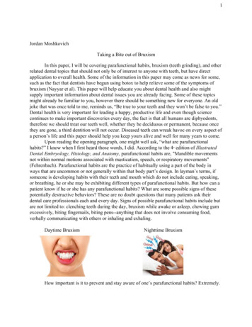
ReviewSpecial Issue: Penile Anomalies in ChildrenTheScientificWorldJOURNAL (2010) 10, 1174–1179TSW UrologyISSN 1537-744X; DOI 10.1100/tsw.2010.112Penile Embryology and AnatomyJenny H. Yiee* and Laurence S. BaskinDepartment of Urology, University of California San FranciscoE-mail: yieejh@urology.ucsf.edu; lbaskin@urology.ucsf.eduReceived March 1, 2010; May 12, 2010; Published June 29, 2010Knowledge of penile embryology and anatomy is essential to any pediatric urologist inorder to fully understand and treat congenital anomalies. Sex differentiation of theexternal genitalia occurs between the 7th and 17th weeks of gestation. The Y chromosomeinitiates male differentiation through the SRY gene, which triggers testiculardevelopment. Under the influence of androgens produced by the testes, externalgenitalia then develop into the penis and scrotum. Dorsal nerves supply penile skinsensation and lie within Buck’s fascia. These nerves are notably absent at the 12 o’clockposition. Perineal nerves supply skin sensation to the ventral shaft skin and frenulum.Cavernosal nerves lie within the corpora cavernosa and are responsible for sexualfunction. Paired cavernosal, dorsal, and bulbourethral arteries have extensiveanastomotic connections. During erection, the cavernosal artery causes engorgement ofthe cavernosa, while the deep dorsal artery leads to glans enlargement. The majority ofvenous drainage occurs through a single, deep dorsal vein into which multiple emissaryveins from the corpora and circumflex veins from the spongiosum drain. The corporacavernosa and spongiosum are all made of spongy erectile tissue. Buck’s fasciacircumferentially envelops all three structures, splitting into two leaves ventrally at thespongiosum. The male urethra is composed of six parts: bladder neck, prostatic,membranous, bulbous, penile, and fossa navicularis. The urethra receives its bloodsupply from both proximal and distal directions.KEYWORDS: penis, urethra, anatomy, embryologyPENILE EMBRYOLOGYExternal genital differentiation into the male sex begins in the 7th week of gestation and is completed by16–17 weeks[1,2,3]. Prior to this, between the 4th and 7th weeks, the mesodermal mesenchyme migrates tothe cranial aspect of the cloacal membrane to form the genital tubercle. The cloacal membrane itself iscomposed of two layers: endoderm and ectoderm. The caudal portion of the cloacal membrane developsinto urogenital folds. These structures are the precursors for external genitalia in both males andfemales[4], Fig. 1.In those with a Y chromosome, the SRY gene signals the differentiation of primitive sex cords intotestes by first signaling the development of Sertoli cells[5]. Sertoli cells then aid in the development ofgerm cells and Leydig cells[6] within the testes. Leydig cells produce testosterone, which is converted todihydrotestosterone to induce external genitalia development.*Corresponding author. 2010 with author.Published by TheScientificWorld; www.thescientificworld.com1174
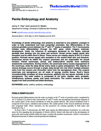
Yiee and Baskin: Penile Embryology and AnatomyATheScientificWorldJOURNAL (2010) 10, 1174–1179Genital TubercleUrogenital FoldsLabioscrotal SwellingsCloacal MembraneAnal FoldsBCUrethra (urogenital folds)Clitoris (genital tubercle)Penis (genital tubercle)Scrotum (labioscrotal folds)Anus (anal folds)Labia Minora(urogenital folds)Labia Majora(labioscrotal folds)Anus (anal folds)FIGURE 1. Human urogenital embryology. (A) Sex undifferentiated precursors prior to 7 weeks gestational age. (B) Male development ingestational weeks 7–17 from precursors shown in (A). (C) Female development in weeks 7–17 from precursors shown in (A).Under the influence of androgens, in males the mesoderm of the genital tubercle enlarges to becomethe corpora cavernosa and glans penis. The endoderm tubularizes in a proximal to distal direction to formthe penile urethra. The ectoderm develops into the penile skin and prepuce, which begins formation at 8weeks. There is some debate as to whether the distal urethra is formed from tubularization of theendoderm, as with the proximal urethra, or from canalization of the ectoderm distally[2,7].1175
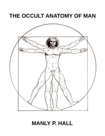
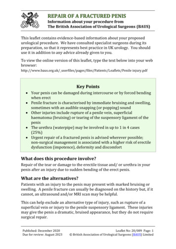
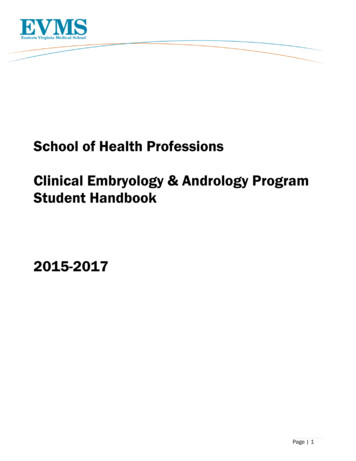
Yiee and Baskin: Penile Embryology and AnatomyTheScientificWorldJOURNAL (2010) 10, 1174–1179PENILE ANATOMYNervesPenile innervation consists of the dorsal, cavernosal, and perineal nerves. Dorsal nerves arising from thepudendal nerves travel within Buck’s fascia, together with the dorsal arteries and veins, to supplysensation to penile skin[8], Fig. 2. Despite its nomenclature, it is important to note that the nerves do notlie directly in the dorsal midline, but rather extend from the 11 and 1 o’clock positions laterally to thejunction of the cavernosa and spongiosum[9,10], Fig. 2. These nerves do not send perforators deepthrough the tunica albuginea to the corpora cavernosa[9]. There is a paucity of nerves at the 12 o’clockshaft position. Therefore, in correction of penile curvature, plication at the 12 o’clock position is the arealeast likely to result in nerve damage.Dorsal Nerves (black)VeinTunica AlbugineaCorpora CavernosaBuck’s FasciaDartos FasciaUrethraCorpus SpongiosumFIGURE 2. Histology of penile anatomy in cross-section. Note that the dorsal nerves start at the 11 and 1 o’clock positionsof the corpora cavernosa and extend around laterally to the cavernosal-spongiosal junction. There is a notable paucity ofnerves at the 12 o’clock position.Like the dorsal nerves, the perineal nerves also arise from the pudendal nerve to supply the ventralshaft skin, the frenulum, and the bulbospongiosus muscle[11], Fig. 3. The cavernosal nerves arise fromthe autonomic pelvic plexus and travel along the periprostatic neurovascular bundle, well known tourologists performing radical retropubic prostatectomies. Underneath the pubic arch, the cavernosalnerves pierce through the corpora cavernosa[12], Fig. 4. Proximal to this point, the cavernosal and dorsalnerves lie within close proximity at the penile hilum and are thought to exchange signal communication,which may have implications on erectile function. As well, there are interactions between perineal anddorsal nerves laterally at the junction of the cavernosa and spongiosum along the penis, which may alsohave implications on erection and ejaculation[13].1176
Yiee and Baskin: Penile Embryology and AnatomyTheScientificWorldJOURNAL (2010) 10, 1174–1179Pubic arch (navy)Dorsal nerves (green)Corpora cavernosa (purple)Glans (yellow)Corpus spongiosum (teal)Perineal nerves (yellow/green)FIGURE 3. Ventral-lateral view: 3-dimensional reconstruction of penile anatomy.Glans penis (yellow)Corpora cavernosa (white)Dorsal nerves (yellow/green)Urethra (light blue)Corpus spongiosum (teal)Cavernosal nerves (pink)Pubic arch (navy)FIGURE 4. Dorsal-lateral view: 3-dimensional reconstruction of penile anatomy.1177
Yiee and Baskin: Penile Embryology and AnatomyTheScientificWorldJOURNAL (2010) 10, 1174–1179VasculatureArteriesThere are three paired main arteries in the penis: cavernosal, dorsal, and bulbourethral. All three arisefrom a shared branch of the internal pudendal artery, which itself arises from the internal iliac artery. Oneach side, the first branching occurs at the bulb of the spongiosum external to the urogenital diaphragmforming the bulbourethral artery, which then lies at the 9 and 3 o’clock positions of the corpusspongiosum. Then the cavernous artery branches to penetrate the corpora cavernosa and the remainder ofthe artery continues as the deep dorsal artery. The deep dorsal artery causes glans enlargement duringerection, whereas the cavernosal arteries cause corporal enlargement. All three arteries communicatedistally near the glans to provide an extensive anastomotic network[8,14].Penile skin derives its supply from a separate origin. Branches of the external pudendal artery supplythe dorsal and lateral aspects of the penis, and branches of the internal pudendal artery supply the ventralpenis and scrotum via the posterior scrotal artery[15]. These branches course in the Dartos fascia andenable pedicled skin flaps to be used in urethral reconstruction.VeinsVenous drainage is not analogous to arterial supply, unlike many other body systems. In contrast to thepaired dorsal arterial system, there exists only one deep dorsal vein that runs alongside the dorsal arteriesand nerves in Buck’s fascia above the tunica albuginea. The deep dorsal vein receives drainage from thedistal two-thirds of the corpora cavernosa via emissary veins and the corpus spongiosum via circumflexveins. Emissary veins are the veins that traverse obliquely through the tunica albuginea, allowing them tobe compressed during erections for penile tumescence. The deep dorsal vein then drains to theperiprostatic plexus[16].Recently, a small pair of dorsal veins have been found that lie just deep to the deep dorsal vein, butabove the tunica albuginea, which independently receive emissary vein drainage. These veins have beentermed cavernosal veins, but do not lie within the corpora cavernosa[17]. Older literature refers to thecavernosal veins as short veins located in the triangle between the proximal crus that drain the proximalone-third of the corpora cavernosa. These veins join with the bulbourethral veins (which drain theproximal spongiosum) to lead into the internal pudendal vein. The penile skin drains via the superficialdorsal vein, which drains into the saphenous vein.Corpora Cavernosa and GlansThe proximal crus of the corpora cavernosa begin as separate structures spread apart under the ischiumbilaterally and move medially under the pubis until they lie adjacent to each other. They are eachenveloped in a fibrous tunica albuginea, which share a septum medially when the crus are adjacent. Thisseptum is perforated, allowing for communication of the cavernosal sinuses in the penis. The distal tips ofthe corpora cavernosa are capped by the glans penis, which is itself a continuation of the corpusspongiosum that surrounds the urethra. Buck’s fascia surrounds the paired corpora cavernosa and corporaspongiosum circumferentially. It splits into two leaves to surround the corpus spongiosum completely onthe ventral aspect of the penis, see Fig. 2. An extension of Buck’s fascia forms the suspensory ligament,which attaches the penis to the underside of the pubis. Buck’s fascia is continuous with deep pelvicmuscle fascia. Superficial to Buck’s fascia is Dartos fascia, which is continuous with Scarpa’s fascia ofthe abdomen and Dartos fascia of the scrotum[8,14].1178
Yiee and Baskin: Penile Embryology and AnatomyTheScientificWorldJOURNAL (2010) 10, 1174–1179UrethraThe male urethra is divided into six parts: bladder neck, prostatic urethra, membranous urethrasurrounded by external sphincter, bulbous urethra proximal to the ischiocavernosus muscle,penile/pendulous urethra distal to the ischiocavernosus muscle, and the fossa navicularis within the distalglans. The corpus spongiosum is erectile tissue akin to corpora cavernosa, but with a thinner tunicaalbuginea. The penile and bulbar urethra lie within the spongiosum. The penile urethra lies in a centrallocation within the spongiosum, whereas the bulbar urethra lies eccentrically closer to the dorsalspongiosum prior to exiting dorsally to become the membranous urethra to join the prostate[15]. Whereasthe condition known as “chordee” or penile curvature was once believed to result from fibrous bands nearthe urethra, no such fibrous tissue has been found in the penile urethra, even in severe cases ofhypospadias[18]. Because of anastomotic communications between the dorsal arteries and thebulbourethral arteries, the urethra receives arterial supply from both distal and proximal directions. Thisenables complete transaction of the urethra without necrosis of the distal .15.16.17.18.Belman, A. and King, L. (1976) Urethra. In Clinical Pediatric Urology. Vol. 1. Kelalis, P., King, L., and Belman, A.,Eds. WB Saunders, Philadelphia. p. 577.Moore, K. and Persaud, T. (1998) Before We are Born: Essentials of Embryology and Birth Defects. WB Saunders,Philadelphia.Baskin, L. (2000) Hypospadias and urethral development. J. Urol. 163, 951–956.Park, J. (2007) Normal development of the urogenital system. In Campbell-Walsh Urology. Vol. 4. Wein, A.,Kavoussi, L., Novick, A., Partin, A., and Peters, C., Eds. Saunders-Elsevier, Philadelphia.Sinclair, A., Berta, P., and Palmer, M. (1990) A gene from the human sex-determining region encodes a protein withhomology to a conserved DNA-binding motif. Nature 346, 240–244.Koopman, P. (2009) The delicate balance between male and female sex determining pathways: potential fordisruption of early steps in sexual development. Int. J. Androl. 32, 1–7.Kurzrock, E., Baskin, L., and Cunha, G. (1999) Ontogeny of the male urethra: theory of endodermal differentiation.Differentiation 64, 115–122.Brooks, J.D. (2007) Anatomy of the lower urinary tract and male genitalia. In Campbell-Walsh Urology. Vol. 1.Wein, A., Kavoussi, L., Novick, A., Partin, A., and Peters, C., Eds. Saunders-Elsevier, Philadelphia.Baskin, L., Erol, A., Li, Y., and Liu, W. (2000) Anatomy of the neurovasuclar bundle: is safe mobilization possible?J. Urol. 164, 977–980.Baskin, L., Erol, A., Li, Y., and Cunha, G. (1998) Anatomical studies of hypospadias. J. Urol. 160, 1108–1115.Yucel, S. (2003) Neuroanatomy of the male urethra and perineum. BJU Int. 92, 624–630.Akman, Y., Liu, W., Li, Y., and Baskin, L. (2001) Penile anatomy under the pubic arch: reconstructive implications.J. Urol. 166, 225–230.Yucel, S. and Baskin, L. (2003) Identification of communicating branches among thedorsal, perineal and cavernousnerves of the penis. J. Urol. 170, 153–158.Redman, J. (1991) Anatomy of the genitourinary system. In Adult and Pediatric Urology. Vol. 1. Gillenwater, J.,Grayhack, J., Howards, S., and Duckett, J., Eds. Mosby, St. Louis.Jordan, G. and Schlossberg, S. (2007) Surgery of the penis and urethra. In Campbell-Walsh Urology. Vol. 2. Wein,A., Kavoussi, L., Novick, A., Partin, A., and Peters, C., Eds. Saunders-Elsevier, Philadelphia.Bella, A., Brant, W., and Lue, T. (2009) Anatomy of erectile function. In Textbook of Erectile Dysfunction. Carson, C.and Wyllie, M., Eds. Informa Healthcare, New York.Hsu, G., Hsieh, C., Wen, H., Chen, Y., Chen, S., and Mok, M. (2003) Penile venous anatomy: an additionaldescription and its clinical implication. J. Androl. 24, 921–927.Erol, A., Baskin, L., Li, Y., and Liu, W. (2000) Anatomical studies of the urethral plate: why preservation of theurethral plate is important in hypospadias repair. BJU Int. 85, 728–734.This article should be cited as follows:Yiee, J.H. and Baskin, L.S. (2010) Penile embryology and anatomy. TheScientificWorldJOURNAL: TSW Urology 10, 1174–1179. DOI 10.1100/tsw.2010.112.1179
MEDIATORSofINFLAMMATIONThe ScientificWorld JournalHindawi Publishing Corporationhttp://www.hindawi.comVolume 2014GastroenterologyResearch and PracticeHindawi Publishing Corporationhttp://www.hindawi.comVolume 2014Journal ofHindawi Publishing Corporationhttp://www.hindawi.comDiabetes ResearchVolume 2014Hindawi Publishing Corporationhttp://www.hindawi.comVolume 2014Hindawi Publishing Corporationhttp://www.hindawi.comVolume 2014International Journal ofJournal ofEndocrinologyImmunology ResearchHindawi Publishing Corporationhttp://www.hindawi.comDisease MarkersHindawi Publishing Corporationhttp://www.hindawi.comVolume 2014Volume 2014Submit your manuscripts athttp://www.hindawi.comBioMedResearch InternationalPPAR ResearchHindawi Publishing Corporationhttp://www.hindawi.comHindawi Publishing Corporationhttp://www.hindawi.comVolume 2014Volume 2014Journal ofObesityJournal ofOphthalmologyHindawi Publishing Corporationhttp://www.hindawi.comVolume 2014Evidence-BasedComplementary andAlternative MedicineStem CellsInternationalHindawi Publishing Corporationhttp://www.hindawi.comVolume 2014Hindawi Publishing Corporationhttp://www.hindawi.comVolume 2014Journal ofOncologyHindawi Publishing Corporationhttp://www.hindawi.comVolume 2014Hindawi Publishing Corporationhttp://www.hindawi.comVolume 2014Parkinson’sDiseaseComputational andMathematical Methodsin MedicineHindawi Publishing Corporationhttp://www.hindawi.comVolume 2014AIDSBehaviouralNeurologyHindawi Publishing Corporationhttp://www.hindawi.comResearch and TreatmentVolume 2014Hindawi Publishing Corporationhttp://www.hindawi.comVolume 2014Hindawi Publishing Corporationhttp://www.hindawi.comVolume 2014Oxidative Medicine andCellular LongevityHindawi Publishing Corporationhttp://www.hindawi.comVolume 2014
TSW Urology ISSN 1537-744X; DOI 10.1100/tsw.2010.112 *Corresponding author. 2010 with author. Published by TheScientificWorld; www.thescientificworld.com 1174 Penile Embryology and Anatomy Jenny H. Yiee* and Laurence S. Baskin Department of Urology, University of California San Francisco E-mail: yieejh@urology.ucsf.edu; lbaskin@urology.ucsf.edu