Transcription
(2021) 18:65Nicolás‑Párraga et al. Virol Jhttps://doi.org/10.1186/s12985-021-01529-9Open AccessRESEARCHHuman DNA decays faster with timethan viral dsDNA: an analysis on HPV16 usingpathology archive samples spanning 85 yearsSara Nicolás‑Párraga1*† , Montserrat Torres1*†, Laia Alemany2,3,7, Ana Félix4, Eugenia Cruz5,Silvia de Sanjosé2,3,6,7, Francesc Xavier Bosch2,3,8,9 and Ignacio G. Bravo1,10,11 on behalf of the RIS HPV TT studygroupsAbstractBackground: Quality of the nucleic acids extracted from Formalin Fixed Paraffin Embedded (FFPE) samples largelydepends on pre-analytic, fixation and storage conditions. We assessed the differential sensitivity of viral and humandouble stranded DNA (dsDNA) to degradation with storage time.Methods: We randomly selected forty-four HPV16-positive invasive cervical cancer (ICC) FFPE samples collectedbetween 1930 and 1935 and between 2000 and 2004. We evaluated through qPCR the amplification within the samesample of two targets of the HPV16 L1 gene (69 bp, 134 bp) compared with two targets of the human tubulin-β gene(65 bp, 149 bp).Results: Both viral and human, short and long targets were amplified from all samples stored for 15 years. In samplesarchived for 85 years, we observed a significant decrease in the ability to amplify longer targets and this differencewas larger in human than in viral DNA: longer fragments were nine times (CI 95% 2.6–35.2) less likely to be recoveredfrom human DNA compared with 1.6 times (CI 95% 1.1–2.2) for viral DNA.Conclusions: We conclude that human and viral DNA show a differential decay kinetics in FFPE samples. The fasterdegradation of human DNA should be considered when assessing viral DNA prevalence in long stored samples, asHPV DNA detection remains a key biomarker of viral-associated transformation.Keywords: Human Papillomavirus 16, Invasive cervical carcinoma, Formalin-Fixed Paraffin Embedded, Viruses, DNA,Degradation, Integrity, Sample storage, StabilityBackgroundIn pathology routine most biopsies and surgical samplesare formalin fixed paraffin embedded (FFPE) to preserve tissue structure and cellular morphology. Biobanksand FFPE sample collections constitute valuable sample*Correspondence: sara.nicolas.parraga@gmail.com; mtorres@iconcologia.net†Sara Nicolás-Párraga and Montserrat Torres have contributed equally tothis work1Infections and Cancer Laboratory, Cancer Epidemiology ResearchProgram, Catalan Institute of Oncology (ICO), Granvia de L’Hospitalet199‑203, 08908 L’Hospitalet de Llobregat, SpainFull list of author information is available at the end of the articlesources because they allow to create and maintain wellarchived, extensive, easy to handle and relatively inexpensive sample repositories to perform large histologicaland molecular retrospective studies [1–3].For molecular epidemiology studies, the informationtargeted is usually nucleic acid sequencing, but tissuepreservation in paraffin blocks is variable and depends ona number of factors in the different steps of the processthat affect quantity and quality of the extracted nucleicacids [4, 5]. One of the most important and studied factors is the type of the fixative chosen for the fixationstep, with formalin being the commonest one. Formalin The Author(s) 2021. Open Access This article is licensed under a Creative Commons Attribution 4.0 International License, whichpermits use, sharing, adaptation, distribution and reproduction in any medium or format, as long as you give appropriate credit to theoriginal author(s) and the source, provide a link to the Creative Commons licence, and indicate if changes were made. The images orother third party material in this article are included in the article’s Creative Commons licence, unless indicated otherwise in a credit lineto the material. If material is not included in the article’s Creative Commons licence and your intended use is not permitted by statutoryregulation or exceeds the permitted use, you will need to obtain permission directly from the copyright holder. To view a copy of thislicence, visit http://creat iveco mmons .org/licen ses/by/4.0/. The Creative Commons Public Domain Dedication waiver (http://creat iveco mmons .org/publi cdoma in/zero/1.0/) applies to the data made available in this article, unless otherwise stated in a credit line to the data.
Nicolás‑Párraga et al. Virol J(2021) 18:65fixation occurs through the formation of methylenebridges between the aldehyde group in the fixative andthe amino groups in nucleic acids and proteins [6]. Overfixation or the use of a fixative molecule with multipleactive groups, e.g. glutaraldehyde, hardens the tissue bycross-linking multiple molecules and can induce physical fragmentation of the nucleic acids during extraction[7, 8]. If unbuffered, the aldehyde group in formalin canundergo chemical disproportionation, so that two molecules of formaldehyde yield one molecule of methanoland one molecule of formic acid. The exposure of DNAto methanol and formic acid can lead to DNA depurination and strand breaks [6, 9]. Overall, DNA damageinduced by fixation conditions can hinder downstreamapplications based on amplification techniques [10, 11].Other important factor for the effects of the fixationprocess on nucleic acid quality is the duration of storageperiod and in this regard the literature is contradictory.While certain studies describe that the use of FFPE specimens stored for several years (i.e. samples stored between10 to 20 years) has only minor effects on subsequentDNA analysis [12, 13], other authors show that the lengthof PCR-amplified fragments and whole genome amplified-fragments decreases with storage time even whencoupled with optimized DNA extraction procedures [14].Persistent infection by oncogenic Human Papillomaviruses (HPVs) is the major risk factor for the development of cervical cancer and is associated with most analand vaginal cancers, as well as with a significant fraction of vulvar, penile and oropharyngeal cancers [15].Most assays for screening and diagnosis of HPVs-relateddiseases rely on the detection of viral nucleic acids andinclude amplification steps, often using consensus ormultiplexed PCR [16]. Although fixation and storageaffect the efficiency of consensus PCR assays for HPVsdetection, FFPE specimens remain a crucial source formolecular epidemiology purposes when fresh clinicalmaterial is unavailable -which is often the case- makingit possible to perform large retrospective studies correlating molecular features with therapeutic response andclinical outcome [17–21].This work was designed to evaluate the differentialimpact of time storage on the quality, quantity and degradation of viral and human DNA employing a quantitative PCR on FFPE invasive cervical cancer samplesHPV16 single infected that had been archived for 15 and85 years.MethodsSample selectionSamples used in this study belonged to a repositoryincluding FFPE of primary invasive squamous cell cervical cancer tissues from hospital pathology archives.Page 2 of 8Among all centers that had supplied samples for the historic retrospective study, we focused on the one singleinstitution that maximized time span [21]. By using a single source, we intended to homogenize sample treatmentand procedures. The procedures employed for FFPEsectioning, DNA purification and HPV DNA detectionand genotyping have been previously described [21–23].Samples had been formalin fixed under standard conditions, using a 10% v/v of a formaldehyde (gas) saturatedsolution in water, corresponding to a final concentration of 3.7–4.0% w/v formaldehyde. As it became a common practice in anatomopathology departments overthe world, from late 1990s this fixative solution was prepared using phosphate buffer saline, to prevent formaldehyde disproportionation that can occur during longtime storage if unbuffered. Briefly, four 5-µm paraffinsections were systematically obtained from each block.The first and last sections were used for histopathological assessment, and the second and third sections wereused for backup and analysis of HPVs DNA, respectively.DNA was released by incubation of the tissue sections for16 h at 56 C with 250 µL buffer (10 mg/mL proteinaseK, 50 mM Tris–HCl, 1.0 mM EDTA, 0.5% Tween 20, pH8.0), followed by 10 min at 95 C to inactivate the protease. HPV DNA detection were performed throughPCR-DEIA using a consensus primers (SPF10) that targetthe L1 gene and amplify a 65 base pairs (bp) fragment.HPV DNA genotyping of positive samples was performed employing LiPA25 system (version 1; DDL Diagnostic Laboratory, Rijswijk, The Netherlands) [24, 25].Samples were stored at 80 C.The selection of the samples for this study wasrestricted to invasive cervical cancer (ICC) cases having tested positive exclusively for the presence of HPV16DNA, this way reducing the possible competitionbetween different HPVs for the primers during amplification step. We chose fifty HPV16-positive ICC samples, 25samples collected between 1930 and 1935, i.e. archivedfor 80–85 years and referred to as 85 years storage, and25 samples between 2000 and 2004, i.e. archived for11–15 years and referred to as 15 years storage. We double-checked as far as possible the actual nature of the fixative used in these samples treated in the 30s. Exhaustivesearches confirmed that 19 of these samples had actuallybeen fixed using formalin solution. However, three of thesamples had been treated with the so-called Zenker solution as fixative, a mixture using heavy cations in an acidenvironment [22] while for three of the samples we couldnot elucidate with certainty the fixative. For this reason,and given that we could not find any additional samplefulfilling the criterion of single-infection by HPV16 fromthis period, we decided to perform the laboratory analyses on all 50 samples, but to run the statistical analyses
Nicolás‑Párraga et al. Virol J(2021) 18:65Page 3 of 8only on using the 19 samples from the 30s for which wecan assure they have been fixed in formalin. Nevertheless, we present the results for the full comparison. Allresults are similar both in trend as well as in significancelevel of the corresponding comparisons.Quantitative PCR assayPrimers were designed through Primer3 plus (http://www.bioin forma tics.nl/cgi-bin/prime r3plu s/prime r3plu s.cgi/) to amplify fragments no longer than 200 bp withsimilar amplicon sizes (differences of 15 bp), meltingtemperatures (around 64 C), and GC content (50%). Forthe detection of HPV16 DNA, we targeted the L1 geneas the most conserved open reading frame at the nucleotide level within Papillomaviruses [26]. For the detection of human DNA we chose tubulin-β gene, which hadbeen used for quality control purposes of the originalFFPE repository [19]. For the tubulin-β gene we verifiedthe conservation of the primer targets in the availableHuman 1000 Genomes (TUBB, hg38 chr6:30,720,352–30,725,422), and the absence of off-target amplificationby means of primer-BLAST (https ://www.ncbi.nlm.nih.gov/tools /prime r-blast /). PCR systems were designed torespectively amplify two fragments of 69 bp and 134 bpwithin L1, and 65 bp and 149 bp in tubulin-β, employing for each gene a common forward primer and two different reverse primers. Primer sequences and ampliconsizes are shown in Table 1.To maximize and improve the DNA yield from thestored samples, a recovery protocol was applied beforeamplification that included a pre-heat step of 60 C during 48 h to facilitate the release of DNA adsorbed to theplastic walls of the tubes. The concentration of DNA wasmeasured with Qubit dsDNA HS Assay kit (Invitrogen,Life Technologies, CA, USA) on a Qubit 3.0 Fluorometer(Invitrogen, Life Technologies, CA, USA) according tomanufacturer’s instructions.SYBR-green based quantitative PCR (qPCR) was performed in 20 µL reaction mix containing FastStart Essential DNA Green Master (Roche Molecular Systems,Branchburg, NJ, USA), 0.3 µM of each forward andreverse primer, and 2 µL of DNA. qPCR amplificationwas performed using the LightCycler 96 Real-TimePCR System (Roche Molecular Systems) programmed for10 min at 95 C, followed by 40 cycles of 10 s at 95 C,10 s at 64 C and 10 s at 72 C. After data acquisition, thecycle threshold value was calculated by determining thepoint at which the fluorescence reached the thresholdlimit of 0.2. Following amplification, melting curve analysis was performed to assess the nature of the PCR product using a melting program with an increase of 2.2 C/sfrom 65 C to 97 C. The standard curve used for quantification of tubulin-β was performed with 7-fold serial1:5 dilutions of human genomic DNA (Roche MolecularSystems) starting with 0.2 mg/mL. For tubulin-β quantification we considered that one cell contains approximately 6 pg of DNA and that a diploid cell contains twocopies of tubulin-β. For L1 quantification, the standardcurve was performed using 7-fold serial 1:5 dilutions ofan international standard for HPV16 (NIBSC, London,UK). According to manufacturer’s instructions HPV16plasmid stock was 1.0 107 genome equivalents/mL. Allsamples and controls were tested in technical triplicates,and were considered positive for the analysis when thequantification cycle (Cq) value for either tubulin-β or L1was below 35 cycles. Quantification of human and viralDNA was expressed as copies per µL.Statistical analysesAnalyses were conducted with R statistical package (version 3.2.5). We calculated Exact McNemar’s test to evaluate the discordant results. We also used Fisher’s exacttests for the comparison of presence/absence of viral andhuman PCR amplification. For quantitative variables,comparisons were performed using the Wilcoxon ranksum in the case of unpaired values and the Wilcoxonsigned-rank test in the case of paired values. All testswere two-tailed and the cut off value for significance wasset at p 0.05.ResultsOverall concentration of DNA retrieved from FFPE samples.Table 1 ce 5′– 3’andviralPCRAmplicon SizeTUBB-FTCC TCC ACT GGT ACA CAG GC—TUBB-R1CAT GTT GCT C TC AGC C TC GG65 bpTUBB-R2CTC C TC T TC GGC C TC C TC AC149 bpHPV16 L1-FAAT AGG GCT GGT RCT GTT GG—HPV16 L1-R1TGC AGT AGA CCC RGA GCC T T69 bpHPV16 L1-R2ATT TGG GCA TCA GAG GTA ACCAT 134 bpThe DNA concentration in samples stored for 15 yearswas around 25% higher than in those stored for 85 years.Respective median values were 5.2 ng/µL (IQR: 3.2–11.0)and 1.2 ng/µL (IQR: 0.6–1.6) (Wilcoxon rank sum test,p 2.6*10–6).Differential quality of viral and human DNA retrievedfrom FFPE samples with storage time.We analyzed first the differential amplification abilityof long and short fragments in human and viral DNAbetween samples with the same storage time. In all
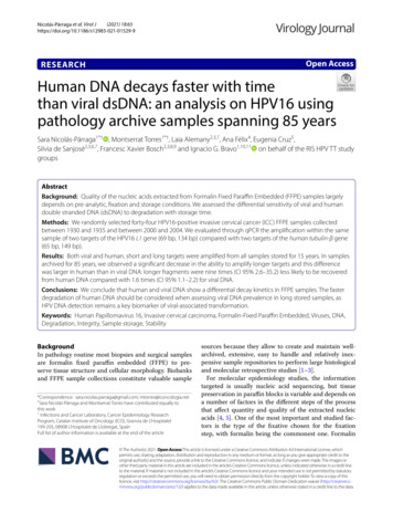
Nicolás‑Párraga et al. Virol J(2021) 18:65Page 4 of 8(100%) samples stored for 15 years, we could successfully amplify the two fragments sizes for both human andviral DNA. In the set of samples archived for 85 years,for both human and viral DNA, when the long amplicon was generated the corresponding short ampliconwas also retrieved. In samples stored for 85 years, overallcongruence between the amplification results obtainedfor human and for HPV16 DNA (Table 2) was low (21%concordance). This weak concordance reflected thathuman DNA amplification of the long fragment was lesslikely than that of the short fragment (respectively 2/19 vs17/19; Exact McNemar test, p 6.1*10–5), while for viralDNA this difference was not observed (12/19 vs 16/19;Exact McNemar test, p 0.125).We analyzed the differential amplification ability ofthe same DNA target in different samples with regards tothe storage time. We did not observe differences betweensamples stored for 85 and for 15 years in the potential forgenerating the short amplification product, neither forhuman DNA (17/19 vs 25/25, Fisher’s Exact test; p 0.18)nor for HPV16 DNA (16/19 vs 25/25, Fisher’s Exact test;p 0.07). In contrast, viral and human DNA respondedvery differently to FFPE to long-term storage regarding the amplification of longer products. Human DNAamplification potential dropped significantly: 25/25 after15 years and 2/19 after 85 years storage (Fisher’s Exacttest; p 2.5*10–10) (Table 2). The relative amplification ofthe long amplicon was thus nine times higher for sampleswith 15 years of storage than for samples with 85 years ofstorage (Risk ratio: 9.5; CI 95% 2.6–35.2). Regarding viralDNA instead, the decrease in amplification of the longproduct with time was far less important: 25/25 after15 years vs 12/19 after 85 years storage (Fisher’s Exacttest; p 1.3*10–3). In this case, the effect of storage timefor the relative amplification was lesser (Risk ratio: 1.6; CITable 2 Score based on amplified fragments in the set ofsamples archived for 85 yearsNo. of samples amplified on human tubulin-βgeneNegativePositive (65 bp)Positive(149 bp)†TotalNo. of samples amplified on HPV16 L1 geneNegative0303Positive (69 bp)2204Positive (134 bp)‡010212Total215219bp: base pairs†All samples named “149 bp” were also positive for both 65 bp and 149 bpamplicons‡All samples named “134 bp” were also positive for both 69 bp and 134 bpamplicons95% 1.1–2.2). Our results show therefore that FFPE longterm storage decreased qualitatively the ability to recoverlonger DNA fragments by means of amplification, andthat the impact of DNA degradation was more importantfor human than for HPV16 DNA.Differential quantification of viral and human DNAretrieved from FFPE samples with storage time.We have further quantitatively validated the results onthe differential potential for amplification by means ofqPCR. In all samples, quantification values for the shortamplification target and for the long target were different for both viral and human DNA (for 15 years storage,Wilcoxon signed rank test, p 1.3*10–4 and p 1.3*10–5,respectively for viral and human targets; for 85 yearsstorage, Wilcoxon signed rank test, p 4.8*10–4 andp 3.2*10–4, respectively for viral and human targets).In both cases, the short fragment was estimated to bearound four times more present than the long fragment. Similarly, for samples archived for 85 years thevalues obtained using the short amplicon were different,approximately forty times more present, compared withthe ones obtained when was amplified the long amplicon,both for human and HPV16. The results revealed a differential DNA quantification for human and HPV affectedby the length of the amplicon, both in samples archivedfor 15 and for 85 years (Figs. 1 and Additional file 1: Fig.S1).DiscussionArchived FFPE specimens are an invaluable source formolecular biological analyses, molecular epidemiologystudies and/or identification of biomarkers. Tissue preservation in paraffin blocks is variable and dependent ofmultiple factors that influence nucleic acid integrity,potentially affecting the results for long-term retrospective analyses. Considering HPVs and the associated cancers, some studies have assessed the usefulness of FFPEsamples and the different viral detection efficiencyregarding distinct techniques [3, 24], while other haveexamined whether the storage period has significanteffect on DNA, RNA or protein retrieval, however, focusing solely in human macromolecules [2]. Additionalresearch studied DNA amplification using differentamplicon sizes [13], albeit without considering the different origin of DNA, i.e. human or viral DNA. In the present work we have tried to combine all these approachesto provide a complete picture on the quality and quantityof viral and human DNA retrieved from samples storedfor 85 and 15 years, and using two sets of different sizedamplicons (ca. 70 bp and ca. 150 bp) implemented indifferent qPCR systems on invasive cervical FFPE samples exclusively containing HPV16 as viral agent. Our
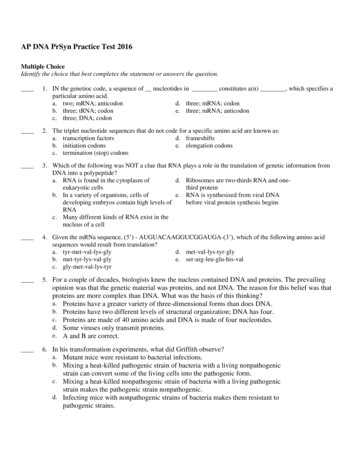
Nicolás‑Párraga et al. Virol J(2021) 18:65Page 5 of 8p-value 1.6*10-4p-value 4.8*10-4p-value 1.3*10-5p-value 3.2*10-42.65 (2.53-2.92)3.31(2.32-3.99)2.83(1.72-3.64)2.03 (1.90-2.27)1.76 (1.16-2.04)p-value 0.052.53(1.61-3.32)p-value 3.3*10-4p-value 0.03Human shortHuman long0.92(0-1.47)p-value 0.07Viral shortViral long15 years of storageHuman shortHuman longViral shortViral long85 years of storageFig. 1 Violin plots comparing the distribution of the quantification values by gene, amplicon size and storage time including only the subset ofsamples fixed in formalin. For each period of time, median values of copies/µL (depicted as L og10) per amplicon either between host and virus orfor the two different amplicons within host and within viruses are compared by means of a Wilcoxon Mann–Whitney test. For each comparison,p-values for the null-hypothesis of non-different median values between the corresponding distributions are indicated. Fragments are representedas follows: “Human short” to represent 65 bp tubulin-β gene amplicon; “Human long” to represent 149 bp tubulin-β gene amplicon; “Viral short” torepresent 69 bp L1 gene amplicon and “Viral long” to represent 134 bp L1 gene amplicon. Each period of time is represented below fragmentsresults show that longer fragments of HPV dsDNA canbe amplified from 80-years old FFPE samples comparedwith human dsDNA. Differential amplification couldreflect increased chemical stability of the episomal, oftensupercoiled viral DNA and/or be related to the largenumber of copies per infected cell of viral DNA either inform of integrated concatemers or of episomes, as discussed below.Our qualitative results for human and viral DNA detection describe higher efficiency and quality of amplification with primers amplifying shorter fragments, in linewith previous reports [25, 27]. Álvarez-Aldana and coworkers amplified human and viral DNA from a set ofFFPE cervical tissues stored for six years. They workedwith 209 and 110 bp fragments in human β-globin geneand with 142 and 96 bp amplicons in HPV16 L1 and E6genes respectively, and they observed higher quality ofdetection in human DNA compared with HPV DNA[25]. The results from this study contrast with ours, butthe origin for the lack of concordance lies probably at theprimers used for viral amplification and at the ill-definedset of samples. Indeed, for the small HPV ampliconthese authors used the GP5 /6 set of generic primers[28], suited for a broad detection of HPVs but not specific enough for assessing sensitivity, and definitely notfor a type-specific qPCR, which has been our choice forprimer design. For the long HPV amplicon the authorsused the TS16 primers, targeting HPV16 [25], but theactual genotype that could have been detected in thelesions had never been identified in forehand, so that anegative result is not necessarily informative. In our case,we have exclusively worked with samples containingHPV16, and our primers were designed with the samedegree of specificity for the human and for the viral targets. In our hands, results from the PCR system on thehuman tubulin-β target are more dependent on the fragment size than on the viral L1 gene. The same amplification pattern for human DNA depending on amplicon sizewas observed by Nakayama and coworkers, who amplified different targets of the human gapdh gene from paraffin samples (spanning from 300 bp to 1350 bp) andreported higher proportion of short amplicons, suggesting less amplification efficiency for larger fragment sizes[29].Regarding differential amplification with storage time,Ademà and coworkers studied FFPE DNA integrity of
Nicolás‑Párraga et al. Virol J(2021) 18:65certain human genes between different storage periodsand observed that DNA integrity decreased in FFPE samples stored with eight years of difference [1]. Our resultsspanning a longer period of time point in the same direction: with longer storage time, human DNA quality, asestimated by qPCR in terms of amplifiable length, is lowerand only a minority of the analyzed samples remainedequally amplifiable for short and long fragments. Furthermore, we observed that the rates of amplification forshort fragments either human or HPV are similar in spiteof the time of storage (85 years of storage vs 15 years ofstorage), whereas for large fragments, we observed different amplification time trends, being more appreciablein human DNA than in viral. Specifically, whereas quality of human DNA was nine times more prone to amplifyfor samples storage 15 years than 85 years, viral DNAonly was less than two times more prone. According tothese time-differential PCR-fragments rates of amplification observed, we suggest that PCR systems designedto amplify DNA fragments 150 bp from FFPE samplesstored more than 70 years, may result in false negativesdue to the decreased sensitivity of the PCR system.Herráez-Hernández and coworkers compared different HPV genotyping systems that include an initial PCRstep of different fragments lengths [30]. They concludedthat there are differences in sensitivity rates as functionof amplicon size, suggesting that systems based on theamplification and genotyping of short HPV DNA fragment size, for instance INNO-LiPA HPV Genotyping(Fujirebio, Belgium) that includes the amplification of65 bp fragment through consensus primers S PF10, present higher sensitivity to detect HPV DNA in FFPE samples than other techniques that work with larger DNAfragment, e.g. HPV2 CLART (Genomica, Spain) or Linear Array Genotyping test (Roche Molecular Systems),both detecting a 450 bp amplicon size. The same amplification pattern was described by Martró and coworkerscomparing two HPV assays (INNO-LiPA HPV Genotyping and F-HPV typing ), which amplify fragmentsof respectively 158 bp and 484 bp in length in the E6/E7region, and reporting higher positivity for the approachusing shorter amplicon size for initial steps [31]. In ourwork, we have additionally compared human and HPVPCR systems, in order to assess dependence of the system performance. We observe that human and viral PCRsystems perform differently for the same sample. Weconfirm that in FFPE samples, especially in those with aprolonged storage, amplification based-genotyping assaysshould use amplicons of around 60–70 bp in order to behighly sensitive. Furthermore, we propose that internalcontrols should not only be focused on human DNA,and if so, they should use amplicons of similar 60–70 bpPage 6 of 8length, as we have observed significant differences ofPCR amplification with the tubulin-β gene, not detectedfor HPV DNA fragments.Relating to the quantifying methodology implementedin our work, Nakayama and colleagues suggested thatqPCR reflects more accurately the degree of DNA fragmentation than other techniques (e.g. UV or fluorescencespectroscopy) [28]. Thus, in our research we analyzed thetrends intra and inter storage time-periods of the quantification values, and we further explored the variance ofthese values when stratifying not only by time, but alsoby the DNA nature and the amplicon size. For all comparisons for the same storage time, amplicon size influences the quantification values, in all cases we obtainedhigher values of quantification for short fragments compared with large. Dietrich and coworkers, developed asemi-nested qPCR system (a unique forward primer anddifferent reverse primers) for the pitx2 human DNA genecovering 13 amplicon lengths ranging from 200 to 850 bpto be employed in FFPE samples collected between 1992and 2011. These authors did not observe viral load variation or qPCR inhibition, reporting Cq values of 40 whenincreasing the fragment length above 200/250 bp [10].Dal Bello and colleagues quantified the viral load employing a qPCR system with the SPF10 consensus primers on173 FFPE samples collected in three periods (1985–87;1995–97 and 2005–07), and they observed that HPVtiters did not present differences attributable to duration in recently stored samples [32]. We propose that forstudies in which the goal is to quantify human (targeting tubulin-β) or HPV DNA (targeting L1) in FFPE samples recently stored or stored 15 years through qPCR,the amplicon sizes should to be considered as one of themain variables to avoid possible under-estimation of theviral load or copies/µL. The obtained data could be helpful in deciding the design of qPCR amplification on FFPEsamples with storage time below 15 years.This study is subject to some a number of limitations.First, the paraffin employed for the embedding processhas moved along the years to lower melting temperaturesand consequently, the DNA purification could be differentially impacted by an inefficient releasing in the initialsteps in the samples archived for 85 years versus thosearchived for 15 years. In addition, the buffered formalinused beyond 2000 has shown higher PCR efficiency ratescompared to unbuffered formalin. The more recent caseswere collected from Portuguese Institutes of OncologyFrancisco Gentil of Coimbra and Lisbon. In spite of thestandardization of the routine sample processing in theinstitution, there could exist slight variations betweenboth cities that could affect the preservation of the DNAin the tissues. A second limitation concerns the targeted
Nicolás‑Párraga et al. Virol J(2021) 18:65genes: only one gene per genome, viral and human. Ourresults of an increased lack of detection for human DNAwith the time of storage suggest that it could be advisable to test multiple human gene to assess the suitabilityof a sample for epidemiological studies. Third, we havedeveloped for this study a novel qPCR system. It was notour aim to exhaustively describing the sensitivity of thisamplification system to detect HPV infections, and wehave thus restrained ourselves to samples that had alreadybeen tested positive by using the very sensitive and wellcharacterized SPF10-DEIA-LiPA25 algorithm. Indeedtwo of the samples from the 1930s that had tested positive with the SPF10 primers were negative with our novelprimer set. Fourth, we have used in our study exclusivelyinvasive cervical cancer samples, with the aim of standardizing the input materi
measured with Qubit dsDNA HS Assay kit (Invitrogen, Life Technologies, CA, USA) on a Qubit 3.0 Fluorometer (Invitrogen, Life Technologies, CA, USA) according to manufacturer's instructions. SYBR-green based quantitative PCR (qPCR) was per- ).