Transcription
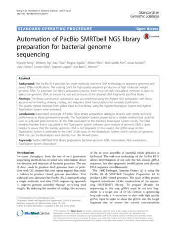
Kong et al. Standards in Genomic Sciences (2017) 12:27DOI 10.1186/s40793-017-0239-1STANDARD OPERATING PROCEDUREOpen AccessAutomation of PacBio SMRTbell NGS librarypreparation for bacterial genomesequencingNguyet Kong1, Whitney Ng2, Kao Thao3, Regina Agulto1, Allison Weis1, Kristi Spittle Kim4, Jonas Korlach4,Luke Hickey4, Lenore Kelly5, Stephen Lappin5 and Bart C. Weimer1*AbstractBackground: The PacBio RS II provides for single molecule, real-time DNA technology to sequence genomes anddetect DNA modifications. The starting point for high-quality sequence production is high molecular weightgenomic DNA. To automate the library preparation process, there must be high-throughput methods in place toassess the genomic DNA, to ensure the size and amounts of the sheared DNA fragments and final library.Findings: The library construction automation was accomplished using the Agilent NGS workstation with Bravoaccessories for heating, shaking, cooling, and magnetic bead manipulations for template purification.The quality control methods from gDNA input to final library using the Agilent Bioanalyzer System and AgilentTapeStation System were evaluated.Conclusions: Automated protocols of PacBio 10 kb library preparation produced libraries with similar technicalperformance to those generated manually. The TapeStation System proved to be a reliable method that could beused in a 96-well plate format to QC the DNA equivalent to the standard Bioanalyzer System results. The DNAIntegrity Number that is calculated in the TapeStation System software upon analysis of genomic DNA is quitehelpful to assure that the starting genomic DNA is not degraded. In this respect, the gDNA assay on theTapeStation System is preferable to the DNA 12000 assay on the Bioanalyzer System, which cannot run genomicDNA, nor can the Bioanalyzer work directly from the 96-well plates.Keywords: PacBio SMRTbell NGS library preparation, Bacterial genomic DNA, Automation, NGS workstation,TapeStation System, BioanalyzerIntroductionIncreased throughput from the use of next generationsequencing methods has revealed new information aboutthe function and structure of bacterial genomes. The useof short reads to produce draft genomes leads to problems with GC content bias and repeat regions that makeit tedious to produce closed genome assemblies. Thistechnical note discusses the PacBio RS II approach usinga single molecule, real-time DNA sequencing approachto improve genome assembly through extra-long readlengths. By reducing the number of contigs, the accuracy* Correspondence: bcweimer@ucdavis.edu1Population Health and Reproduction Department, School of VeterinaryMedicine, University of California-Davis, Davis, CA, USAFull list of author information is available at the end of the articleof the de novo assembly of bacterial whole genomes isfacilitated. The real-time technology of the PacBio RS IIallows determination of not only the full, closed, gDNAsequence, but also epigenetic modifications and plasmidDNA sequence simultaneously.The 100K Pathogen Genome Project [1] is using thePacBio 10 kb SMRTbell Template Preparation kit toproduce 1,000 closed genomes. The scale of this projectrequired automation of the construction of the sequencing (SMRTbell ) library. To prepare libraries forsequencing in this way, gDNA must be cut into fragments to a target size of 10 kb. Critical to generatinglong sub-reads, it is important to start with high qualitygDNA input in order to shear the gDNA into the targetfragment size to ensure the correct concentrations The Author(s). 2017 Open Access This article is distributed under the terms of the Creative Commons Attribution 4.0International License (http://creativecommons.org/licenses/by/4.0/), which permits unrestricted use, distribution, andreproduction in any medium, provided you give appropriate credit to the original author(s) and the source, provide a link tothe Creative Commons license, and indicate if changes were made. The Creative Commons Public Domain Dedication o/1.0/) applies to the data made available in this article, unless otherwise stated.
Kong et al. Standards in Genomic Sciences (2017) 12:27during library construction to react properly with theconcentrations of reagents in each of the given steps. Gelelectrophoresis is a low-resolution traditional method withsizing against a ladder and determining concentration onan agarose gel by comparing peak density to a standard,and since it cannot be automated, is not suitable for a project of this size. Another way to measure size and concentration is to use the Agilent 2100 Bioanalyzer with theDNA 12000 assay, but the instrument only runs 12 samples at a time and cannot be automated. We will discussthe automation of preparation of libraries with theSMRTbell Template Preparation kit as well as analysis ofgDNA, fragmented DNA and the final libraries ready forsequencing with both the Agilent electrophoresis platform: Agilent 2100 Bioanalyzer System using the DNA12000 assay and the Agilent TapeStation System using thegenomic DNA ScreenTape and matching reagents.ProcedureCampylobacter jejuni, Listeria monocytogenes, Vibrio fluvialis and Salmonella enterica serovar. Enteritidis werecultured in appropriate culture medium and growingcondition listed in Tables 1 and 2. Bacteria were culturedon the appropriate agar and pellets were made for extraction. DNA was extracted from the cell pellets usinga kit and clean-up was accomplished with a spin column[2–4]. Absorbance ratios at 260/280 and 260/230 weremeasured with a NanoDrop 2000 UV-vis spectrophotometer (Thermo Fisher Scientific, Waltham MA). AQubit 2.0 Fluorometer (Q32866) was used with a QubitdsDNA HS Assay Kit (Q32854, both from Invitrogen,Carlsbad CA) to measure the gDNA concentration andconfirm DNA input of 10 μg before shearing. The initialevaluation of the quantity and size distribution of thepurified gDNA was with the Agilent 2200 TapeStationNucleic Acid System (G2965AA) controlled by Agilent2200 TapeStation Software A.01.05, using the AgilentGenomic DNA ScreenTape (5067–5365) and the AgilentGenomic DNA Reagents (5067–5366) with samplesdrawn from a 96-well plate [5, 6]Genomic DNA was sheared using the Covaris g-TUBEdevice (520079) according to the manufacturer specifications [7]. After fragmentation, DNA was evaluated withTable 1 Organisms used in this studyPage 2 of 10the TapeStation System with the Genomic DNA assay andalso with the Agilent 2100 Bioanalyzer System with theAgilent DNA 12000 assay (5067–1508) [8, 9]. Both ofthese methods have minimal sample consumption and return both sizing and quantitation. The sheared gDNAsample input was normalized for all samples between 1–5μg into library construction for PacBio SMRTbell 10 kbLibrary Preparation.The SMRTbell Template Preparation kit from PacificBiosciences (Menlo Park CA) was used on the AgilentNGS Workstation (G5522A, Agilent Technologies, SantaClara CA). The workflow to construct the final DNAlibraries for sequencing is shown in Fig. 1 and involvedautomation of these steps:1.2.3.4.Determination of the quality of the gDNAFragment gDNA using a Covaris g-TUBE deviceQC the sizing and adjust the concentrationRepair DNA damage and repair ends offragmented DNA5. Purify the DNA6. Blunt-end ligate using blunt adapters7. Purify template for submission to a sequencerIn Fig. 2, A (Post Shearing Clean-up) and B (10kbLibrary Prep Runset Dual SPRI) are two of the VWorksprotocol graphical user interfaces that help with theNGS Workstation setup and deck layout to optimize theuse of reagent volumes. This interface allows the user toview the progress of the procedure. In Fig. 2c, the Exceltemplate assists with laying out the reagent amounts andcalculations, and provides a record of each batch ofreagents preparation and lot numbers.With the automation, this workflow takes about 7 h forpost-shearing clean-up and library construction. Once thePacBio 10 kb library is made, the final library was confirmed with the Agilent 2200 TapeStation with theGenomic DNA ScreenTape assay and the Agilent 2100Bioanalyzer System with the Agilent DNA 12000 assay todetermine the size of the library. Libraries are quantifiedusing a Qubit 2.0 Fluorometer (Q32866) with a QubitdsDNA HS Assay Kit (Q32854, both from Invitrogen,Carlsbad CA) to measure the library concentration before
Kong et al. Standards in Genomic Sciences (2017) 12:27Page 3 of 10Table 2 gDNA quality, average shearing size and average final library for each bacteriumsubmission to the sequencing facility. The sequencingfacility anneals sequencing primer and binds polymeraseto the SMRTbell templates before loading the library ontothe PacBio RS II.DiscussionThe genomic DNA isolated from four model organismswith a range of GC content were made into librariesprepared on the Agilent NGS Workstation with PacBioSMRTbell Template Preparation kit for sequencing onthe PacBio RSII. Finished sequences showed GC contentvery close to the known GC content, thus showing thisprocess produced minimal bias (Table 1).For the best results to produce genomic sequences, itis important the starting material be relatively free oforganics and protein, and be at least 50 kilobases toinsure long fragments can be obtained for sequencing.The microbes used are listed in Table 1 and include fourgenera of varying length and GC content. The organismswere cultured and genomic DNA was extracted followedby spin column clean-up. The quality of the gDNA wasmeasured with the NanoDrop and the 260/280 nm andthe 260/230 nm ratios were calculated. The 260/280 nmratio and 260/230 nm ratio of 1.8 was the requirementfor further use of each extraction. The Agilent 2200TapeStation System with the Genomic DNA assay wasused to assess size and concentration of each sample asFig. 1 PacBio SMRTbell Template Preparation Workflow for PacBio RS II system. PacBio SMRTbell Template Preparation Workflow for PacBio RS IIsystem. This workflow is used to prepare libraries from fragmented and concentrated DNA using Covaris g-TUBE and concentrated using theAMPure magnetic beads before following PacBio SMRTbell 10 kb Library Preparation procedures
Kong et al. Standards in Genomic Sciences (2017) 12:27Page 4 of 10Fig. 2 VWorks protocols and Excel workbook for PacBio Library Preparation. VWorks protocols and Excel workbook for PacBio Library Preparationmethod provide an interactive, visual layout for the end user. a Post Shearing Cleanup Form. b 10 kb Library Prep Runset Dual SPRI Form. cPacBio Library Excel Workbook
Kong et al. Standards in Genomic Sciences (2017) 12:27Page 5 of 10Fig. 3 Quantitation of Genomic DNA. Electropherogram (a) and gel image (b) of high molecular weight gDNA from Agilent 2200 TapeStationusing the Genomic DNA ScreenTape System. Campylobacter (green), Listeria (blue), Vibrio (aqua), and Salmonella (red). Green lines at the bottom ofthe gel image are internal standards added to permit quantitation. Lower marker is not shown in the electropherogram
Kong et al. Standards in Genomic Sciences (2017) 12:27Page 6 of 10Fig. 4 Appearance of sheared DNA from Agilent 2100 Bioanalyzer analysis. Representative electropherogram (a) and virtual gel (b) are used forvisual inspection (generated with the Agilent 2100 Bioanalyzer system with the DNA 12000 Kit) of sheared bacterial genomic DNA with averageshearing size for Campylobacter (green, 10 kb), Listeria (blue, 13.5 kb), Vibrio (aqua,11.6 kb), and Salmonella (red, 17 kb). Peaks near 35 are the lowermarker internal standard for the DNA 12000 kit. A typical electropherogram using the Agilent Bioanalyzer 2100 DNA 12000 kit shows the lowermarker at 35 s and the upper marker at 90 s. The sheared DNA and the red upper marker, seen in the gel image, co-elute together
Kong et al. Standards in Genomic Sciences (2017) 12:27Page 7 of 10Fig. 5 Appearance of sheared DNA from Agilent 2200 TapeStation analysis. Representative electropherogram (a) and virtual gel (b) of shearedbacterial genomic DNA was generated with the Agilent 2200 TapeStation genomic DNA Kit with the average shearing size for Campylobacter(green, 16 kb), Listeria (blue, 12 kb), Vibrio (aqua, 14 kb), and Salmonella (red, 20 kb). Green lines at the bottom of the gel image are internalstandards added to permit quantitation. Lower marker is not shown in the electropherogram
Kong et al. Standards in Genomic Sciences (2017) 12:27Page 8 of 10Fig. 6 Appearance of DNA libraries from Agilent 2100 Bioanalyzer analysis. Representative electropherogram (a) and virtual gel (b) used for visualinspection (generated with the Agilent 2100 Bioanalyzer system with the DNA 12000 Kit) of DNA libraries sizes prepared for sequencing with thePacBio SMRTbell 10 kb Template Preparation Kit on the Agilent NGS Workstation. A typical electropherogram using the Agilent bioanalyzer 2100DNA 12000 kit shows the lower marker at 35 s and the upper marker at 90 s. The DNA libraries and the upper marker co-elutes with each other,the sharper peak is the upper marker, shown in red on the gel image. The average library sizes are: Campylobacter (green, 9.1 kb), Listeria(blue, 9.5 kb), Vibrio (aqua, 10 kb), and Salmonella (red, 15 kb)
Kong et al. Standards in Genomic Sciences (2017) 12:27Page 9 of 10Fig. 7 Appearance of DNA libraries from Agilent 2200 TapeStation analysis. Representative electropherogram (a) and virtual gel (b) of DNAlibraries sizes (generated with the Agilent 2200 TapeStation DNA genomics Kit) prepared for sequencing with the PacBio SMRTbell 10kb TemplatePreparation Kit on the Agilent NGS Workstation. The average library size for Campylobacter (green, 16 kb), Listeria (blue, 12 kb), Vibrio (aqua, 14 kb),and Salmonella (red, 20 kb) is displayed on the software screen. Green lines at the bottom of the gel image are internal standards added to permitquantitation. Lower marker is not shown in the electropherogram
Kong et al. Standards in Genomic Sciences (2017) 12:27shown in Fig. 3, where an electropherogram overlay andvirtual gel images are shown for the four model organisms, together with the DIN calculated by the TapeStationsoftware. The DNA Integrity Number (DIN) helped establish a cut-off for the suitability of the gDNA for furtherwork and can be useful for library construction.Following qualification of the gDNA, the next step is toshear the gDNA into the target fragment size required forlibrary construction using a Covaris g-TUBE deviceaccording to manufacturer instructions. It is important tocheck the fragment size and the DNA amount prior toproceeding with the library construction. Traditionally,this has been done with the Agilent 2100 Bioanalyzersystem with the DNA 12000 kit and these results areshown in Fig. 4 as overlaid electropherograms and avirtual gel image together with the sizing ladder provided.The DNA 12000 kit uses both a lower and an uppermarker as internal standard. For these samples with a target size of 10 kb, the DNA fragments usually run togetherwith the upper marker, which can be easily seen on the gelimage since it is shown in red. In the electropherogramview, the upper marker is the sharp peak at 90 s. The Agilent 2200 TapeStation System with the gDNA ScreenTapeassay can qualify the fragment size too, and this is shownin Fig. 5. The assay has a larger range to quantify genomicDNA larger than 12 kb with no upper marker and can rundirectly out of a 96 well plate. It is important to determinethe correct sizing, in order for the sequencing facility toproperly load the libraries on the sequencer.Libraries are made following the PacBio SMRTbell 10kbLibrary Preparation on the Agilent NGS Workstation andtraditionally confirmed with the Agilent 2100 BioanalyzerSystem with the DNA 12000 kit, shown in Fig. 6. Thus,with SMRTbell templates around 10 kb in size, it’s difficultto determine the correct sizing for those libraries as theseconstructs also run with the upper marker shown in redon the virtual gel images. Since the Agilent 2200 TapeStation System can size larger fragments up to 60 kb, it candetermine the size more accurately, as shown in Fig. 7.ConclusionThe PacBio SMRTbell 10 kb Library preparation kit can beused with automation such as the Agilent Bravo to preparemicrobial libraries with minimal GC bias. QC of the starting DNA and the required fragment preparation with theCovaris g-TUBE can be done with the Agilent 2200 TapeStation and the gDNA ScreenTape assay directly from the96 well plates used by the Bravo to prepare the libraries.AbbreviationsDIN: DNA integrity number; gDNA: Genomic DNA; NGS: Next generationsequencing; SMRT: Single molecule, real-timeAcknowledgementsWe gratefully acknowledge the technical assistance provided by Kerry Le,Sum Leung, Christina Kong, Lucy Cai, Alvin Leonardo, Vivian Lee, SurenePage 10 of 10Foutouhi and Patrick Ancheta. We thank the 100K Pathogen GenomeSequencing Project for providing the cultures to conduct the study.FundingFunding provided to BCW (NIH - 1R01HD065122-01A1; NIH - U24-DK097154;AGILENT TECHNOLOGIES THOUGHT LEADER AWARD, FDA - 5U01FD003572-04).Availability of data and materialsAll data was analyzed during this study are included in this published article.Authors’ contributionsNK isolated DNA, conducted experiments, analyzed TapeStation data, andwrote the manuscript; WN and KT conducted experiments and analyzedTapeStation data; RA & AW isolated DNA; KSK, JK and LH provided technicalassistance with library preparation; LK conceived of experiments, analyzeddata, and wrote the manuscript; SL provided programming of theautomation to run the protocols; BCW conceived of experiments, analyzeddata, and wrote the manuscript. All authors read and approved the finalmanuscript.Competing interestsAgilent Technologies provided test instruments and initial funding to BCW.Pacific Biosciences provided PacBio SMRTBell 10 kb Library Preparation Kitand sequencing.Consent for publicationNot applicable.Ethics approval and consent to participateNot applicable.Publisher’s NoteSpringer Nature remains neutral with regard to jurisdictional claims inpublished maps and institutional affiliations.Author details1Population Health and Reproduction Department, School of VeterinaryMedicine, University of California-Davis, Davis, CA, USA. 2Genentech, S. SanFrancisco, CA, USA. 3University of California-San Francisco, San Francisco, CA,USA. 4Pacific Biosciences, Menlo Park, CA, USA. 5Agilent Technologies, Inc.,Santa Clara, CA, USA.Received: 2 July 2016 Accepted: 26 February 2017References1. 100K Pathogen Genome Project. 2013 [cited 2016 June 30]; Available from:http://www.100kgenomes.org.2. Kong N, et al. Production and analysis of high molecular weight genomicDNA for NGS pipelines using Agilent DNA extraction kit (p/n 200600). 2013.doi:10.13140/RG.2.1.2961.4807.3. QIAamp DNA Mini Kit. 2016 [cited 2016 June 30]; Available from: .4. Greenspoon SA, et al. QIAamp spin columns as a method of DNA isolationfor forensic casework. J Forensic Sci. 1998;43(5):1024–30.5. Agilent Genomic DNA ScreenTape System Quick Guide (p/n G2964-90040).2016 [cited 2016 June 30]; Available from: ic/ScreenTape gDNA QG.pdf.6. Agilent 2200 TapeStation User Manual (p/n G2964-90002). 2016 [cited 2016June 30]; Available from: ic/G2964-90002 TapeStationPalpatine USR EN.pdf.7. Covaris. Covaris USER MANUAL: g-TUBE 2012 [cited 2016 June 30]; Availablefrom: http://covarisinc.com/wp-content/uploads/pn 010154.pdf.8. Agilent 2100 Bioanalyzer User Manual (p/n G2946-90004). 2016 [cited 2016June 30]; Available from: lic/G2946-90004 Vespucci UG eBook (NoSecPack).pdf.9. Agilent Technologies, I. Agilent DNA 7500 and DNA 12000 Kit Quick StartGuide. 2013 [cited 2016 June 30]; Available from: ic/G2938-90025 DNA7500-12000 QSG.pdf.
Genomic DNA ScreenTape assay and the Agilent 2100 Bioanalyzer System with the Agilent DNA 12000 assay to determine the size of the library. Libraries are quantified using a Qubit 2.0 Fluorometer (Q32866) with a Qubit dsDNA HS Assay Kit (Q32854, both from Invitrogen, Carlsbad CA) to measure the library concentration before