Transcription
Turnier et al. Arthritis Research & Therapy (2017) 19:242DOI 10.1186/s13075-017-1444-4RESEARCH ARTICLEOpen AccessUrine S100 proteins as potential biomarkersof lupus nephritis activityJessica L. Turnier1*, Ndate Fall1, Sherry Thornton1, David Witte2, Michael R. Bennett3, Simone Appenzeller4,Marisa S. Klein-Gitelman5, Alexei A. Grom1 and Hermine I. Brunner1AbstractBackground: Improved, noninvasive biomarkers are needed to accurately detect lupus nephritis (LN) activity. Thepurpose of this study was to evaluate five S100 proteins (S100A4, S100A6, S100A8/9, and S100A12) in both serumand urine as potential biomarkers of global and renal system-specific disease activity in childhood-onset systemiclupus erythematosus (cSLE).Methods: In this multicenter study, S100 proteins were measured in the serum and urine of four cSLE cohorts andhealthy control subjects using commercial enzyme-linked immunosorbent assays. Patients were divided into cohortson the basis of biospecimen availability: (1) longitudinal serum, (2) longitudinal urine, (3) cross-sectional serum, and (4)cross-sectional urine. Global and renal disease activity were defined using the Systemic Lupus Erythematosus DiseaseActivity Index 2000 (SLEDAI-2K) and the SLEDAI-2K renal domain score. Nonparametric testing was used for statisticalanalysis, including the Wilcoxon signed-rank test, Kruskal-Wallis test, Mann-Whitney U test, and Spearman’srank correlation coefficient.Results: All urine S100 proteins were elevated in patients with active LN compared with patients with activeextrarenal disease and healthy control subjects. All urine S100 protein levels decreased with LN improvement,with S100A4 demonstrating the most significant decrease. Urine S100A4 levels were also higher with proliferative LNthan with membranous LN. S100A4 staining in the kidney localized to mononuclear cells, podocytes, and distal tubularepithelial cells. Regardless of the S100 protein tested, serum levels did not change with cSLE improvement.Conclusions: Higher urine S100 levels are associated with increased LN activity in cSLE, whereas serum S100 levels donot correlate with disease activity. Urine S100A4 shows the most promise as an LN activity biomarker, given itspronounced decrease with LN improvement, isolated elevation in urine, and positive staining in resident renal cells.Keywords: Systemic lupus erythematosus, Lupus nephritis, Biomarker, S100 protein, S100A4BackgroundChildhood-onset systemic lupus erythematosus (cSLE) isa complex inflammatory autoimmune disease of unknownetiology that carries a risk of irreversible damage in multiple organ systems. Up to 80% of children with cSLE willdevelop lupus nephritis (LN), and LN persists as a leadingcause of morbidity and mortality in patients with cSLE[1–4]. Current noninvasive laboratory and clinical measures of LN activity are not sensitive or specific enough toreliably measure the course of LN [5, 6], supporting the* Correspondence: jessica.turnier@cchmc.org1Department of Rheumatology, Cincinnati Children’s Hospital Medical Center,MLC 4010, 3333 Burnet Avenue, Cincinnati, OH 45229, USAFull list of author information is available at the end of the articleneed for novel noninvasive biomarkers. S100 proteinshave the potential to fill this unmet need.S100 proteins are emerging as reliable biomarkers ofdisease activity in a multitude of pediatric inflammatorydiseases, including systemic juvenile idiopathic arthritisand inflammatory bowel disease [7, 8]. These proteinsrepresent a diverse group of calcium-binding proteins,and mainly the heterodimer S100A8/9 (also known asMRP8/14 or calprotectin) and S100A12 have been studied to date in inflammatory diseases. S100A8/9 andS100A12 are expressed predominantly by phagocytesand serve as danger or damage-associated molecularpatterns upon extracellular release, triggering continuedinflammation [7]. The Author(s). 2017 Open Access This article is distributed under the terms of the Creative Commons Attribution 4.0International License (http://creativecommons.org/licenses/by/4.0/), which permits unrestricted use, distribution, andreproduction in any medium, provided you give appropriate credit to the original author(s) and the source, provide a link tothe Creative Commons license, and indicate if changes were made. The Creative Commons Public Domain Dedication o/1.0/) applies to the data made available in this article, unless otherwise stated.
Turnier et al. Arthritis Research & Therapy (2017) 19:242Elevated serum levels of S100A8/A9 have been described in several adult SLE populations and correlatewith disease activity [9, 10]. In particular, S100A8/9 expression is increased on interferon-producing plasmacytoid dendritic cells of patients with SLE with active disease[11]. Intrarenal S100A8/9 messenger RNA (mRNA) levelswere also recently shown to predict progression to chronickidney disease in an adult LN cohort [12].Whereas S100A8/9 and S100A12 are predominantlyphagocyte-specific proteins, S100A4 (also known asfibroblast-specific protein 1 or metastasin) is morewidely expressed and has demonstrated correlation withdisease activity in various forms of glomerulonephritis[13]. S100A4 also has proposed roles in enhancement ofcell motility, dysregulation of cell death, and progressionof fibrosis that may be important for pathogenesis of LNand SLE in general [14, 15]. Similarly to S100A4,S100A6 functions to influence cell motility throughcytoskeletal changes and can cause dysregulated apoptosis, which is known to be a contributor to development of autoimmunity in SLE.Combined assessment of select S100 proteins in bothserum and urine in relation to the status and course ofcSLE has not been reported and was the focus of thisstudy. The purpose of this study was to evaluate fiveS100 proteins (S100A4, S100A6, S100A8/9, andS100A12) for their relationship to renal and extrarenalcSLE activity. We hypothesized that serum S100 proteinswould correlate with cSLE disease activity in general andthat urine S100 levels would reflect LN activity.MethodsStudy populationFor this study, we used banked serum and urine samples.For the purpose of analysis, four groups were defined:1) Longitudinal serum (Cohort Ls) (n 47): Patients hadserum samples collected at two time points, onceduring an active disease visit and once at animproved visit.2) Longitudinal urine (Cohort Lu) (n 39): Patients hadurine samples collected at the time of active LN andalso at the time of improved LN.3) Cross-sectional serum (Cohort Xs) (n 100): Patientshad a single serum sample collected from visits withactive extrarenal disease activity only (n 34), activeLN (n 41), or low global disease activity (n 25).4) Cross-sectional urine (Cohort Xu) (n 96): Patientshad a single urine sample collected from visits withactive extrarenal disease activity only (n 18), activeLN (n 52), or low global disease activity (n 26).The samples from active disease visits of patients inCohorts Ls and Lu were also included in the respectivePage 2 of 11cross-sectional cohorts. Patients could also be includedin both the serum and urine longitudinal cohorts if bothserum and urine from an active and improved diseasevisit were present.This study was approved by the Cincinnati Children’sInstitutional Review Board with a waiver of consent andincluded patients from nine pediatric rheumatology centers.All patients met American College of Rheumatology classification criteria for SLE [16, 17] and had disease onset priorto 18 years of age. Patients were excluded if an active infection was present at the time of the visit, because infectionmay elevate S100 levels and become a potential confounder. Control serum and urine samples were obtainedfrom healthy children enrolled in the Cincinnati GenomicControl Cohort [18, 19].Assessment of disease activityFor patients with cSLE, both extrarenal and renal diseaseactivity were assessed using the Systemic Lupus Erythematosus Disease Activity Index 2000 (SLEDAI-2K) [20].The SLEDAI-2K is a validated disease activity measurefor cSLE with a total score of 0–105, consisting of 24weighted items grouped into organ-specific domains.The SLEDAI-2K renal domain (SLEDAI-R) score incorporates four items: urinary casts (heme granular or redblood cell), hematuria ( 5 red blood cells/high-powerfield), proteinuria ( 0.5 g/24 h), and pyuria ( 5 whiteblood cells/high-power field, excluding infection). Eachitem is scored a 4 if present, leading to a range of 0–16with 0 indicating no LN activity [20]. The gold standardfor diagnosis and monitoring of LN activity is a renal biopsy, with findings interpreted as suggested by the 2003International Society of Nephrology/Renal Pathology Society (ISN/RPS) classification [21]. Active features of inflammation on biopsy are classified by the NationalInstitutes of Health activity index (NIH-AI), with a rangeof 0–24 and 0 signifying no active LN features [22].In this study, an active disease visit was defined by aSLEDAI-2K score 8, and an improvement of cSLE wasdefined by a decrease in SLEDAI-2K score by 6. A lowglobal disease activity visit was defined by an extrarenalSLEDAI-2K score 4. Those patients with active diseasewere classified as having active LN if they had aSLEDAI-R score 4, and an improvement in LN wasdefined by a decrease in SLEDAI-R score by 4.Clinical data collectionDemographic and clinical information was collected onall patients at the time of visits, including age at time ofdiagnosis, disease duration, global SLEDAI-2K score andindividual organ-specific disease scores, laboratory data,current medications, blood pressure, and estimatedglomerular filtration rate (eGFR; calculated using the original Schwartz method) [23, 24]. Anti-double-stranded
Turnier et al. Arthritis Research & Therapy (2017) 19:242(ds)DNA status was categorized by positivity or negativity.More detailed information was additionally collected regarding LN history in Cohorts Lu and Xu, including ISN/RPS class of LN [21], date of renal biopsy, and NIH-AI onbiopsy.S100 proteins in serum and urineSerum was obtained from whole blood collected inserum separator tubes and stored frozen at 80 C.Urine was also collected and stored frozen at 80 C.S100 protein levels were measured in duplicate in theserum and urine with commercially available enzymelinked immunosorbent assays (BÜHLMANN MRP8/14;BÜHLMANN Laboratories, Schönenbuch, Switzerland;CircuLex S100A12/EN-RAGE, CircuLex S100A4, andCircuLex S100A6; MBL International, Nagano, Japan)per the manufacturers’ instructions.S100A4 immunohistochemistryImmunohistochemistry (IHC) was performed using 4-μmsections from four paraffin-embedded LN biopsy specimens and two control specimens from 21 and 28 week gestation kidneys. A polyclonal rabbit antihuman S100A4antibody was used at a 1:75 dilution as per the manufacturer’s instructions (Dako, Carpinteria, CA, USA). Thestains were run on the Roche BenchMark ULTRA instrument (Ventana Medical Systems, Tucson, AZ, USA) priorto the antibody (incubated for 32 minutes) and pretreatedwith Roche Mild Cell Conditioning 2 (CC2, citrate;Ventana Medical Systems). The slides were then detected with the Roche ultraView detection kit andcounterstained with Roche Hematoxylin and BluingReagent (Ventana Medical Systems).Statistical analysisStatistical analysis was performed using Prism version7.01 (GraphPad Software, La Jolla, CA, USA) and Excel2016 (Microsoft, Redmond, WA, USA) software. Medianvalues and IQRs of continuous variables and number/percent of categorical values were calculated as descriptive statistics. Patient samples with a nondetectable S100value were assigned a value of 0 for the purpose of statistical analysis. Serum and urine S100 levels were analyzedusing nonparametric testing given a nonnormal distribution of S100 values. Both nonnormalized urine S100values and urine S100 values normalized to the visit urinecreatinine were evaluated. Raw urine S100 values are reported in the Results section below because analyses wereunchanged with normalized S100 values.For analysis of the change in S100 and laboratoryvalues between paired samples in Cohorts Ls and Lu, theWilcoxon signed-rank test was applied. The KruskalWallis test and Mann-Whitney U test were used to evaluate for differences between S100 levels among patientPage 3 of 11groups in Cohorts Xs and Xu and also between patientswith different clinical categorizations in all active samples.Extrarenal disease activity was not corrected for inpatients with active LN. Spearman’s rank correlation coefficients (rs) were calculated to assess the relationshipsbetween different S100 protein levels, SLEDAI-2K scores,SLEDAI-R scores, and standard laboratory tests.ResultsClinical and demographic data for all cohortsAdditional file 1: Table S1 summarizes patient characteristics from Cohorts Xs and Xu. Patients in both cohortshad a similar age at diagnosis, with a median of 13–15years. Overall, patients in Cohort Xs had longer diseaseduration at the time of sample collection than patientsin Cohort Xu (5–8 years compared with 2–2.5 years). Inboth cohorts, the total SLEDAI-2K score was 12–13 foractive visits and 2 for low disease activity visits. No patients had active renal involvement in the low disease activity groups of either cohort, as per the given diseaseactivity definition.Table 1 provides a summary of the patient characteristics from active visits in Cohorts Ls and Lu. Both cohortswere diverse and had serologically active disease. Similarto Cohorts Xs and Xu, the median disease duration was3 years at the time of urine sample collection and 5 yearsat serum sample collection. The median time betweenactive and improved visits in Cohort Lu was 16 months(IQR 7–28), and that for Cohort Ls was 11 months (IQR6–21.5). The majority of patients in both cohorts had bothextrarenal and renal involvement. Nearly all patients hadbiopsy-proven LN in Cohort Lu, with the exception of twopatients. The median SLEDAI-R score for an active andimproved LN visit in Cohort Lu was 12 (IQR 8–12) and 0,respectively. Only 2 of the 39 patients in Cohort Lu had aneGFR 75 ml/minute/1.73 m2.Serum S100 levels in patients with cSLEWe observed no differences in any of the serum S100levels when comparing cSLE patients with active LN, active extrarenal SLE only, or low disease activity (Fig. 1).Serum S100A6, S100A8/9, and S100A12 levels were allhigher in patients with active extrarenal SLE than inhealthy control subjects (Fig. 1b–d). Serum S100A8/9 andS100A12 levels were also higher in patients with active LNthan in healthy control subjects (Fig. 1c, d). Patients withcSLE with low disease activity had serum S100A6, S100A8/9, and S100A12 levels comparable to those of healthy control subjects. Serum S100A4 levels were detectable in only5 of the 100 cSLE patient samples tested and were undetectable in healthy control subjects. Serum S100 levelswere similarly not found to differ between active andimproved visits of patients with cSLE, regardless ofSLE (extrarenal, renal) disease activity (Fig. 2a, c, e, g).
Turnier et al. Arthritis Research & Therapy (2017) 19:242Page 4 of 11Table 1 Demographic and clinical data on patients with childhood-onset systemic lupus erythematosus from Cohorts Ls and LuCohort Ls (n 47)Cohort Lu (n 39)CharacteristicActive SLE serumActive LN urineAge at diagnosis, years15 (10.5–17.5)14 (12–16)Disease duration, years5 (3–9)3 (1–4.5)Female sex, n (%)41 (87.2)31 (79.5)White race, n (%)27 (57.4)18 (46.2)Hispanic ethnicity, n (%)18 (38.3)6 (15.4)Total SLEDAI-2K score13 (10–17)17 (12–20)Active renal involvement, n (%)24 (51.1)39 (100)Biopsy-proven, n (%)14a (93.3)37 (94.9)SLEDAI-R score8 (4–12)12 (8–12)aISN/RPS class of LN, III/IV/V, n (%)3/9/6 (21.4/64.3/42.9)5/16/12 (13.5/43.2/32.4)eGFR 75 ml/minute/1.73 m2, n (%)1a (3.3)2 (5.1)46 (97.9)38 (97.4)Oral or intravenous steroids40 (85.1)35 (89.7)Hydroxychloroquine41 (87.2)32 (82)Other immunosuppressant26 (55.3)27 (69.2)Active extrarenal involvement, n (%)Medications, n (%)Laboratory testsPositive anti-dsDNA, n (%)18a (60)31 (79.5)C3, mg/dl83.6a (59.5–114.8)65 (45.4–93.3)C4, mg/dl10.6a (7–16.2)8.6 (4.5–11.1)Random urine protein/creatinine0.26a (0.12–1.8)2.1 (1.1–5.3)Active urinary sediment, n (%)a14 (46.7)32 (82.1)Abbreviations: Cohort Ls Longitudinal serum cohort, Cohort Lu Longitudinal urine cohort, eGFR Estimated glomerular filtration rate, ISN/RPS International Society ofNephrology/Renal Pathology Society, LN Lupus nephritis, SLEDAI-2K Systemic Lupus Erythematosus Disease Activity Index 2000, SLEDAI-R Systemic LupusErythematosus Disease Activity Index 2000 renal domain score, SLE Systemic lupus erythematosusAll table values are expressed as median (IQR) for continuous variables and number (percent) for categorical variablesaNote that certain data only available for SLE patients with serum from Cincinnati Children’s Hospital Medical Center (n 30), not Brazilian patients (n 17).Percentages in table were calculated from patients with available values for these characteristicsUrine S100 levels in patients with cSLE with and withoutLN from cross-sectional cohortUrine levels of all tested S100 proteins were higher withactive LN than with active extrarenal SLE only (Fig. 3).Differences in urine levels between active renal versusactive extrarenal SLE only were most pronounced forS100A4 level (median [IQR] 8.35 ng/ml [1.7–19.4] inactive LN versus 0.63 ng/ml [0.3–1.9] in active extrarenal SLE only; p 0.0001) (Fig. 3a). In urine samples, onlyS100A4 and S100A6 levels differed between patientswith active LN and patients with cSLE with low diseaseactivity, whereas urine S100A8/9 and S100A12 levelstrended toward significance. All urine S100 levels inCohort Xu were higher in patients with active LN thanin healthy control subjects. Urine S100A6, S100A8/9,and S100A12 levels were all also higher in patients withcSLE with low disease activity than in healthy controlsubjects. Urine S100A4 levels were comparable betweenpatients with cSLE with low disease activity and healthycontrol subjects.Urine S100 levels in patients with cSLE with LN fromlongitudinal cohortAll urine S100 levels differed significantly between activeand improved visits in patients with cSLE with LN.Figure 2 demonstrates urine S100 values in the pairedsamples from Cohort Lu, with healthy control subjectsalso depicted for comparison. Refer to Additional file 2:Table S2 for urine S100 values in patients with cSLE fromactive and improved visits.With improvement of LN (active versus improved visit),urine S100 levels decreased (Fig. 2 b, d, f, h). The mostpronounced difference with changing LN activity was observed for urine S100A4 levels (median change6.38 ng/ml; p 0.0001) (Fig. 2b). The median urineS100A4 levels were 7.35 ng/ml (IQR 1.6–23.9) for
Turnier et al. Arthritis Research & Therapy (2017) 19:242Page 5 of 11abcdFig. 1 Comparison of serum S100 levels in patients with childhood-onset systemic lupus erythematosus (cSLE) from the cross-sectional serumcohort, including those with active lupus nephritis (LN), active extrarenal SLE only, and low disease activity, as well as healthy control subjects. aSerum S100A4. b Serum S100A6. c Serum S100A8/9. d Serum S100A12. The horizontal lines on each plot represent median S100 values. Differences inserum S100 levels between patient groups were assessed using the Kruskal-Wallis and Mann-Whitney U testsactive LN visits, 0.97 ng/ml (IQR 0.4–2.2) for improved LN visits, and 0.32 ng/ml (IQR 0–0.6) forhealthy control subjects.Urine S100A4 levels in all patients with active LN by LNclass and degree of LN activityUrine S100A4 levels were higher with increased levels ofLN activity (SLEDAI-R) (Fig. 4a). The median urineS100A4 level for a SLEDAI-R score of 4 was 1.62 ng/ml(IQR 0.95–13.2), for a SLEDAI-R score of 8 it was5.68 ng/ml (IQR 1.97–14.6), and for a SLEDAI-R scoreof 12 it was 14.08 ng/ml (IQR 6.9–23.97). A statisticallysignificant difference was present between patientswith LN with a SLEDAI-R score of 4 versus those witha SLEDAI-R score 12 (p 0.032 by Mann-Whitney Utest). Urine S100A4 levels also differed among ISN/RPS classes of LN (Fig. 4b). Median S100A4 urinelevels were significantly higher among those with classIII/IV LN than among those with class V LN (median[IQR] 12.3 ng/ml [2.8–31.5] versus 4.2 ng/ml [0.89–58.4]; p 0.03).For those patients with LN with renal biopsiesdone within 2 months of urine sample collection (n 25), there was no statistically significant association of urine S100A4 levels with NIH-AI (rs 0.18,p 0.4). Likewise, no association was observed between urine S100A6, S100A8/9, or S100A12 leveland NIH-AI.
Turnier et al. Arthritis Research & Therapy (2017) 19:242Page 6 of 11UrineSerump 0.0001bUrine S100A4 (ng/mL)86421.00.80.60.40.20.050403020104 LNHeal 50100500p 0.007520001500Urine S100A6 (ng/mL)1000500400300200)30 6040200olEImprActoviveedSLSLESerum S100A6 (ng/mL)c8006004002002SLESerum S100A4 (ng/mL)10eaef100000Urine S100A8/9 00400002000050004000300020001000)30 LNolESLSLeivctALN0ESerum S100A8/9 (ng/mL)p 0.01980000hp 5010050010080604020)30controImprovl(n A1000olSLEUrine S100A12 (ng/mL)2000SLESerum S100A12 (ng/mL)gFig. 2 Serum and urine S100 levels in patients with childhood-onset systemic lupus erythematosus (cSLE) from longitudinal serum and urinecohorts. a Serum S100A4. b Urine S100A4. c Serum S100A6. d Urine S100A6. e Serum S100A8/9. f Urine S100A8/9. g Serum S100A12. h UrineS100A12. The horizontal line represents the median S100 value on each plot. The Wilcoxon signed-rank test was used to assess for a significantchange between active and improved LN visits
Turnier et al. Arthritis Research & Therapy (2017) 19:242acPage 7 of 11bdFig. 3 Comparison of urine S100 levels in patients with childhood-onset systemic lupus erythematosus (cSLE) from cross-sectional urine cohort,including those with active lupus nephritis (LN), active extrarenal SLE only, and low disease activity, as well as healthy control subjects. (a UrineS100A4. b Urine S100A6. c Urine S100A8/9. d Urine S100A12. Patients in the active extrarenal SLE only and low disease activity categorizations atthe time of sample collection with a prior history of LN are signified by open symbols. The horizontal line on each plot represents the medianS100 value. Note that the scale of the y-axis is broken to allow visualization of all patient values. Differences in urine S100 levels between patientgroups were assessed using the Kruskal-Wallis and Mann-Whitney U testsAssociation of urine S100 levels with clinical diseaseactivity measures and other S100 protein levels in allpatients with active LNAll urine S100 protein levels had a weak positive association with SLEDAI-R score in patients with active LN (for S100A4, rs 0.34, p 0.014; for S100A6,rs 0.22, p 0.12; for S100A8/9, rs 0.31, p 0.03; forS100A12, rs 0.4, p 0.003). Urine S100A4 levelsshowed a moderate correlation with urine S100A6(rs 0.46, p 0.001), urine S100A8/9 (rs 0.47, p 4.3 10 4) and urine S100A12 levels (rs 0.55, p 2.2 10 5).Association of urine S100A4 levels with standardlaboratory measures of disease activity in all patientswith active LNUrine S100A4 levels showed a weak negative correlationwith both C3 and C4 levels (C3, rs 0.28, p 0.05; C4,
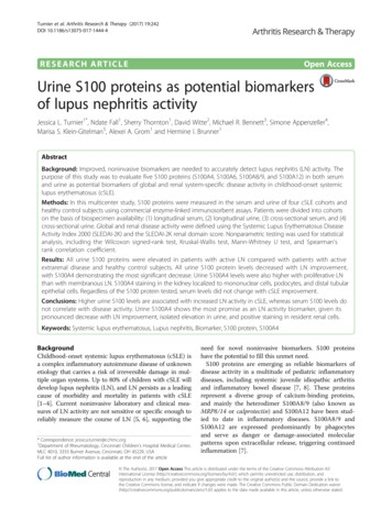
Turnier et al. Arthritis Research & Therapy (2017) 19:242Page 8 of 11rs 0.32, p 0.03) but lacked association with proteinuria (rs 0.08, p 0.57). There was no difference in urineS100A4 levels based on anti-dsDNA positivity ornegativity.S100A4 immunohistochemistry in renal biopsies ofpatients with LN versus control subjectsBoth patients with LN and control subjects demonstrated positive nuclear and cytoplasmic S100A4 stainingin renal biopsy specimens (Fig. 5). Staining was presentin podocytes, mononuclear cells, and distal tubular epithelial cells of both LN and control biopsies. WhereasLN biopsy specimens had a moderate S100A4-positivemononuclear cell infiltrate, control specimens had onlyrare S100A4-positive mononuclear cells. The mononuclear cell staining pattern was primarily intranuclear.Proximal tubular epithelial cells and mesangial cells werepredominantly negative.Fig. 4 Urine S100A4 levels from all patients with active lupus nephritis(LN) in longitudinal urine and cross-sectional urine cohorts comparedaccording to (a) LN activity as defined by Systemic Lupus ErythematosusDisease Activity Index 2000 renal domain (SLEDAI-R) score and (b)International Society of Nephrology/Renal Pathology Society (ISN/RPS)class of LN. a The median and IQR S100A4 levels are depicted. Themedian urine S100A4 level for a SLEDAI-R score of 4 was 1.62 ng/ml (IQR0.95–13.2), increasing to 5.68 ng/ml (IQR 1.97–14.6) for a SLEDAI-Rscore of 8 and 14.08 ng/ml (IQR 6.9–23.97) for a SLEDAI-R score 12. Astatistically significant difference was present between patients with LNwith a SLEDAI-R score of 4 and those with a SLEDAI-R score 12 (p 0.032). b The median is represented by the horizontal line on each plot.Differences between urine S1004 values in class III/IV versus class V LNwere assessed using the Mann-Whitney U testDiscussionWe found elevated levels of urine S100 proteins with active LN, and urine S100 levels also demonstrated responsiveness to change in LN activity, with S100A4 showingthe most pronounced differences. Conversely, S100 levelsin the serum did not correlate with extrarenal or renal disease activity in cSLE, although serum S100A8/9 andS100A12 levels were higher in patients with active SLEthan in healthy control subjects. To determine expressionof S100A4 locally in the kidney, IHC was performed,which demonstrated positive staining for S100A4 in inflamed LN kidneys with expression in podocytes, distaltubular epithelial cells, and mononuclear cells.S100 proteins represent a heterogeneous group ofcalcium-binding proteins that exhibit diverse functionsand cell-specific expression patterns. In particular, S100A4has been proposed to have roles in enhancing cell motility,promoting cell growth, mediating epithelial-mesenchymaltransition (EMT), inhibiting cell death, and influencingthe progression of fibrosis [14, 15]. Although S100A4 iswell known in the oncology literature as a marker of metastatic tumor progression, it has not been studied as extensively in autoimmune diseases. Research to date, however,does support that S100A4 has both biologic plausibilityas a biomarker of LN and a potential role in diseasepathogenesis.Urine S100A4 levels have previously been described tocorrelate with disease activity in various forms of crescentic glomerulonephritis [13]. S100A4 has also shownprognostic value as a biomarker in immunoglobulin Anephropathy, with the number of S100A4-positive cellson biopsy found to predict renal failure and corticosteroid responsiveness [25, 26]. Although prior data onS100A4 in patients with SLE are lacking, S100A4 mRNA
Turnier et al. Arthritis Research & Therapy (2017) 19:242Page 9 of 11Fig. 5 S100A4 immunohistochemistry in lupus nephritis (LN) and control biopsy specimens. LN kidney with nuclear and cytoplasmic staining in(a) podocytes and mononuclear cells, (b) distal tubular epithelial cells and control kidney with nuclear and cytoplasmic staining in (c) podocytesand mononuclear cells, and (d) distal tubular epithelial cells. Original magnification 20was found to be upregulated in microdissected glomeruliof an MRL/lpr mouse model of LN [27].The presence of positive S100A4 protein staining inresident renal cells further establishes biologic relevancefor this novel biomarker of LN activity. IHC results fromthis study are in line with those of prior studies localizing S100A4 staining to podocytes and cellular crescentsin antineutrophil cytoplasmic antibody-associated glomerulonephritis [13]. Given that IHC did not demonstratea distinct differential S100A4 expression pattern betweenLN kidneys and those of control subjects, it remains undefined which cell type upregulates S100A4 expressionin LN. One possibility is that resident renal cells increaseproduction of S100A4 during a diseased state such asLN, with S100A4 levels increasing proportionally to LNactivity. The isolated elevation of S100A4 in LN urineand lack of correlation with extrarenal disease activity orproteinuria lend support to this theory. In fact, a priorstudy demonstrated S100A4 expression in 86% of detached urine podocytes detected in patients with diabeticnephropathy [28]. Given the proposed role of S100A4 inEMT, S100A4 may play a role in dedifferentiation ofpodocytes prior to their detachment and loss in theurine.Alternatively, S100A4 may be upregulated in inflammatory cells in LN, more in line with the known expressionpatterns of S100A8/9 and S100A12. In fact, biopsy specimens of patients with LN were found to have a highernumber of S100A4-positive mononuclear cells than thoseof control subjects. S100A8/9 protein expression has beenshown to correlate with inflammatory activity on renalbiopsy in patients with varied etiologies of nephritis [29].This does not necessarily equate to increased inflammatory cell infiltration. In fact, studies on S100A8/9 in glomerulonephritis suggest that increased S100A8/9 expressionactually reflects a change in the immune cell phenotypeversus a simple increase in the number of infiltrating immune cells within the kidney [29]. S100A4 may also fundamentally alter the behavior of immune cells central tothe pathogenesis of LN.Although S100 proteins have been studied predominantly in the serum, urine S100A8/9 h
analysis, including the Wilcoxon signed-rank test, Kruskal-Wallis test, Mann-WhitneyU test, and Spearman's rank correlation coefficient. Results: All urine S100 proteins were elevated in patients with active LN compared with patients with active extrarenal disease and healthy control subjects.