Transcription
Miller and Wright Neural Development(2021) ARCH ARTICLEOpen AccessNeuronal Dystroglycan regulates postnataldevelopment of CCK/cannabinoid receptor1 interneuronsDaniel S. Miller1 and Kevin M. Wright2*AbstractBackground: The development of functional neural circuits requires the precise formation of synaptic connectionsbetween diverse neuronal populations. The molecular pathways that allow GABAergic interneuron subtypes in themammalian brain to initially recognize their postsynaptic partners remain largely unknown. The transmembraneglycoprotein Dystroglycan is localized to inhibitory synapses in pyramidal neurons, where it is required for theproper function of CCK interneurons. However, the precise temporal requirement for Dystroglycan duringinhibitory synapse development has not been examined.Methods: In this study, we use NEXCre or Camk2aCreERT2 to conditionally delete Dystroglycan from newly-born oradult pyramidal neurons, respectively. We then analyze forebrain development from postnatal day 3 throughadulthood, with a particular focus on CCK interneurons.Results: In the absence of postsynaptic Dystroglycan in developing pyramidal neurons, presynaptic CCK interneurons fail to elaborate their axons and largely disappear from the cortex, hippocampus, amygdala, andolfactory bulb during the first two postnatal weeks. Other interneuron subtypes are unaffected, indicating thatCCK interneurons are unique in their requirement for postsynaptic Dystroglycan. Dystroglycan does not appear tobe required in adult pyramidal neurons to maintain CCK interneurons. Bax deletion did not rescue CCK interneurons in Dystroglycan mutants during development, suggesting that they are not eliminated by canonicalapoptosis. Rather, we observed increased innervation of the striatum, suggesting that the few remaining CCK interneurons re-directed their axons to neighboring areas where Dystroglycan expression remained intact.Conclusion: Together these findings show that Dystroglycan functions as part of a synaptic partner recognitioncomplex that is required early for CCK interneuron development in the forebrain.Keywords: Dystroglycan, Cannabinoid receptor, Cholecystokinin, Interneuron, Synapse, ApoptosisBackgroundProper function of neural circuits requires precise connections between specific populations of excitatory pyramidal and inhibitory neurons. GABAergic interneuronsare a highly diverse group of neurons that control brain* Correspondence: wrighke@ohsu.edu2Vollum Institute, Oregon Health & Science University, VIB 3435A, 3181 SWSam Jackson Park Road, L474, Portland, OR 97239-3098, USAFull list of author information is available at the end of the articlefunction by synchronizing and shaping the activity ofpopulations of excitatory pyramidal neurons (PyNs) [1–5]. In mice and humans, the majority of interneurons inthe cortex and hippocampus are produced in the medialand caudal ganglionic eminences (MGE and CGE) of theventral forebrain, and migrate long distances to theirfinal destinations [6–8]. The importance of interneuronsfor brain function is underscored by their involvementin a wide variety of neurodevelopmental and The Author(s). 2021 Open Access This article is licensed under a Creative Commons Attribution 4.0 International License,which permits use, sharing, adaptation, distribution and reproduction in any medium or format, as long as you giveappropriate credit to the original author(s) and the source, provide a link to the Creative Commons licence, and indicate ifchanges were made. The images or other third party material in this article are included in the article's Creative Commonslicence, unless indicated otherwise in a credit line to the material. If material is not included in the article's Creative Commonslicence and your intended use is not permitted by statutory regulation or exceeds the permitted use, you will need to obtainpermission directly from the copyright holder. To view a copy of this licence, visit http://creativecommons.org/licenses/by/4.0/.The Creative Commons Public Domain Dedication waiver ) applies to thedata made available in this article, unless otherwise stated in a credit line to the data.
Miller and Wright Neural Development(2021) 16:4neurological disorders including autism, schizophrenia,seizures, and Alzheimer’s disease [9–12].The proper integration of inhibitory interneurons intoneural circuits during development relies on multipleprocesses such as proliferation, migration, axon guidance, cell death, synaptic target selection, synapse formation (synaptogenesis) and synaptic maintenance.Although much progress has been made in identifyingcandidate molecules that regulate inhibitory synaptogenesis, our understanding of how molecularly defined subtypes of inhibitory interneurons initially identify specificpostsynaptic target cells is lacking [13, 14]. One prominent hypothesis for explaining how diverse interneuronsubtypes recognize one another during synapse development is the “molecular code” hypothesis, whereby different cell types use unique pairs or complexes of celladhesion molecules to select their target cells [15–18].Cell adhesion molecules are ideally suited to regulatesynaptic target recognition due to their large diversityand presence at pre- and postsynaptic membranes. Several recent studies support the idea that cell adhesionmolecules are key players in regulating subcellular targeting and synaptic specificity [19–22]. Although manyfamilies of cell adhesion molecules have been implicatedin controlling synapse development, they are often involved in multiple aspects of neural circuit development,making it difficult to determine their precise role in mediating synaptic specificity.Dystroglycan is a cell adhesion molecule widelyexpressed throughout the body including the developingand adult brain. Dystroglycan is extensively glycosylated,and mutations in at least 19 genes that participate insynthesizing and elongating specific O-mannose sugarchains on Dystroglycan result in a form of congenitalmuscular dystrophy called dystroglycanopathy, characterized by muscle weakness and neurological defects ofvarying severity [23–25]. Dystroglycan (Dag1) isexpressed by multiple cell types in the developing nervous system, including neuroepithelial cells, astrocytes,oligodendrocytes, and excitatory neurons [26, 27]. Lossof Dystroglycan function in the nervous system phenocopies the most severe forms of dystroglycanopathy, andcauses multiple structural brain and retinal abnormalities due to its indirect role in regulating neuronal migration and axon guidance [28–33]. However, someindividuals with milder forms of dystroglycanopathy exhibit cognitive impairments even in the absence of detectable brain malformations, suggesting a possible rolefor Dystroglycan at later stages of brain development including synaptogenesis [34, 35]. In PyNs, Dystroglycan ishighly concentrated on the cell body and proximal dendrites where it is a major postsynaptic component of inhibitory synapses (Fig. 1A [27, 36–38]). However,because of its importance in early aspects of brainPage 2 of 21development, the role of Dystroglycan at synapses hasremained obscure. Using a mouse genetic approach toselectively delete Dystroglycan from PyNs, a recent studyshowed that Dystroglycan is required for the formationand maintenance of CCK interneuron (CCK IN)synapses in adult animals, but its specific role in theearly development of these interneurons has not beenexamined [39].In this study, we show that postsynaptic Dystroglycanon PyNs is required for the proper development of presynaptic CCK INs throughout the forebrain. In micelacking Dystroglycan in PyNs, CCK INs fail to elaboratetheir axons during the first postnatal week and arelargely absent by P10. CCK INs were not rescued bygenetic deletion of Bax suggesting that CCK INs mayundergo Bax-independent cell death or fail to differentiate in the absence of Dystroglycan. Some remainingCCK INs retarget their axons into the striatum, whereDystroglycan expression is retained, suggesting that Dystroglycan functions to allow CCK INs to recognizetheir synaptic partners. Collectively, these results demonstrate that Dystroglycan is a critical regulator ofCCK IN development.ResultsCCK interneurons are largely absent in mice lackingDystroglycan from pyramidal neuronsTo investigate the role of neuronal Dystroglycan in forebrain development, we used a conditional genetic approach to delete Dystroglycan selectively from pyramidalneurons (PyNs). We crossed Dystroglycan conditionalmice (Dag1Flox/Flox) with NexCre driver mice to deleteDystroglycan in all postmitotic excitatory neurons of theforebrain except Cajal-Retzius cells, beginning at E12.5[40–43]. Control (NexCre;Dag1F/ ) and conditionalknockout mice (NexCre;Dag1F/ ) are hereafter referred toas Dag1Control and Dag1cKO mice, respectively (Fig. 1B).We verified the recombination specificity of the NexCreline by crossing it with a reporter mouse that expressesmCherry in the nuclei of Cre-recombined cells(R26LSL-H2B-mCherry [44]). mCherry nuclei were detectedin excitatory neurons of the forebrain including the cortex, hippocampus, amygdala, and nucleus of the lateralolfactory tract (nLOT) (Fig. S1A). Importantly,mCherry nuclei did not overlap with markers for interneurons (CB1R, PV, Calbindin) or astrocytes (GFAP),confirming the specificity of the NexCre mouse (Fig. S1B,C). In Dag1Control mice, Dystroglycan staining was observed as puncta concentrated primarily on the cell bodies and proximal dendrites of PyNs, as well as bloodvessels (Fig. 1C). In Dag1cKO mice, Dystroglycan stainingwas absent from PyNs but was still present on bloodvessels, confirming the specificity of the conditionalknockout.
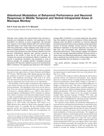
Miller and Wright Neural Development(2021) 16:4Page 3 of 21Fig. 1 Neuronal Dystroglycan is not required for pyramidal neuron migration. (a) Schematic of Dystroglycan on pyramidal neurons. Inset showsthe structure of Dystroglycan and sugar chain moieties present on the extracellular subunit. (b) Mouse breeding scheme for generating pyramidalneuron-specific Dag1 conditional knockout mice using NexCre driver mice. (c) Immunostaining for Dystroglycan in the hippocampal CA1 region ofP30 Dag1Control mice (left panel) shows punctate Dystroglycan protein on the soma and proximal dendrites of pyramidal neurons, whereasDag1cKO mice (right panel) lack perisomatic staining. Asterisks denote Dystroglycan staining on blood vessels which is retained in Dag1cKO mice.(d) Coronal sections from P15 Dag1Control and Dag1cKO cortex were immunostained for upper layer marker CUX1 (L2–4). (e) Coronal sections ofthe cortex from P30 Dag1Control and Dag1cKO mice crossed with a Thy1YFP reporter mouse to sparsely label layer 5–6 pyramidal neurons (green)and stained for Calbindin (magenta) to label layer 2–3 pyramidal neurons. (f) Coronal sections of the hippocampus from P30 Dag1Control andDag1cKO mice crossed with a Thy1YFP reporter mouse to label excitatory neurons (green) in the CA regions and dentate gyrusDeletion of Dystroglycan from neuroepithelial cellsresults in disrupted neuronal migration, axon guidance, and dendrite development in the brain, spinalcord and retina [28–33]. In contrast, deletion of Dystroglycan from PyNs with NexCre did not affect overallbrain architecture, consistent with previous results[32]. Cortical lamination in Dag1cKO mice was normalbased on CUX1 immunostaining of layer 2–4 PyNsand labeling of layer 5–6 and hippocampal PyNs witha Thy1GFP-H transgenic line (Fig. 1D-F). Therefore,neuronal Dystroglycan is not required for PyN migration in the forebrain.Forebrain interneurons (INs) are a remarkably diverse population, with multiple molecularly andmorphologically distinct IN subtypes forming synapsesonto different subcellular domains of PyNs [2, 45,46]. Since Dystroglycan is localized to inhibitorysynapses on the soma and dendrites of PyNs, we examined whether IN development is affected inDag1cKO mice. We performed immunostaining with apanel of molecular markers that label IN subpopulations in the hippocampus of adult mice (Fig. 2). InDag1Control mice, parvalbumin (PV) and somatostatin(SOM) positive INs, which label the majority of interneurons that originate from the medial ganglioniceminence (MGE), were abundant throughout thehippocampus. The distribution of PV and SOM cellbodies and their synaptic targeting patterns appeared
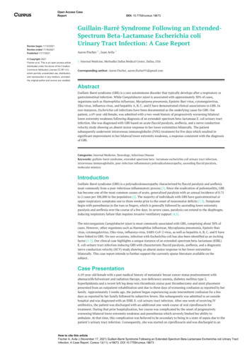
Miller and Wright Neural Development(2021) 16:4Page 4 of 21Fig. 2 CCK interneurons are selectively reduced in mice lacking Dystroglycan from pyramidal neurons. (a-b) Immunostaining for medialganglionic eminence (MGE)-derived interneuron markers (green) parvalbumin (PV) (a) and somatostatin (SOM) (b) show normal innervation of thehippocampus in P30 Dag1Control and Dag1cKO mice. Insets (yellow boxed regions) show enlarged images of the CA1. (c-d) Immunostaining forcaudal ganglionic eminence (CGE)-derived interneuron markers (green) Calretinin (c), and CB1R (d) show normal innervation of Calretinininterneurons in Dag1Control and Dag1cKO mice, whereas CB1R is largely absent from the CA regions of Dag1cKO mice. Insets (yellow boxed regions)show enlarged images of the CA1. (e) Immunostaining for CB1R in hippocampal CA1 (top) and CA3 (bottom) of P30 Dag1Control and Dag1cKOmice. (f) Quantification of CB1R pixels for each CA layer of the CA1 and CA3 shows a significant reduction in CB1R staining in Dag1cKO mice (*P 0.05, unpaired two-tailed Student’s t-test; n 4 mice/genotype). Data are presented as mean values s.e.m. Data are normalized to Dag1Controlsignal in each CA layer. CA layers: SO, stratum oriens; SP, stratum pyramidale; SR, stratum radiatum
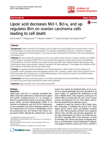
Miller and Wright Neural Development(2021) 16:4the same in Dag1cKO mice, suggesting these populations are unaffected by the loss of Dystroglycan (Fig.2A, B).We next stained the hippocampus for IN subtypes thatoriginate from the caudal ganglionic eminence (CGE).The distribution and synaptic targeting of Calretinin interneurons, which target other INs as well as PyN dendrites, appeared normal (Fig. 2C [47, 48]). In contrast,we found a dramatic reduction in cannabinoid receptor1 (CB1R) staining in the hippocampus, which labels theaxon terminals of cholecystokinin (CCK) INs (Fig. 2D[49–51]). CB1R terminals were significantly reduced inall CA subregions (Fig. 2E, F). In both the CA1 andCA3, the magnitude of the reduction varied by layer.CB1R terminals were most strongly reduced ( 95%) inthe stratum pyramidale (SP) where CCK INs form basket synapses onto PyN cell bodies, and more moderatelyPage 5 of 21reduced in the stratum radiatum (SR) and stratumoriens (SO) where CCK/CB1R INs synapse onto PyNdendrites (Fig. 2E, F). In contrast, CB1R terminals wereabundant in the dentate gyrus of Dag1cKO mice (Fig. S2).This is likely because NexCre recombination is restrictedto the outer third of granular layer neurons (Fig. S1C[41]).The loss of CB1R staining in the hippocampus ofDag1cKO mice could reflect either downregulation ofCB1R expression or a loss of CCK INs. To distinguishbetween these possibilities, we examined whether otherindependent markers of CGE-derived CCK INs weresimilarly reduced. These include NECAB1, a calciumbinding protein that specifically labels CCK IN cellbodies (Fig. 3A) [52], and VGLUT3, a vesicular glutamate transporter enriched at CCK IN synapses (Fig. 3C)[53–55]. Both NECAB1 cell bodies and VGLUT3 Fig. 3 Cell body and synaptic markers for CCK interneurons are reduced in Dag1cKO mice. (a) Immunostaining showing the co-localization ofCB1R (green) and NECAB1 (magenta) in CCK interneurons. Insets (yellow boxed regions) show enlarged images of the CA1 and CA3. (b)Immunostaining for NECAB1 (green) shows a reduction of NECAB1 interneurons in the hippocampus of P30 Dag1cKO mice. Insets (yellow boxedregions) show enlarged images of the CA1 and CA3. (c) Immunostaining of hippocampal sections from VGLUT3Cre mice crossed with a Lox-STOPLox-tdTomato (Ai9) reporter mouse showing the co-localization of CB1R (green) and VGLUT3 (magenta) in a subset of CCK interneurons. Insets(yellow boxed regions) show enlarged images of the CA1 and CA3. (d) Immunostaining for VGLUT3 (green) shows a reduction of CCK interneuron synaptic terminals in the hippocampus of P30 Dag1cKO mice. Insets (yellow boxed regions) show enlarged images of the CA1 andCA3. CA layers: SO, stratum oriens; SP, stratum pyramidale; SR, stratum radiatum
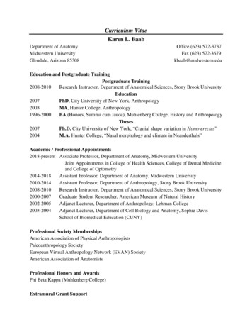
Miller and Wright Neural Development(2021) 16:4synaptic terminals were reduced in the hippocampus ofDag1cKO mice (Fig. 3B, D). Based on the loss of all threemarkers, we conclude that CCK INs are largely absentfrom the hippocampus of Dag1cKO mice.In addition to the hippocampus, Dystroglycan ispresent in PyNs of the cortex, amygdala, and nucleusof the lateral olfactory tract (nLOT) [27], which allreceive extensive innervation from CCK INs [56–58]. Therefore, we assessed whether deletion of Dystroglycan from PyNs affects CCK INs and their terminals in these forebrain regions. We first performedimmunostaining for CB1R on sagittal sections fromDag1Control and Dag1cKO mice. CB1R terminals werelargely absent throughout the entire forebrain ofPage 6 of 21Dag1cKO mice (Fig. 4A, B). Next, we stained P30Dag1Control and Dag1cKO mice for NECAB1 and CB1Rto label the cell bodies and terminals of CCK INs,respectively (Fig. 4C-E). In Dag1Control mice,NECAB1 cell bodies were numerous and CB1R innervation was extensive in the cortex, amygdala, andnLOT. In contrast, NECAB1 cell bodies were dramatically reduced, and CB1R staining was almostcompletely absent in all regions of Dag1cKO mice (Fig.4C-E). In each region, a few NECAB1 cell bodiesremained in Dag1cKO mice, and these co-localizedwith CB1R. Therefore, Dystroglycan expressed in PyNsis required broadly in the developing forebrain for theproper integration of CCK INs.Fig. 4 CCK interneurons are reduced throughout the forebrain of mice lacking Dystroglycan from pyramidal neurons. (a-b) Sagittal sections fromP60 Dag1Control;Ai9 (a) and Dag1cKO;Ai9 mice (b) immunostained for CB1R (green; right panels) and tdTomato/Ai9 (magenta; middle panels). InDag1cKO;Ai9 mice, CB1R staining is lacking in all the forebrain regions where NexCre drives recombination in excitatory neurons (tdTomatoexpression, middle panels) including the cortex (CTX), hippocampus (HC), and olfactory bulb (OB). Note the absence of tdTomato signal in thestriatum (STR) and midbrain (MB), which are not targeted by NexCre. (c-e) Immunostaining for CB1R (green) and NECAB1 (magenta) in the cortex(c), amygdala (d), and nucleus of the lateral olfactory tract (e) shows the reduction of CCK interneuron markers in the forebrain of P30 Dag1cKOmice (right panels). Enlarged images (yellow boxed regions) show individual NECAB1 cell bodies (magenta) co-localized with CB1R (green)
Miller and Wright Neural Development(2021) 16:4Postnatal development of CCK interneurons is impairedin the forebrains of Dag1cKO miceOur results showing that deletion of Dystroglycan fromPyNs leads to a reduction in CCK IN innervation isconsistent with previous work [39]. However, the temporal onset of this phenotype has not been determined.During embryonic development, CCK INs generated inthe caudal ganglionic eminence (CGE) begin populatingthe hippocampus around E14.5 [59, 60] (Fig. 5A). Atpostnatal ages, CCK INs settle into their final positionswithin the hippocampus and initially extend axonsthroughout the hippocampal layers before refining theirprojections to form characteristic basket synapses ontoPyN somas (Fig. 5B, D) [61–63]. We first examined thedevelopment of CB1R terminals in Dag1Control miceduring the first two postnatal weeks (P3-P15), as CB1Rstaining is largely absent from CCK INs before birth[64–67]. At early postnatal ages (P3-P5), the majority ofCB1R terminals were observed in the stratum radiatum(SR) layer of the hippocampus, where immature PyNdendrites are located (Fig. 5B, D). Between P5 and P10,CB1R terminals increased in the stratum pyramidale(SP) where PyN cell bodies are located. From P15through adulthood (15 months), CB1R terminals became progressively concentrated in the SP.Next, we examined CB1R terminal development inDag1cKO mice. At P3, the earliest age we were able toconclusively identify CCK INs, CB1R staining wasalready reduced in the hippocampus of Dag1cKO mice.This reduction persisted throughout the period of postnatal development and into adulthood, as late as 15months (Fig. 5C, E, F). To further confirm this finding,we stained the hippocampus for VGLUT3, an independent synaptic marker for CCK IN terminals that is upregulated during early postnatal ages (Fig. S3A). InDag1Control mice, VGLUT3 terminals increased in thehippocampus during the first two postnatal weeks, andshowed a similar pattern of innervation as CB1R staining (Fig. S3B). In contrast, VGLUT3 terminals were reduced at all ages examined in Dag1cKO mice (Fig. S3B).PV staining, which increases between P10 and P15 [68],was unaltered in Dag1cKO mice compared with controls(Fig. S3C).We next determined whether the reduction ofCCK INs in the cortex, amygdala, and nLOTfollowed the same developmental time course as thehippocampus. In Dag1Control mice, CB1R terminalsgradually increased in density in all regions betweenP3 and P15, and remained stable beyond this age intoadulthood (15 months) (Fig. 6). In contrast, CB1R terminals in Dag1cKO mice failed to elaborate duringthe first two postnatal weeks, and remained sparse inadult animals. Collectively, these results demonstratethat Dystroglycan in PyNs is critical during the firstPage 7 of 21two postnatal weeks for the development and integration of CCK INs throughout the forebrain.Post-developmental maintenance of CCK interneuronsdoes not require DystroglycanInhibitory synaptogenesis increases between P5-P15, andis largely complete by P30 [20, 22, 54]. Therefore, wewanted to assess whether deletion of Dystroglycan afterinhibitory synapse formation impairs the maintenance ofCCK INs. To achieve temporal control of Dystroglycandeletion from PyNs, we generated mice expressingtamoxifen-inducible Cre recombinase under the controlof an excitatory neuron-specific promoter Camk2a, (Calcium/calmodulin-dependent protein kinase II alpha[69]). Control (Camk2aCreERT2;DGF/ ;Ai9) or induciblecKO (Camk2aCreERT2;DGF/ ;Ai9) mice were administeredtamoxifen at P23 via oral gavage, which induced recombination in the majority of PyNs in the hippocampus(Fig. 7A, B). We then analyzed CB1R innervation 6weeks later at P65. No differences were found betweenthe Dag1 inducible-cKO and controls, suggesting thatDystroglycan is not required for the post-developmentalmaintenance of CCK INs (Fig. 7C, D).Blocking Bax-dependent cell death does not rescue CCK interneurons in Dag1cKO miceThe number of PyNs and INs in the forebrain is tightlyregulated during early postnatal development, with excess or inappropriately connected neurons pruned byBax-dependent apoptosis [70–73]. PyN apoptosis islargely complete by P5, followed by IN apoptosis whichpeaks at P7-P9. We hypothesized that in the absence ofPyN Dystroglycan, CCK INs are unable to recognizetheir postsynaptic targets and are therefore eliminatedby apoptosis. We tested this hypothesis by generatingDag1Ctrl and Dag1cKO mice that lack either one(Dag1Ctrl;BaxCtrl and Dag1cKO;BaxCtrl) or both copies ofBax (Dag1Ctrl;BaxKO and Dag1cKO;BaxKO) to block apoptosis (Fig. 8A). Deletion of Bax from control mice(Dag1Ctrl;BaxKO) did not alter CB1R innervation in theCA1 subregion of the hippocampus (Fig. 8B-C, F). Inline with our previous results, Dag1cKO;BaxCtrl micelacking one copy of Bax had a similar reduction inCB1R terminals as Dag1cKO mice (Fig. 8D, F). Surprisingly, we found that complete deletion of Bax inDag1cKO mice (Dag1cKO;BaxKO) was not sufficient to rescue CB1R innervation (Fig. 8E, F). Staining for an additional CCK IN synapse marker (VGLUT3) furtherconfirmed this result (Fig. S4). Finally, we examinedwhether deletion of Bax could rescue CB1R innervationin the cortex, amygdala, and the nucleus of the lateral olfactory tract (nLOT) of Dag1cKO mice (Fig. S5). In all regions examined, CB1R terminals were reduced in micelacking Dystroglycan (Dag1cKO;BaxCtrl). Similar to our
Miller and Wright Neural DevelopmentFig. 5 (See legend on next page.)(2021) 16:4Page 8 of 21
Miller and Wright Neural Development(2021) 16:4Page 9 of 21(See figure on previous page.)Fig. 5 Postnatal development of CCK interneurons is impaired in the hippocampus of Dag1cKO mice. (a) Timeline of interneuron developmentalmilestones including interneuron migration, cell death, and inhibitory synapse formation. (b-c) Immunostaining for CB1R (green) in thehippocampus of Dag1Control mice (b) shows a progressive increase in CCK interneuron axon terminals from P3-P15. In contrast, CB1R axonterminals are diminished at all ages in Dag1cKO mice (c). Asterisks (P3 and P5) denote the presence of CB1R immunoreactivity in pyramidal neuronaxons at early postnatal ages. Yellow boxes (b, c) indicate approximate locations of high magnification images in (d-e). High magnification (20X),single channel images (gray) of CB1R axon terminals in the CA1 of Dag1Control (d) and Dag1cKO mice (e) from P3–15 months. Dotted white linesindicate the position of the pyramidal cell layer (SP). SO, stratum oriens; SP, stratum pyramidale; SR, stratum radiatum. f Quantification of CB1Rpixels in hippocampal CA1 layers from Dag1Control (gray) and Dag1cKO (pink) mice shows significantly reduced CB1R staining at all ages examined(*P 0.05, unpaired two-tailed Student’s t-test; n 3–4 mice/genotype). Data are presented as mean values s.e.m. Data are normalized toDag1Control signal in each CA layerobservations in the hippocampus, deleting both copiesof Bax (Dag1cKO;BaxKO) was not sufficient to rescueCB1R innervation in the cortex, amygdala, or nLOT(Fig. S5). Collectively, these results suggest that loss ofCB1R innervation in the absence of PyN Dystroglycanis not due to CCK INs undergoing Bax-dependentapoptosis.CCK interneurons inappropriately innervate the striatumof Dag1cKO miceDuring embryonic development, CCK INs are produced in and migrate through the caudal ganglionic eminence (CGE), one of two ventral forebrain regions thatultimately develop into the striatum. Expression of Dystroglycan in striatal neurons is retained in Dag1cKO mice,Fig. 6 Postnatal development of CCK interneurons is impaired in the forebrain of Dag1cKO mice. (a-c) Immunostaining for CB1R (green) andHoechst (magenta) shows the progressive innervation of the cortex (a), amygdala (b), and nucleus of the lateral olfactory tract (c) of Dag1Control(left panels) mice by CCK interneurons from P3-P15. CB1R staining is decreased in all regions of Dag1cKO mice (right panels) at all ages examinedfrom P3–15 months
Miller and Wright Neural Development(2021) 16:4Page 10 of 21Fig. 7 Post-developmental maintenance of CCK interneurons does not require Dystroglycan. (a) Breeding scheme and experimental approachfor generating tamoxifen-inducible Dystroglycan conditional knockout mice. Dag1Ctrl;Camk2aCreERT2;Ai9 and Dag1icKO;Camk2aCreERT2;Ai9 mice weretreated with tamoxifen (5 mg/ml) at P23 and brains were collected for immunohistochemistry 6 weeks later at P65. (b) Single channel images oftdTomato staining in the hippocampus show the recombination pattern in PyNs. Insets show enlarged view of tdT pyramidal neurons in theCA1. (c) Immunostaining for CB1R terminals (green) and tdTomato signal (magenta) in the hippocampus of P65 Dag1Ctrl;Camk2aCreERT2;Ai9 (leftpanels) and Dag1icKO;Camk2aCreERT2;Ai9 mice (right panels) shows that the deletion of Dystroglycan in adult PyNs does not affect CB1R terminalmaintenance. (d) Quantification of CB1R pixels in hippocampal CA1 of Dag1Ctrl;Camk2aCreERT2;Ai9 (gray) and Dag1icKO;Camk2aCreERT2;Ai9 (pink) mice(n.s. not significant, unpaired two-tailed Student’s t-test; n 3 mice/genotype). Data are presented as mean values s.e.m. Data are normalizedto Dag1Control signal in each layer. SO, stratum oriens; SP, stratum pyramidale; SR, stratum radiatumas they are not targeted by NexCre (Fig. 4A-B [39, 41]).We therefore examined CB1R innervation of the striatum in Dag1Control and Dag1cKO mice. In Dag1Controlmice, CB1R innervation in the striatum was present, butsparse compared with neighboring regions of the cortex(Fig. 9A) [74, 75]. In contrast, CB1R innervation in thestriatum of Dag1cKO mice was noticeably increased (Fig.9C, I). The lateral regions of the striatum closest to thecortex exhibited dense CB1R innervation, which decreased towards the medial striatum. Global deletion ofBax from Dag1Control or Dag1cKO mice did not alter thepattern of CB1R innervation in the striatum (Fig. 9B, D).Examination of the developmental timecourse ofCB1R innervation in the striatum revealed sparseCB1R terminals at P10 in both Dag1Control andDag1cKO mice (Fig. 9E), which increased in Dag1cKOmice compared with controls between P15 and P30(Fig. 9F-G). This coincides with the period of CB1R innervation of forebrain targets in Dag1Control mice.Occasionally, CB1R cell bodies could be seen in thecortex near the striatal boundary, with their axon terminals projecting into the striatum (Fig. 9H). Theseresults suggest that some CCK INs in the cortex ofDag1cKO mice may redirect their axons into the
Miller and Wright Neural Development(2021) 16:4Page 11 of 21Fig. 8 Constitutive deletion of Bax does not rescue CB1R terminals in the hippocampus. (a) Breeding scheme for deletion of Bax in Dag1Controland Dag1cKO mice; the four genotypes analyzed and their abbreviations are shown to the right. (b-e) Coronal sections of the hippocampusstained for CB1R (gray) from (b) Dag1Control;BaxCon
ent hypothesis for explaining how diverse interneuron subtypes recognize one another during synapse develop-ment is the "molecular code" hypothesis, whereby differ-ent cell types use unique pairs or complexes of cell adhesion molecules to select their target cells [15-18]. Cell adhesion molecules are ideally suited to regulate