Transcription
Journal of Athletic Training2002;37(4):406–412q by the National Athletic Trainers’ Association, Incwww.journalofathletictraining.orgAssessment of the Injured Anklein the AthleteScott A. LynchPennsylvania State University, Hershey, PAScott A. Lynch, MD, provided conception and design; acquisition and analysis and interpretation of the data; and drafting,critical revision, and final approval of the manuscript.Address correspondence to Scott A. Lynch, MD, Section of Sports Medicine, Department of Orthopedics and Rehabilitation,H089, MS Hershey Medical Center, PO Box 850, Hershey, PA 17033-0850. Address e-mail to slynch@psu.edu.Objective: To present appropriate tools to assist in the assessment and evaluation of ankle injuries in athletes.Data Sources: A MEDLINE search was performed for the years1980–2001 using the terms ankle injuries and ankle sprains.Data Synthesis: Ankle sprains are the most common injuriessustained by athletes. In order to render appropriate treatment,a proper evaluation must be made. Assessment of ankle injuries includes obtaining a good history of the mechanism of injury, a thorough physical examination, and judicious use of ra-diographic evaluation and special tests. I will outline techniquesfor diagnosing the most common ankle injuries among athletes.Conclusions/Recommendations: In order to provide appropriate treatment, the examiner must differentiate among injuriesto the lateral ankle-ligament complex, subtalar joint, deltoid ligament, and syndesmosis. It is important to realize that injurycan occur to any or all of these structures simultaneously.Key Words: ankle sprain, syndesmosis, deltoid ligament,subtalar jointILATERAL ANKLE-LIGAMENT SPRAINSAND INSTABILITYt is estimated that one ankle sprain occurs per 10 000 persons per day.1–3 Ankle sprains are the most common sportsinjury,4,5 accounting for 10% to 15% of sport-related injuries,6 and are responsible for 7% to 10% of all emergencyroom visits.7 Most of these injuries occur in persons under 35years of age.8 Findings from a recent study9 suggested thatwomen are more at risk for minor ankle sprains than men.Injuries to the lateral-ligament complex caused by ankle inversion are the most common ankle sprains.6Isolated lateral ankle sprains must be differentiated fromother sprains. Subtalar-joint sprains often occur with lateralankle-ligament sprains but can occur as isolated injuries. Isolated subtalar sprains are difficult to diagnose but usually respond well to nonoperative treatment.Isolated medial ankle sprains are relatively uncommon, withmost deltoid injuries occurring in combination with lateralmalleolus fractures or syndesmosis injuries.10 However, isolated injury to the deltoid ligament can occur during an eversion injury in which the body rolls over an everted foot. Theanterior fibers of the deltoid are most commonly injured.10Isolated syndesmosis injuries, often referred to as ‘‘high’’ankle sprains, are also relatively uncommon, although they areprobably underreported in the literature.11 More often, syndesmosis injuries are associated with an injury to the anteriorpart of the deltoid ligament or fractures of the medial or lateralmalleoli (or both).12 The mechanism of injury is combinedforced external rotation, dorsiflexion, and axial loading of theankle.13 The anterior tibiofibular ligament is the usual site ofinjury in isolated sprains.14 Isolated partial tears can be treatednonoperatively,13 but complete syndesmosis ruptures carry ahigh risk for chronic pain, arthrosis, or ankle instability andare best treated surgically.14406Volume 37 Number 4 December 2002The main lateral soft tissue stabilizers of the ankle are theligaments of the lateral ligamentous complex: the anterior talofibular ligament (ATFL), the calcaneofibular ligament (CFL),and the posterior talofibular ligament (PTFL). In the neutralposition, especially when coupled with compressive loads during weight bearing, the bony architecture of the ankle jointgreatly assists with stability.15 As the foot goes into plantarflexion, thereby dissociating the bony talar contribution to talocrural stability, the ligamentous structures assume a greaterrole in providing stability and are more susceptible to injury.The ATFL is a small thickening of the tibiotalar capsule.When the foot is in plantar flexion, the ligament courses parallel to the axis of the leg.16,17 Because most sprains occurwhen the foot is in plantar flexion, this ligament is most frequently injured in inversion sprains. The CFL and PTFL areless commonly injured.18,19 Rupture of these ligaments typically occurs in more severe injuries, as the inversion forcecontinues posteriorly around the ankle after the ATFL issprained. Isolated injuries of the CFL can occur when the ligament is under maximum strain with the foot in dorsiflexionbut are infrequent. Isolated injuries of the PTFL are extremelyrare. Most injuries to the PTFL occur with very severe anklesprains in which both the ATFL and CFL have been torn, andthe forces continue around the lateral aspect of the ankle. Broström18 found that isolated, complete rupture of the ATFL waspresent in 65% of all ankle sprains. A combined injury involving the ATFL and the CFL occurred in 20% of his patients.The extent of tissue damage that occurs during an injurydepends on the direction and magnitude of the forces and theposition of the foot and ankle during the trauma. Ankle sprains
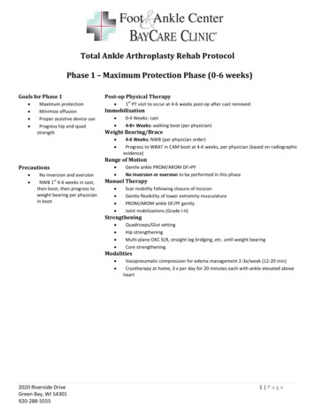
Figure 1. Typical ankle-inversion injury. Note the plantar-flexed ankle.occur significantly more often in athletes who have had previous ankle sprains.20 Pes cavus, rearfoot varus, tibial varus,and previous trauma are factors that may contribute to ankleinversion injury, although none of these have been scientifically verified as contributing factors.Figure 2. Anterior drawer test. The ankle is held between neutraland 108 of plantar flexion, and the calcaneus is pulled anteriorlywhile the tibia is held stable.EvaluationThe most common mechanism of injury is an athlete who‘‘rolled’’ over the outside of his or her ankle (Figure 1). Thisusually occurs as either a noncontact injury or when the athletelands from a step or jumps onto an opponent’s foot with aninverted foot. The foot is usually plantar flexed at the time ofthe injury. Many patients state that they have heard something‘‘snap’’ during the trauma; however, feeling a tearing sensationor hearing a snap does not appear to correlate with the severityof the injury.8 The main site of pain and swelling is usuallylocalized to the lateral side of the ankle over the ATFL. Several hours after the injury, generalized swelling and pain canmake the evaluation more difficult and less reliable. Most patients have pain and discomfort when trying to ambulate onthe injured extremity. Ecchymosis can occur 24 to 48 hoursafter the injury. The discoloration is usually worst along thelateral side but can also occur in the retrocalcaneal bursal areaand along the heel because of the potential space available forswelling and the pooling effect of gravity. It is important thatthe entire leg, ankle, and foot be examined to ensure that noother injuries have occurred. With tenderness over the midshaft of the fibula or medial-side tenderness and swelling, theexaminer should be suspicious of fracture or more significantinjury.Clinical stability tests for ligamentous disruption are bestperformed between 4 and 7 days after the injury, when theacute pain and swelling are diminished and the patient is ableto relax during the examination.21 The anterior drawer test ismore specific for assessing the integrity of the ATFL, and thetalar tilt test is more specific for detecting injury to the CFL.These findings are best recorded as differences between theankles (assuming the opposite ankle is uninjured), but the testscan still be difficult to interpret, and the results often varygreatly among examiners.22,23 Caution must be exercised ininterpreting these tests, but a positive test can help to confirmthe diagnosis in a patient with a suspicious history.18,24,25The anterior drawer test evaluates ATFL integrity by theamount of anterior-talar displacement that can be produced inthe sagittal plane. To perform this test, the patient should beFigure 3. Talar tilt test. The calcaneus and talus are grasped as aunit and tilted into inversion. The tibia is held stable with the anklein neutral dorsiflexion.sitting with the knee flexed to relax the calf muscles and prevent the patient from actively guarding against the examiner.The examiner grasps the heel firmly in one hand and pullsforward while holding the anterior aspect of the distal tibiastable with the other hand (Figure 2). The sensitivity of thetest can be improved by placing the ankle in 108 of plantarflexion.26 Increased anterior translation of the talus with respect to the tibia is a positive sign and indicates a tear of theATFL, particularly if the translation is significantly differentfrom the opposite side. However, how much translation isphysiologically normal is the subject of disagreement: it hasbeen reported to be anywhere from 2 mm to 9 mm.27,28 Therefore, it is better to compare the amount of pathologic anteriorlaxity with the normal side. This analysis by the examiner issubjective, and agreement among observers varies.The talar tilt test is defined as the angle produced by thetibial plafond and the dome of the talus in response to forcefulinversion of the hindfoot. The talar tilt test is performed withthe ankle in the neutral position. The examiner holds the heelstable while trying to invert the heel with respect to the tibia(Figure 3). It is important to try to grasp the talus and calca-Journal of Athletic Training407
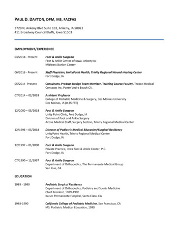
neus as a unit to limit subtalar motion during the test. As inthe anterior drawer examination, the results from the talar tilttest are difficult to interpret, with reports indicating normalvalues between 58 and 238,29,30 but as a general rule, morethan 108 difference from the normal side is considered abnormal.31A new testing device developed by Kirk et al32 applies standardized loads for both the anterior drawer and talar tilt tests.At an anterior force of 111 N (25 lbs) and a torque of 16 Nm,the mean anterior-drawer translation was 5.9 mm, and themean talar-tilt translation was 518. The device has not yet beenadopted into widespread use.Radiographic AnalysisClinical guidelines for determining the necessity of radiographs have been developed to limit the number of radiographs. These guidelines carry tremendous potential for costsavings. The Ottawa Ankle Rules (OAR) are the commonlyused criteria for predicting which patients require radiographicimages.33 Radiographs are only required for those patientswith (1) tenderness at the posterior edge or tip of the medialor lateral malleolus; (2) inability to bear weight (4 steps) eitherimmediately after the injury or in the emergency room; or (3)pain at the base of the fifth metatarsal. Following these rulesprovided nearly 100% sensitivity for detecting fractures whilesignificantly reducing the number of unnecessary radiographs.33 Standard radiographs, if necessary, should includeanteroposterior (AP), lateral, and mortise views. The mortiseview is an AP view with the tibia internally rotated by 158 to208. This position allows evaluation of the syndesmosis andassessment of mortise disruption. In the mortise view, the talusshould be equidistant from both malleoli.Stress radiography for acute injuries will not change thetreatment protocol and is generally not indicated. These techniques are more often used for research purposes or to differentiate between mechanical instability and functional instability in patients with chronic ankle problems. Specializedinstruments have been developed to apply standardized loadsduring the stress radiographs. The anterior-drawer stress radiograph is more sensitive for ATFL insufficiency, and the talartilt stress radiograph is more sensitive for CFL integrity. However, the amount of displacement that represents a pathologiccondition is variable. The most commonly used criteria for theanterior-drawer stress test are those of Karlsson,31 who definedabnormal laxity as an absolute anterior displacement of 10 mmor a side-to-side difference of more than 3 mm (Figure 4).Abnormal talar tilt is even more controversial due to the largevariation in ‘‘normal’’ talar tilt, which is reported to be from08 to 278.19,31,34,35 A talar tilt of 158 more than the normalside correlated with a complete double-ligament rupture(ATFL and CFL).19 As a general rule, a finding of more than108 greater than the normal side is considered abnormal (Figure 5).28If the results of the 2 stress-radiographic images are combined, the sensitivity of the tests increases to 68%, but thespecificity falls to 71%21; therefore, it is difficult to recommend routine use of stress radiography.Ankle-joint arthrography is a sensitive and specific diagnostic test for ligament ruptures,36,37 as shown by Lahde etal,22 who studied 7000 ankle arthrographies performed over a15-year period. But they also found limitations of arthrography: it is reliable only within the first 24 to 48 hours, cannot408Volume 37 Number 4 December 2002Figure 4. Anterior-drawer stress test. A, schematic drawing, and B,radiograph. (Copyright 2002 by the Hughston Sports MedicineFoundation, Inc).
drawback to this system is that, unless the injury is treatedsurgically, objective evidence of injury to each ligament islacking. Finally, because of the problems of these grading systems, a classification based on clinical severity has been used.This system has 3 clinical grades: grade I (mild), grade II(moderate), and grade III (severe).16,17 A grade I injury involves little swelling and tenderness, minimal or no functionalloss, and no mechanical joint instability. A grade II injury hasmoderate pain, swelling, and tenderness over the involvedstructures; some joint motion is lost, and joint instability ismild to moderate. A grade III injury is a complete ligamentrupture with marked swelling, hemorrhage, and tenderness;function is lost, and joint motion and instability are markedlyabnormal. Grading of ankle sprains remains a largely subjective interpretation, and agreement among independent observers varies.Differential DiagnosisFigure 5. Talar-tilt stress radiograph. (Copyright 2002 by theHughston Sports Medicine Foundation, Inc.)quantify the severity of ligament damage, and is an invasiveprocedure. Proper interpretation of arthrographic images requires a full understanding of the variant and natural leakageof contrast. Arthrography is a valuable research tool, but it israrely indicated for clinical use because it does not change thetreatment protocol.Similarly, magnetic resonance imaging (MRI) and computed tomography (CT) scanning are rarely necessary for typicalacute ankle sprains because the results do not affect the treatment protocol. Gaebler et al19 compared intraoperative findings with MRI results in 25 patients who had a talar tilt greaterthan 158 and found that MRI was reliable in diagnosing lateralligament injuries. Magnetic resonance imaging and CT scanning have been useful for identifying osteochondral injuriesthat may mimic, or occur in conjunction with, chronic lateralankle instability.38Grading Lateral Ankle-Ligament SprainsSeveral lateral ankle-ligament grading systems have beenused. This makes comparison in the literature difficult, asmany authors did not state which grading system they used.The traditional grading system for ligament injuries focuses ona single ligament, with a grade I injury representing a microscopic injury without stretching of the ligament on a macroscopic level. A grade II injury has macroscopic stretching, butthe ligament remains intact. A grade III injury is a completerupture of the ligament.31 Applying this grading system to lateral ankle-ligament sprains addresses only the status of theATFL and ignores injury to either the CFL or PTFL. Someauthors have thus resorted to grading lateral ankle-ligamentsprains by the number of ligaments injured.18,19,24 The majorOther problems can mimic, or be coupled with, lateral ankle-ligament sprains. Fractures of the ankle are often associated with ankle-ligament injuries.12 In particular, the examination should focus on potential fractures of the lateral,medial, and posterior malleolus; proximal fibula; lateral orposterior process of the talus; anterior process of the calcaneus; fifth metatarsal; navicular or other midtarsal bones; andchildren’s epiphyseal separations.Patients with stress fractures about the ankle joint may present with a different type of history but similar symptoms. Inparticular, a transverse, proximal diaphyseal fracture of thefifth metatarsal bone (Jones fracture) can mimic an acute lateral ankle sprain.33 This is particularly true when an acutefracture occurs through an area of previous stress reaction thatmay have had minimal or no symptoms. The distal fibula,medial malleolus, calcaneus, navicular, and metatarsals arealso prone to stress fracture.Osteochondral fractures or osteochondritis dissecans of thetalar dome or the tibial plafond can occur with lateral ankleligament sprains.38 These fractures can become chronic problems, with continued pain and recurrent instability episodes.If plain radiographs are negative despite continued pain, abone scan, CT scan, or MRI may be helpful to evaluate forthis lesion.38 Arthroscopy is the definitive test for the diagnosisand treatment of these fractures.Athletes with sprains of the subtalar joint or midfoot ligaments can present with a similar history.39 In particular, thedorsal calcaneocuboid ligament, bifurcate ligament, cervicalligament, and interosseous talocalcaneal ligament are prone toinjury.Subluxation or dislocation of the peroneal tendons can mimic an ankle sprain.40 However, these injuries typically occurby a violent dorsiflexion and pronation moment of the ankleinstead of the typical inversion injury of lateral-ligament injuries.40SUBTALAR-JOINT SPRAINS AND INSTABILITYThe incidence of subtalar sprains is unknown, mainly dueto the difficulty of assessing these injuries and the commonassociation with lateral ankle-ligament sprains. They are probably more common than appreciated. Meyer et al39 studiedsubtalar arthrograms in 40 patients with acute ankle sprainsand found that 17 (43%) had subtalar-ligament injury. Fortu-Journal of Athletic Training409
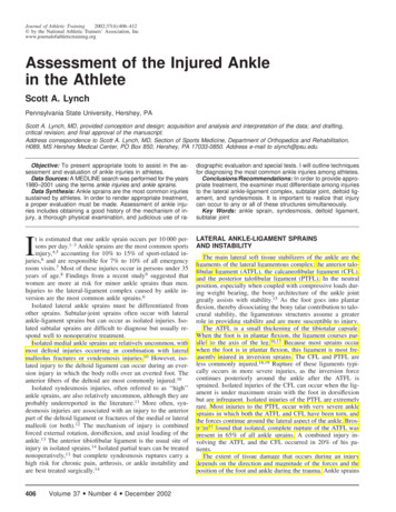
nately, the incidence of chronic ankle problems is low, withsubtalar instability present in only about 10% of patients whopresent with chronic lateral ankle-ligament instability.41 Patients with acute subtalar sprains seem to do well with nonoperative treatment similar to that used for acute lateral ankleligament sprains. However, since the definition and diagnosisof subtalar sprains are not agreed upon in the literature, thisis difficult to prove.Acute symptoms of subtalar sprains are similar to, and canoccur with or be masked by, lateral ankle-ligament sprains.Tenderness over the subtalar joint is characteristic but can bedifficult to differentiate from the tibiotalar joint because of theclose proximity and the swelling that obscures the anatomy.Clinical evaluation of subtalar instability is very difficultand unreliable. An evaluation of the change in angle betweenthe heel and the tibia with passive inversion and eversion ofthe heel can be made by comparing this angle with that on theuninjured side,41 but the sensitivity and specificity of this testis unknown.Radiographs should be obtained as per the Ottawa anklerules.33 Subtalar stress radiographs,42 subtalar arthrography,39or stress tomography41 can show increased motion and differentiate between subtalar and talocrural motion; however, mostof these injuries can be effectively treated by rehabilitation, sospecialized studies are usually unnecessary. If surgery is considered, stress radiographs may be helpful in planning the surgery.Classification of Subtalar-Joint SprainsSubtalar-joint sprains are classified by the injury mechanismand the degree of ligamentous damage.39 The injury can occurin either plantar flexion or dorsiflexion. Forceful supinationwith the foot in plantar flexion tears the ATFL (and possiblythe cervical ligament), followed by either disruption of theCFL and lateral capsule (type 1) or tearing of the interosseoustalocalcaneal ligament (type 2). When the ankle is in dorsiflexion, the ATFL is not under tension and remains uninjured.This type of injury tears the CFL, the cervical ligament, andthe interosseous talocalcaneal ligament (type 3). A type 4 subtalar sprain is a rupture of all lateral and medial capsuloligamentous components of the posterior tarsus. This injury occursas the foot moves from dorsiflexion to plantar flexion whileforceful hindfoot supination occurs.39Figure 6. Radiographic measurements of deltoid and syndesmosisinjuries. (Adapted with permission from Harper MC. The deltoidligament: an evaluation of need for surgical repair. Clin Orthop.1988;226:156–158; Lippincott Williams & Wilkins.10)to the lateral side of the foot, placing a large abduction forceonto the ankle and deltoid ligament. Because the forces required to injure the strong deltoid ligament are so great, theinjury usually continues through the syndesmosis by the stronglever action of the lateral malleolus on the lateral aspect of thetalus.12DELTOID LIGAMENT TEARSIn Broström’s18 series of 281 acute ankle sprains, medialside ankle sprains constituted only 3%. Nearly all of the medial-side injuries were partial ligament tears. Complete deltoidligament ruptures most often occur in combination with anklefractures. In Harper’s10 review of 42 patients with completedeltoid ligament ruptures, all had other injuries. In the anklefracture classification described by Lauge-Hansen,12 a deltoidligament tear or medial malleolar fracture occurs as the injurypattern continues around the ankle in a circular fashion. The3 most characteristic mechanisms of injury of the deltoid ligament are pronation-abduction, pronation-external rotation,and supination-external rotation of the foot.12,43,44 The firstcomponent describes the position of a planted foot, and thesecond term indicates the relative motion of the foot as the legrotates about the planted foot. So, in the pronation-abductioninjury, the foot is planted in pronation as the upper body falls410Volume 37 Number 4 December 2002EvaluationDeltoid ligament injuries cause pain, tenderness, and swelling on the medial side of the ankle. A defect may be palpablebelow the medial malleolus in complete ruptures. If a deltoidligament injury is present, it is extremely important to evaluatethe ankle for a syndesmosis sprain or fracture. The entire fibula, including the proximal third and proximal tibiofibularjoint, must also be palpated to rule out complete syndesmosisdisruption.For medial-side injuries, radiographs are necessary to evaluate the bony structures and syndesmosis. The minimum radiographic series includes AP, lateral, and mortise views. Ifthere is any suspicion for a proximal fibular fracture, AP andlateral radiographs of the entire tibia and fibula should be taken. If the deltoid ligament and syndesmosis are both completely disrupted, the medial clear space between the medial talus
and the lateral border of the medial malleolus will be widenedto 4 mm or more (Figure 6).45,46 However, isolated deltoidruptures do not cause widening of the medial clear space because the lateral malleolus holds the talus in position. Similarly, syndesmosis injuries without deltoid tears do not havemedial joint-space widening. In this case, the inferior tibiofibular joint must be carefully evaluated for syndesmosis injury.Eversion-stress radiographs, arthrography, or MRI may behelpful in difficult cases, but the diagnosis can most often bemade with clinical examination and plain radiographs.TIBIOFIBULAR SYNDESMOSIS TEARS: ‘‘HIGH’’ANKLE SPRAINSPartial or complete rupture of the syndesmosis ligamentcomplex can cause diastasis of the inferior tibiofibular joint.47Isolated complete syndesmosis injuries are rare, and relativelylittle information exists in the literature about ankle diastasisin the absence of fracture. Fritschy48 reported only 12 casesof complete isolated syndesmosis ruptures in a series of morethan 400 ankle-ligament ruptures. All 12 injuries were causedby a sudden external rotation of the ankle that caused the talusto pry the fibula laterally, thus opening the distal tibiofibulararticulation.Isolated partial syndesmosis injuries occur with some frequency, and their incidence is probably underreported in theliterature. It is important to recognize that syndesmosis involvement generally increases recovery time 2- or 3-fold overthat for a lateral ankle-ligament sprain. Early diagnosis of thesyndesmosis sprain can help to give the athlete and coachesrealistic expectations of return to play. Nussbaum et al13 suggested that the expanse of the syndesmosis tenderness waspredictive of recovery time, with more syndesmosis tendernesscorrelating with more playing days missed. However, it ismuch more common for the injury to be associated with afracture or deltoid ligament injury (or both).12 With ankle fractures, the frequency of syndesmosis ruptures is related to thetype and level of associated fibular fractures.12 Syndesmosisinjuries are more common as the level of the fibular fracturerises above the level of the ankle joint, as predicted by theLauge-Hansen12 injury-mechanism classification of ankle fractures. In this classification scheme, ligamentous injuries orfractures occur as the rotatory forces continue around the anklein a circular fashion.12Figure 7. Dorsiflexion and external-rotation stress test for syndesmosis injury. The tibia is held stable while the foot is dorsiflexedand externally rotated.and osseous avulsions and to evaluate syndesmosis widening.In athletes with possible syndesmosis widening or proximalfibular tenderness, AP and lateral films of the entire tibia andfibula are necessary to rule out a Maisonneuve fracture. Acceptable radiographic guidelines that indicate syndesmosis diastasis are controversial, and measurements can be affectedgreatly by the amount of tibial rotation. The most commonlyused guidelines are a joint-space widening of greater than 5mm or a tibiofibular overlap of less than 10 mm, both as measured on the AP view. Other authors prefer to use the ratio ofmeasurements to the fibular width.50 Ninety percent predictiveintervals for a normal relationship were a tibiofibular overlapto-fibular width ratio greater than 24% and a tibiofibular clearspace-to-fibular width ratio of less than 44%, both as measuredon the AP radiograph.50 Stress radiographs with the foot inexternal rotation in both dorsiflexion and plantar flexion maydemonstrate the diastasis.48 Magnetic resonance imaging hasnow become the test of choice for evaluating the syndesmosisin difficult cases.51EvaluationCONCLUSIONSPain and tenderness are located primarily on the anterioraspect of the syndesmosis and interosseous membrane. Activeand passive external rotation of the foot is painful. The external-rotation test is performed by externally rotating the footwith the ankle in dorsiflexion (Figure 7), which stresses thesyndesmosis by levering the talus against the lateral malleolus.Patients with a syndesmosis injury have pain over the anteriorinferior tibiofibular ligament and joint. The squeeze test is performed by compressing the midshaft of the tibia and fibulatogether. If a syndesmosis injury is present, the patient haspain at the inferior tibiofibular joint. Biomechanical studieshave confirmed distal tibiofibular motion during the squeezetest.49Radiographs should be taken according to the Ottawa anklerules as outlined in the previous sections.33 Anteroposterior,lateral, and mortise views may be needed to exclude fracturesIn order to provide appropriate treatment after an athletesprains an ankle, a thorough evaluation is necessary. Thisshould include the mechanism of injury, physical examination,and appropriate radiographic studies and special tests. The injury can affect the lateral ankle-ligament complex, the subtalarjoint, the deltoid ligament, or the syndesmosis or any combination of these structures simultaneously. Defining the extentof injury allows the clinician to institute the proper treatmentregimen in preparation for the athlete’s safe return to sport.REFERENCES1. Brooks SC, Potter BT, Rainey JB. Treatment for partial tears of the lateralligament of the ankle: a prospective trial. Br Med J (Clin Res Ed). 1981;282:606–607.2. McCulloch PG, Holden P, Robson DJ, Rowley DI, Norris SH. The valueof mobilisation and non-steroidal anti-inflammatory analgesia in the man-Journal of Athletic Training411
22.23.24.25.26.agement of inversion injuries of the ankle. Br J Clin Pract. 1985;2:69–72.Ruth C. The surgical treatment of injuries of the fibular collateral ligaments of the ankle. J Bone Joint Surg Am. 1961;43:229–239.Lassiter TE Jr, Malone TR, Garrett WE Jr. Injury to the lateral ligamentsof the ankle. Orthop Clin North Am. 1989;20:629–640.McConkey JP. Ankle sprains, consequences and mimics. Med Sport Sci.1987;23:39–55.MacAuley D. Ankle injuries: same joint, different sports. Med Sci SportsExerc. 1999;31(7 suppl):409–411.Viljakka T, Rokkanen P. The treatment of ankle sprain by bandaging andantiphlogistic drugs. Ann Chir Gynaecol. 1983;72:66–70.Nilsson S. Sprains of the lateral ankle ligaments, part II: epidemiologicaland clinical study with special reference to different forms of conservativetreatment. J Oslo City Hosp. 1983;33:13–36.Hosea TM, Carey CC, Harrer MF. The gender issue: epidemiology ofankle injuries in athletes who participate in basketball. Clin Orthop. 2000;372:45–49.Harper MC. The deltoid ligament: an evaluation of need for surgicalrepair. Clin Orthop. 1988;226
Journal of Athletic Training 407 Figure 1. Typical ankle-inversion injury. Note the plantar-flexedan-kle. Figure 2. Anterior drawer test. The ankle is held between neutral and 108 of plantar flexion, and the calcaneus is pulled anteriorly while the tibia is held stable. Figure 3. Talar tilt test. The calcaneus and talus are grasped as a