Transcription
PRESENTSDr. Mufa T. Ghadiali is skilled in all aspects of General Surgery.His General Surgery Services include:General SurgeryAdvanced Laparoscopic SurgerySurgical OncologyGastrointestinal SurgeryHernia SurgeryEndoscopyAnatomy Of The Foot And AnkleMultimedia Health EducationDisclaimerThis movie is an educational resource only and should not be used tomanage Orthopaedic Health. All decisions about management of theFoot and Ankle must be made in conjunction with your Physician or alicensed healthcare provider.Mufa T. Ghadiali, M.D., F.A.C.SDiplomate of American Board of Surgery6405 North Federal Hwy., Suite 402Fort Lauderdale, FL 33308Tel.: 954-771-8888Fax: 954- 491-9485www.ghadialisurgery.com
Anatomy Of The Foot And AnkleMultimedia Health EducationMULTIMEDIA HEALTH EDUCATION MANUALTABLE OF CONTENTSCONTENTSECTION1 . ANATOMYa. Ankle & Foot Anatomyb. Soft Tissue Anatomy2 . BIOMECHANICSwww.ghadialisurgery.com
Anatomy Of The Foot And AnkleMultimedia Health EducationUnit 1:AnatomyIntroductionThe foot and ankle in the human body work together to provide balance, stability,movement, and Propulsion.This complex anatomy consists of:26 bones33 jointsMusclesTendonsLigamentsBlood vessels, nerves, and soft tissueIn order to understand conditions that affect the foot and ankle, it is important tounderstand the normal anatomy of the foot and ankle.AnkleThe ankle consists of three bones attached by muscles, tendons, and ligaments that connectthe foot to the leg. In the lower leg are two bones called the tibia (shin bone) and the fibula.These bones articulate (connect) to the Talus or ankle bone at the tibiotalar joint (ankle joint)allowing the foot to move up and down.The bony protrusions that we can see and feel on the ankle are:Lateral Malleolus: this is the outer ankle bone formed by the distal end of the fibula.Medial Malleolus: this is the inner ankle bone formed by the distal end of the tibia.Tibia (shin bone)(Refer fig.1)Tibia(shin bone)(Fig.1)www.ghadialisurgery.com
Anatomy Of The Foot And AnkleMultimedia Health EducationUnit 1:AnatomyFibulaFibula(Refer fig.2)(Fig.2)Talus(Refer fig.3)Talus(Fig.3)Lateral Malleolus(Refer fig.4)LateralMalleolus(Fig.4)Medial Malleolus(Refer m
Anatomy Of The Foot And AnkleMultimedia Health EducationUnit 1:2:GastritisLessonsAnatomyHindfootThe foot can be divided into three anatomical sections called the hindfoot, midfoot, andforefoot.The hindfoot consists of the Talus bone or ankle bone and the calcaneous boneor heel bone. The calcaneous bone is the largest bone in your foot while the talus bone isthe highest bone in your foot. The calcaneous joins the Talus bone at the subtalar jointenabling the foot to rotate at the ankle. The hindfoot connects the midfoot to the ankle atthe transverse tarsal joint.Talus(Refer fig.6)Talus(Fig.6)Calcaneus(Refer fig.7)Calcaneus(Fig.7)MidfootThe midfoot contains five tarsal bones: the navicular bone, the cuboid bone, and 3cuneiform bones. It connects the forefoot to the hindfoot with muscles and ligaments.The main ligament is the plantar fascia ligament. The midfoot is responsible for formingthe arches of your feet and acts as a shock absorber when walking or running. Themidfoot connects to the forefoot at the five tarsometatarsal joints.www.ghadialisurgery.com
Anatomy Of The Foot And AnkleMultimedia Health EducationUnit 1:2:GastritisLessonsAnatomyNavicular(Refer fig.8)Navicular(Fig.8)Cuboid(Refer fig.9)Cuboid(Fig.9)Cuneiform Bones(Refer fig.10)CuneiformBones(Fig.10)ForefootThe forefoot consists of your toe bones, called phalanges, and metatarsal bones, the longbones in your feet. Phalanges connect to metatarsals at the ball of the foot by jointscalled phalange metatarsal joints.Each toe has 3 phalange bones and 2 joints, while the big toe contains two phalangebones, two joints, and two tiny, round sesamoid bones that enable the toe to move upand down. Sesamoid bones are bones that develop inside of a tendon over a bonyprominence.The first metatarsal bone connected to the big toe is the shortest and thickest of themetatarsals and is the location for the attachment of several tendons. This bone isimportant for its role in propulsion and weight bearing.www.ghadialisurgery.com
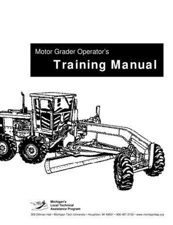
Anatomy Of The Foot And AnkleMultimedia Health EducationUnit 1:AnatomyPhalanges(Refer fig.11)Phalanges(Fig.11)Metatarsal(Refer fig.12)Metatarsal(Fig.12)Soft Tissue AnatomyOur feet and ankle bones are held in place and supported by various soft tissues.CartilagelShiny and smooth, cartilage allowssmooth movement where two bonescome in contact with each other.Cartilage(Refer fig.13)(Fig.13)www.ghadialisurgery.com
Anatomy Of The Foot And AnkleMultimedia Health EducationUnit 1:AnatomyTendonsTendons are soft tissue that connectsmuscles to bones to provide support. TheAchilles tendon, also called the heel cord,is the largest and strongest tendon in thebody. Located on the back of the lowerleg it wraps around the calcaneous, orheel bone. When inflamed it causes avery painful condition called Achillestendonitis and can make walking almostimpossible due to the Pain.Tendons(Fig.14)(Refer fig.14)LigamentsLigaments are strong rope like tissue thatconnects bones to other bones and helphold tendons in place providing stabilityto the joints. The plantar fascia is thelongest ligament in the foot, originatingat the calcaneous, heel bone, andcontinuing along the bottom surface ofthe foot to the forefoot.Ligaments(Fig.15)It is responsible for the arches of the foot and provides shock absorption. A common causeof heel pain in adults, plantar fasciitis can occur when repetitive micro tears occur in theplantar fascia from overuse. Ankle sprains, the most commonly reported injury to the footand ankle area, involve ligament strain, and usually occur to the talo-fibular ligament and thecalcaneo-fibular ligament.(Refer fig.15)MusclesMuscles are fibrous tissue capable of contracting to cause body movement. There are20 muscles in the foot and these are classified as intrinsic or extrinsic. The intrinsic musclesare those located in the foot and are responsible for toe movement. The extrinsic musclesare located outside the foot in the lower leg.(Refer fig.16)www.ghadialisurgery.com
Anatomy Of The Foot And AnkleMultimedia Health EducationUnit 1:The gastrocnemius or calf muscle is thelargest of these and assists withmovement of the foot. Muscle strainsoccur usually from overuse of the musclein which the muscle is stretched withoutbeing properly warmed up.(Refer fig.16)AnatomyMuscles(Fig.16)BursaeBursae are small fluid filled sacs thatdecrease friction between tendons andbone or skin. Bursae contain special cellscalled synovial cells that secrete alubricating fluid. When this fluidbecomes infected, a common painfulcondition known as Bursitis can develop.Bursae(Fig.17)(Refer fig.17)www.ghadialisurgery.com
Anatomy Of The Foot And AnkleMultimedia Health EducationUnit 2:BiomechanicsBiomechanics of Foot & AnkleBiomechanics is a term to describe movement of the body. The ankle joint by itself permitstwo movements:Plantar flexionPointing the foot downward. Thismovement is normally accompanied byinversion of the foot.Plantar flexion(Refer fig.18)(Fig.18)DorsiflexionRaising the foot upward. This movementis normally accompanied by eversion ofthe foot.The foot (excluding the toes) alsopermits two movements:(Refer fig.19)Dorsiflexion(Fig.19)InversionTurning the sole of the foot inward.(Refer fig.20)Inversion(Fig.20)EversionTurning the sole of the foot outward.(Refer fig.21)Eversion(Fig.21)www.ghadialisurgery.com
Anatomy Of The Foot And AnkleMultimedia Health EducationUnit 2:BiomechanicsThe toes allow four different movements:Plantar flexionBending the toes towards the sole of thefoot.Plantar flexion(Refer fig.22)(Fig.22)DorsiflexionBending the toes towards the top of thefoot.Dorsiflexion(Refer fig.23)(Fig.23)AbductionSpreading the toes apart. This movementnormally accompanies plantardorsiflexion.Abduction(Refer fig.24)(Fig.24)AdductionBringing the toes together. Thismovement normally accompanies plantarflexion.Adduction(Refer fig.25)(Fig.25)www.ghadialisurgery.com
Anatomy Of The Foot And AnkleMultimedia Health EducationUnit 2:3:BiomechanicsDisclaimerDisclaimerThis movie is an educational resource only and should not be used to manage OrthopaedicHealth. All decisions about management of the Foot and Ankle must be made inconjunction with your Physician or a licensed healthcare providerwww.ghadialisurgery.com
Anatomy Of The Foot And AnkleMultimedia Health EducationYOUR SURGERY DATEREAD YOUR BOOK AND MATERIALVIEW YOUR VIDEO /CD / DVD / WEBSITEPRE - HABILITATIONARRANGE FOR BLOODMEDICAL CHECK UPADVANCE MEDICAL DIRECTIVEPRE - ADMISSION TESTINGFAMILY SUPPORT REVIEWPhysician's Name :Patient’s Name :Physician's Signature:Patient’s Signature:Date :Date :www.ghadialisurgery.com
Anatomy Of The Foot And Ankle Unit 1: Anatomy Phalanges (Refer fig.11) (Fig.11) Phalanges Metatarsal (Refer fig.12) (Fig.12) Metatarsal Sof t Tissue Anatomy Our feet and ankle bones are held in place and supported by various soft tissues. Cartilagel (Refer fig.13) (Fig.13) Cartilage Shiny and smooth, cartilage allows smooth movement where two bones