Transcription
Open AccessAustin Journal of Anesthesia and AnalgesiaResearch ArticleCan An Embolectomy Catheter Become a One StopSolution for Lung Isolation in One Lung Ventilation?Jain A*Department of Anesthesiology, Gandhi Medical College,Bhopal, Madhya Pradesh, IndiaAbstract*Corresponding author: Anuj Jain, Department ofAnesthesiology, Gandhi Medical College, Bhopal, MadhyaPradesh, IndiaReceived: July 03, 2020; Accepted: August 13, 2020;Published: August 20, 2020One Lung Ventilation (OLV) is often required for surgeries in the thorax;patients may belong to any age group. If the patients are adult, the options forlung isolation are many, but handicap arises when one is dealing with paediatricpatients. While dealing with pediatric patients, Embolectomy Catheters (EC)may be quite useful as they are available in a wide range of sizes, makingthem appropriate for lung isolation even in neonates. Besides, ECs can beimprovised to achieve selective lobar separation in patients who have borderlinelung function and are at risk for intraoperative hypoxemia. Though EC is aversatile device, it is underutilized; only a few low-quality pieces of evidenceare available. Among the people who have used ECs for lung isolation, thetechnique of insertion had been variable. This article is an attempt to review themethods of lung isolation and explore the possibility of EC becoming a universaldevice for lung isolation.BackgroundOne Lung Ventilation (OLV) is usually indicated in thoracic,oesophageal, vascular and non- thoracic surgical settings in perioperative period. A variety of surgeries require OLV; an indication canbe to improve the exposure of intrathoracic structures or to preventsoiling of the healthy lung with contaminant from the affected lung.Now a days, Embolectomy Catheters (ECs) are becoming morepopular for One Lung Ventilation (OLV) because of their easyavailability even in resource limited health care facilities. The purposeof this review is to provide an update on emerging evidence anddefining the role of ECs in OLV, both in paediatric and adult patients.Challenges of OLV in paediatric patients and availablelung isolation techniquesOLV in paediatric patients is a challenge to all anaesthesiologists;smaller the airway, greater is the challenge. Antero-Posterior(AP) diameter of a neonatal trachea, in both males and females isjust 4.3mm; at 15 years, the size increases to 14mm in both sexes,thereafter the trachea in female stops growing, while in males it growsto a size of 16-18mm at 19years (Table 1).In adults, the Lung Isolation Techniques (LITs) most commonlyused are Double Lumen Tubes (DLTs); less common options include,Bronchial Blockers (BB), Univent Tubes (UT), and EmbolectomyCatheters (EC).Double lumen tube: DLT is most popular, but it is not availablebelow 26 Fr [1]. A generic interpretation of a 26Fr DLT is two 4mmETTs joined together, implying thereby that in order to accommodateeven the smallest DLT the tracheal diameter has to be more than8mm and the bronchial diameter has to be more than 4mm. So, aDLT is fit for paediatric patients of age more than 8 years, weighingmore than 30kg or height more than 130 centimeters; recommendedsize varies with age and human race too [2]. According to Brodsky,appropriate size of DLT may be determined either by directAustin J Anesthesia and Analgesia - Volume 8 Issue 3 - 2020ISSN : 2381-893X www.austinpublishinggroup.comJain. All rights are reservedmeasurement of bronchial diameter in a chest radiograph or bypredicting left Bronchus Diameter (BD) based on Tracheal Diameter(TD) according to the formula, LBD (0.4 TD) 3.3 [3].Marraro-bilumen tube: These bilumen tubes can be used insmaller paediatric patients; smallest tube is made of two 2.0 mm tubesmolded together. The sizes may go up to two 3-3.5mm tubes moldedtogether, making it appropriate for 2-3 years age group. AlthoughMarraro tubes may be used in neonates, it is usually unavailable andmost anaesthesiologists are unaware that such tubes even exist.Univent Tube (UT): A UT with an Internal Diameter (ID) of3.5mm has an External Diameter (ED) of 7.5-8.0mm, while the onewith 4.5mm ID have an ED of 8.5-9.0mm. The large ED limits its usein Asian paediatric population smaller than 4-6years of age [4].Bronchial blockers: Arndt’s BB are available in 5,7 and 9Fr andare appropriate for 6month to 6 years, 6 years to 14 years and 14-18years age groups respectively [5]. BBs have a low pressure cuff and acoaxial channel for suctioning, which allows active deflation of lung.Apart from its high cost and less availability, the major disadvantageof BBs is that the loop guide wire (which is essential for placement)once removed cannot be reinserted, rendering it useless if need forrepositioning arises. In a RCT by Narayanaswamy, the misplacementrate of Arndt’s blocker (the most popular among the 3 BBs) washigher compared to other two BBs [6].Endobronchial Single Lumen Tube (SLT): SLTs can be used forlung isolation in emergency situations like contralateral pneumothorax[7]. Some anaesthesiologists also insert two SLTs, one in trachea andother in the bronchus. Endobronchial SLT has a tendency to progressto the right side due to the more inline orientation of the rightbronchus. It can be of concern, if surgery needs to be performed inthe right hemithorax. In order to overcome this problem, Chengod etal., [8]. Suggested left endobronchial intubation using a SLT that wascurved towards the left side (with a stylet). However, this approachmay not be appropriate up to 2 years of age as the diameter of the leftCitation: Jain A. Can An Embolectomy Catheter Become a One Stop Solution for Lung Isolation in One LungVentilation?. Austin J Anesthesia and Analgesia. 2020; 8(3): 1092.
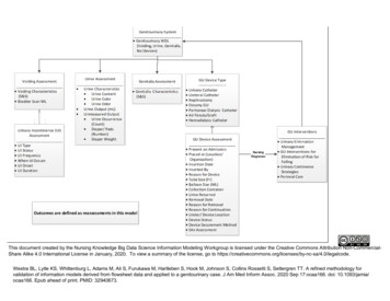
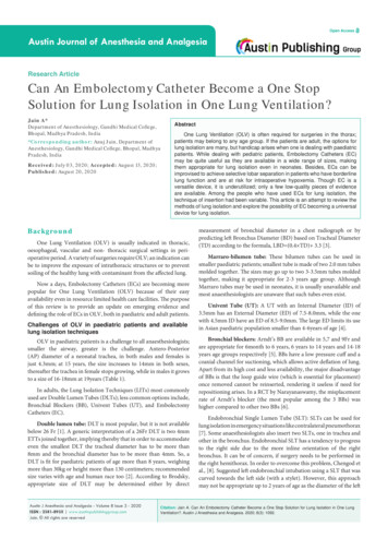
Jain AAustin Publishing GroupAge of the PatientP aedi at ri cappropriate size DLT availableappropriate size DLT not availableorpatient having a limited mouth opening.orn eed fo r s el ect iv e l o b ar i s ol a ti o n d u e t o p o o r l u n gperform fiberoptic bronchoscopy tofunctionrule out a tracheal bronchus (TB)perform direct laryngoscopyNo TBTB presentin s ert an ap p ro p ri at e si zed EC ( wit h a g u id e wi re)through the glottis with distal portion bent toPerform DLT insertion, keepinginsert DLT in usual manner*approximately 15-20 degree .tracheal cuff above the TBPass appropriate size FOB through the ETT.under FOB vision steer the EC into the desired bronchus by twisting the extraoral part.inflate EC cuff with minimum volume of saline or air so that blanching of bronchial mucosa does not occur.check for isolation of the desired lung or lobe by auscultationposition the patient and recheck for isolation after positioning.leave the lung to deflate on its own if the EC being inserted is not a Thru lumencatheter (provide approx. 15minute more than would normally be needed whena DLT is being used.Slow suction to be applied for active deflation ofthe lung if a Thru lumen catheter has been inserted.*Measure the approximate angle of each main stem bronchus from the trachea.Figure 1: Proposed algorithm for lung isolation for OLV, with the inclusion of EC.main bronchus is small to accommodate an appropriate for age SLT(Table 1).Embolectomy catheters: ECs although invented for a differentpurpose, is a versatile equipment, which can be improvised to effectOLV. EC was invented by Thomas Fogarty in 1963; patent for it wasacquired in 1969. EC was first used by Charles Dotter in 1965 for firstballoon angioplasty. There are few evidences in scientific literatureto suggest the use of ECs for OLV but the technique of EC insertionvaries in most of the cases with no consensus regarding the methodof insertion (Table 2). ECs come in a variety of sizes; 2 and 3Fr ECs,Submit your Manuscript www.austinpublishinggroup.comdo not have a guidewire in them (hence, lack the required stiffness),making it difficult to steer them for endobronchial placement. ECs ofhigher sizes have a guidewire and may be used for lung isolation. ForOLV, EC is usually used in conjunction with ETT, but may be usedalong-with DLTs also.Technical review of the ECs as LITs: LITs have used invarious ways, but the quality of evidence regarding itsuse still remains poorCo-axial vs Parallel techniques: ECs may be passed coaxiallyin the ETT or may be passed externally, parallel to the ETT; choiceAustin J Anesthesia and Analgesia 8(3): id1092 (2020) - Page - 02
Jain AAustin Publishing GroupTable 1: Age wise dimensions of the airways as by Szelloe et al [17], in comparison with calculated size of ETT.Age(yr)Smallest Mean AP TD (mm)Mean Right BD (mm)Mean Left BD (mm)Predicted size of cuffed endotracheal tube [3.5 age(yr)/4]mm0-16.0 (2.9-7.7)3.9 (2.4-6.0)3.4 (2.1-5.7)3.51-27.0 (4.3-9.2)4.9 (3.8-7.5)4.2 (2.7-6.6)42-47.9 (4.1-10.3)6.1 (3.0-7.9)5.5 (2.7-8.5)4-4.54-67.5 (5.1-10.9)6.1 (4.4-7.8)5.3 (4.1-6.6)4.5-5.06-88.8 (5.7-11.0)6.8 (4.3-9.0)6.0 (4.5-10.1)5.0-5.58-109.5 (7.4-12)7.0 (5.5-9.4)6.6 (4.9-9.1)5.5-6.010-1210.8 (10.2-15.8)8.2 (6.6-10.0)7.5 (5.6-11.1)6.0-6.512-1411.7 (10.3-15.8)8.5 (7.3-10.5)8.1 (6.5-10.0)6.5-714-1613.0 (8.9-16.8)9.8 (6.5-12.7)9.2 (6.2-11.5)Table 2: Literature review with regards to use of Embolectomy catheters in lung isolation.S.noAuthorTOPYOPRegionE or IToSAgeSize of EC(in F)FOBusedLung isolated1Amar et al [18]CR2001USAE, IEsophageal surgeryA8YRL2Asai et al [19]CR2000JapanILung SurgeryP3Y3Kamra et al [20]CS (N 27)2017IndiaELung surgeryPVariableY4Bakshi et al [21]CR2013IndiaELung surgeryP3YR5Shimamoto et al[22]YLCR2004JapanILung surgeryP4 (Throughlumen)P5-MarYR(4) L(6)P5YL4,5YR(selective rightupper lobe)YL5Ho et al [23]CS N 102008ChinaE, ILung surgery, Mediastinalsurgery, Thoracic Spine surgery,Oesophageal surgery.6Mohan v et al [24]CR2002IndiaILungBaidya et al [25]7R(2) L(2)CS (N 4)2015IndiaELungP8Vretzakis et al [26] CS (N-61)2005GreeceILungA9Umoroglu et al [27]CR2017TurkeyLung lavageP10Peeters et al [28]CR2015BelgiumILung surgeryP3YL11Sharma et al [29]CR2014IndiaILung surgeryA4YR (middle lobe)12Cho et al [30]CR2010KoreaELung surgeryP3YR13Sutton et al [31]CS (N 11)2012USAELing surgeryP5YR(3) L(8)14Chaitanya et al [32]CR2016IndiaELung surgeryP4YL15Mathews andSanders [33]CR2001UKILung surgeryA5yR16Morikawa et al [34]CS (N 3)2002JapanILung surgeryA5YR and LAbbreviations: CR: Case Report; CS: Case Series; N: Number of Patients; YOP: Year of Publication; E: Extraluminal; I: Intraluminal; TOS: Type of Surgery; A: Adults;P: Paediatric; Y: Yes; N: No; R: Right; L: Left.depends on relative size of ETT and EC. Ho et al., passed EC bothextra and intraluminal to the ETT; in wider ETT (ID, more than4.5mm) the stem of EC was passed intraluminal and the distal endwas made extraluminal through the murphy eye [9]. In thinnerETT, the stem of the EC was extraluminal and distal end becameintraluminal through the murphy eye. When the distal end was madeintraluminal, the bevel of the ETT was trimmed obliquely to decreasethe resistance to gas flow; this practice needs to be questioned, dueto the safety concerns that may arise. Asai et al., made a hole in theETT near the connector end and passed a 3F EC through it [19].This practice, seems good in the absence of proprietary three wayairway connector, may become problematic if the EC size needs tobe changed and more so it can used only with wider diameter ETTs.Sutton et al [30]. Successfully used EC extraluminally in 9 patients,of age less than 6months. Mohan et al. [23], tried using a 5F ECSubmit your Manuscript www.austinpublishinggroup.comextraluminally and had to abandon the procedure as they could notnegotiate the EC “smoothly”. They succeeded when they tried puttingthe EC intra-luminally. Park et al., inserted EC for lung isolation inadult patients with subglottic stenosis; they passed the EC within thetube [10].Important consideration while deciding whether to put EC, intraor extraluminally is the size of EC relative to the size of ETT; careshould be taken not to occlude more than 50% of the cross sectionalarea (i.e. 70% of the diameter) of the ETT. However, occlusion ofeven 50% CSA may compromise ventilation and oxygenation inrelatively sick patients. As seen in Table 2, the balloon diameter of aEC (Fogarty Catheter - Edwards Life sciences, Irvine, CA, USA) canvary anywhere from 1.4 mm to 14 mm, thereby making it appropriatefor even the smallest of the airways. Since the diameter of EC is alsosmall compared to the ETT, it is less likely to cause any obstruction/Austin J Anesthesia and Analgesia 8(3): id1092 (2020) - Page - 03
Jain AAustin Publishing GroupTable 3: Dimension of Fogarty EC along with the dimension of its balloon in deflated and inflated state.Catheter size (Fr)Diameter of catheter body(mm) Size in Fr/3 size in mmInflated balloon diameter (mm)Deflated balloon 2.66alteration to airflow. According to our experience, passing the ECextraluminally although not as smooth as passing it intra-luminal,does give two important advantages. Firstly, unlike the intraluminalapproach, this approach need not be changed according to size of theETT. Secondly, ETT cuff when inflated anchors the EC in position,preventing misplacement of EC; besides, no specialized three-wayairway adapter is required.Kamra et al., [11] successfully used a rigid bronchoscope to guideEC placement for OLV. Rigid bronchoscopy would require a deepplane of anesthesia which may not be appropriate when anticipating adifficult airway situation. More so, rigid bronchoscope is unavailablein the airway armamentarium of anesthesiologist. Mathews et al.,achieved OLV by inserting EC in a tracheotomised patient, throughthe tracheostomy tube [32].Utility of ECs in unconventional situation: EC may provideadded advantage in conditions where the lung functions arecompromised. Morikawa et al. [33], inserted two 8F ECs, in orderto obtain selective lobar collapse in 3 patients, who had a poorlung function. Vretzakis et al., evaluated oxygenation in patientundergoing right lung surgery with DLT in situ and found that 13.7percent patients had oxygenation problem during OLV [25]. Outof these, oxygenation issues could be resolved with conventionaltechniques only in 50 percent patients; other half of the patients weremanaged with selective lobar ventilation preserving strategy. In orderto achieve this, they passed EC through the tracheal lumen of DLTand selectively blocked bronchial opening (upper lobe ventilation waspreserved by them, in all the cases).Bent vs straight ECs: Ramakrishna et al., bent the tip of ECto make it steerable into one of the bronchus [31]. In our practice,we have also found the techniques of bending the distal part of ECuseful, in cases requiring selective lobar isolation. We have proposedin detail, the techniques of OLV using a EC in Figure 1. The otherauthors did not describe whether they bent the EC or not.EC insertion, whether FOB guidance is necessary: As peravailable literature, all the users (Table 3), placed EC under FOBguidance, except Munir et al., [12] who considered FOB guidedplacement optional. In our view, performing bronchoscopy ismandatory in all the cases of lung isolation, firstly, to rule out atracheal bronchus [13] and secondly, inserting an EC blindly with astylet predisposes the airway to injury [14,15].Disadvantages of ECs: ECs although versatile, also have certainimportant disadvantages. Since ECs, have low volume and highpressure cuffs they are likely to cause mucosal ischemia if inflated athigh pressure for prolonged duration.EC with a hollow core: Commonly available ECs, do not haveSubmit your Manuscript www.austinpublishinggroup.coma hollow core and cannot be used for active deflation of the lung.ECs having a hollow core are available, they are called as FogartyThru-Lumen embolectomy catheter (Edwards Life Sciences, Irvine,CA, USA). Shimamato et al., [16] used a thru lumen EC and usedit successfully to aspirate blood and sputum from the blocked lung.Based on the available literature,ECs with or without a thru lumen seems to be the most versatileamong all the LITs, especially in paediatric patients.ConclusionThe omnipresence of ECs in health care facilities and, a wide rangeof available sizes make ECs suitable to be used for OLV in pediatricpatients. When inserted parallel to the ETT (extraluminally) andguided by flexible fiberscope, they can be used without any specializedadapters. Specialized adapters shall be required if the EC is insertedintraluminally. Chances of misplacement of an EC inserted parallelto the ETT are comparatively lower, as the cuff of the ETT tendsto keep the catheter body pressed against the tracheal wall, therebyreducing telescoping and misplacements. The problem of delayedlung deflation can be resolved by using the thru lumen ECs or byallowing extra time for lung deflation to occur by resorption. Though,there is no high-grade scientific evidence of EC’s clinical use in OLV,but, author’s experience of using ECs in OLV has been excellent, andthere is low-grade scientific evidence also to suggest their use in OLVespecially in the setting of where situations necessitating selectivelobe isolation, to improve oxygenation. Therefore, we can concludethat because of their omnipresence in health care facilities, cheapness,and versatility in terms of different sizes, the ECs have excellentpotential to become one stop solution for OLV. However, the authorsalso suggest that there should be a high powered Randomized controltrial study (feasibility of such a study is trying) to generate high-gradescientific evidence.References1. M Letal, M Theam. Paediatric lung isolation.BJA Education. 2017; 17: 57-62.2. Chow MY, Liam BL, Lew TW, Chelliah RY, Ong BC. Predicting the size of adouble-lumen endobronchial tube based on tracheal diameter. Anesth Analg.1998; 87: 158-60.3. Brodsky JB, Lemmens HJ. Left double-lumen tubes: Clinical experience with1,170 patients. J Cardiothorac Vasc Anesth. 2003; 17: 289-98.4. Hammer GB, Brodsky Jb, Redepath JH, Cannon WB. The Univent tube forsingle-lung ventilation in paediatric patients. Paediatr Anaesth. 1998; 8: 55-7.5. One lung ventilation & bronchial blocker guideline. 2019.6. Narayanaswamy M, McRae K, Slinger P, Dugas G, Kanellakos GW, RoscoeA, et al. Choosing a lung isolation device for thoracic surgery: A randomizedtrial of three bronchial blockers versus double-lumen tubes. Anesth Analg.2009; 108: 1097-101.7. Baraka A, Dajani A, Maktabi M. Selective contralateral bronchial intubationAustin J Anesthesia and Analgesia 8(3): id1092 (2020) - Page - 04
Jain Ain children with pneumothorax or bronchopleural fistula. Br J Anaesth. 1983;55: 901-4.8. Chengod S, Chandrasekharan AP, Manoj P. Selective left bronchial intubationand left-lung isolation in infants and toddlers: analysis of a new technique. JCardiothorac Vasc Anesth. 2005; 19: 636-41.9. Ho AM-H, Karmakar MK, Critchley L a. H, Ng SK, Wat C-Y. Placing thetip of the endotracheal tube at the carina and passing the endobronchialblocker through the Murphy eye may reduce the risk of blocker retrogradedislodgement during one-lung anaesthesia in small children†. British Journalof Anaesthesia. 2008; 101: 690-3.10. Park HP, Bahk JH, Park JH, Oh YS. Use of a Fogarty catheter as a bronchialblocker through a single-lumen endotracheal tube in patients with subglotticstenosis. Anaesth Intensive Care. 2003; 31: 214-6.11. Kamra SK, Jaiswal AA, Garg AK, Mohanty MK. Rigid BronchoscopicPlacement of Fogarty Catheter as a Bronchial Blocker for One Lung Isolationand Ventilation in Infants and Children Undergoing Thoracic Surgery: ASingle Institution Experience of 27 Cases. Indian J Otolaryngol Head NeckSurg. 2017; 69: 159-71.12. Muhammad A. Munir, Jehad I. Albataineh, Muhammad Jaffar. An AlternativeWay to Use Fogarty Balloon Catheter forPerioperative Lung Isolation.Anesthesiology. 2003; 99: 240.13. Lee DK, Kim YM, Kim HZ, Lim SH. Right upper lobe tracheal bronchus:anesthetic challenge in one-lung ventilated patients -A report of three cases-.Korean J Anesthesiol. 2013; 64: 448-50.14. Schwartz DE, Yost CS, Larson MD. Pneumothorax complicating the use of aUnivent endotracheal tube. Anesth Analg. 1993; 76: 443-5.15. Campos JH. An Alternative Way to Use Fogarty Balloon Catheter forPerioperative Lung Isolation. Anesthes. 2003; 99: 241-241.16. Use T, Shimamoto H, Fukano T, Sumikawa K. Single lung ventilation in apediatric patient using a Fogarty catheter with a hollow center. Masui. 2004;53: 69-71.17. Szelloe P, Weiss M, Schraner T, Dave MH. Lower airway dimensions inpediatric patients-Acomputed tomography study. Pediatr Anesth. 2017; 00:1-7.18. Amar D, Desiderio DP, Bains MS, Wilson RS. A Novel Method of One-lungIsolation Using a Double Endobronchial Blocker Technique. Anesthes. 2001;95: 1528-30.19. Asai T, Ikeda S, Shingu K. Insertion of a Fogarty Catheter through anEndotracheal Tube for One-lung Ventilation: A New Method. Anesthes. 2000;93: 909-909.20. Kamra SK, Jaiswal AA, Garg AK, Mohanty MK. Rigid BronchoscopicPlacement of Fogarty Catheter as a Bronchial Blocker for One Lung Isolationand Ventilation in Infants and Children Undergoing Thoracic Surgery: ASingle Institution Experience of 27 Cases. Indian J Otolaryngol Head NeckSurg. 2017; 69: 159-71.Austin Publishing GroupBronchial Tear.Bombay Hospital Journal. 2009; 51: 547-548.22. Use T, Shimamoto H, Fukano T, Sumikawa K. [Single lung ventilation in apediatric patient using a Fogarty catheter with a hollow center. Masui. 2004;53: 69-71.23. Ho AM-H, Karmakar MK, Critchley L a. H, Ng SK, Wat C-Y. Placing thetip of the endotracheal tube at the carina and passing the endobronchialblocker through the Murphy eye may reduce the risk of blocker retrogradedislodgement during one-lung anaesthesia in small children†. British Journalof Anaesthesia. 2008; 101: 690-3.24. MohanVirender K, Darlong, Vanlal M, KashyapLokesh, MishraSailesh K,Gupta Kalpana. Fiberoptic-Guided Fogarty Catheter Placement Using theSame Diaphragm of an Adapter with in the Single-Lumen Tube in Children.Anesthesia & Analgesia. 2002; 95: 1241-1242.25. Baidya DK, Pawar DK, Maitra S, Bajpai M, Panda SS. Novel Maneuver forEndobronchial Fogarty Embolectomy Catheter Placement for Lung Isolationin Infants: An Experience of Four Cases. Eur J Pediatr Surg. 2015; 25: 541-3.26. Vretzakis G, Dragoumanis C, Papaziogas B, Mikroulis D. Improvedoxygenation during one-lung ventilation achieved with an embolectomycatheter acting as a selective lobar endobronchial blocker. Journal ofCardiothoracic and Vascular Anesthesia. 2005; 19: 270-2.27. Umuroglu T, Altintas M, Abdullayev T, Kiyan G, Ayanoglu HO. An AlternativeLung Isolation Technique in Paediatric Pulmonary Alveolar Proteinosis.Turkish Journal of Anesthesia and Reanimation. 2017; 45: 310-2.28. Peeters Y, de Velde MV, Neyrinck AP, Vermeylen K. Approach toone lung ventilation during the surgical resection of an intrathoracicganglioneuroblastoma in a three-year-old child: A case report and review ofthe literature. 2014; 5.29. Sharma A, Sinha S, Mehta Y, Khanna S, Khandelwal S, Khan A. Anovel technique to prevent endobronchial spillage during video assistedthoracoscopic lobectomy. Annals of Cardiac Anaesthesia. 2014; 17: 164.30. Ah Reum Cho, Kyung Hoon Kim, Sang Wook Shin, Jung Min Hong,and Hee Young Kim. Anesthetic management of a neonate with giantbronchopulmonary sequestration.Anesth Pain Med. 2010; 5: 351-354.31. Christopher J. Sutton, Ayman Naguib, Suvikram Puri, Collin J. Sprenker,Enrico M. Camporesi. One-lung ventilation in infants and small children:blood gas values.J Anesth. 2012; 26: 670-4.32. K Ramakrishna Chaitanya, P Ravikanth, Suhasini Gazula, NagarjunaThakur. Pediatric single lung ventilation for lobectomy: Anesthetic challenge.CHRISMED Journal of Health and Research. 2016; 3: 131-133.33. Matthews AJ, Sanders DJ. Single-lung ventilation via a tracheostomy using afibreoptically-directed ‘steerable’ endobronchial blocker. Anaesthesia. 2001;56: 492-3.34. Morikawa T, Sugiura H, Kaji M, Sasamura Y, Okushiba S, Kondo S, et al.Availability of lobe-selective bronchial blockade for video-assisted thoracicsurgery. Surg Endosc. 2002; 16: 327-30.21. Bakhshi RG. One Lung Ventilation Using Fogarty’s Catheter in a Case ofSubmit your Manuscript www.austinpublishinggroup.comAustin J Anesthesia and Analgesia 8(3): id1092 (2020) - Page - 05
depends on relative size of ETT and EC. Ho et al., passed EC both extra and intraluminal to the ETT; in wider ETT (ID, more than . 4.5mm) the stem of EC was passed intraluminal and the distal end was made extraluminal through the murphy eye [9]. In thinner ETT, the stem of the EC was extraluminal and distal end became