Transcription
Roosink et al. Journal of NeuroEngineering and Rehabilitation 2015, THODOLOGYJNERJOURNAL OF NEUROENGINEERINGAND REHABILITATIONOpen AccessReal-time modulation of visual feedback onhuman full-body movements in a virtual mirror:development and proof-of-conceptMeyke Roosink1, Nicolas Robitaille1, Bradford J McFadyen1,2, Luc J Hébert1,2,3,4, Philip L Jackson1,5,Laurent J Bouyer1,2 and Catherine Mercier1,2*AbstractBackground: Virtual reality (VR) provides interactive multimodal sensory stimuli and biofeedback, and can be apowerful tool for physical and cognitive rehabilitation. However, existing systems have generally not implementedrealistic full-body avatars and/or a scaling of visual movement feedback. We developed a “virtual mirror” thatdisplays a realistic full-body avatar that responds to full-body movements in all movement planes in real-time, and thatallows for the scaling of visual feedback on movements in real-time. The primary objective of this proof-of-conceptstudy was to assess the ability of healthy subjects to detect scaled feedback on trunk flexion movements.Methods: The “virtual mirror” was developed by integrating motion capture, virtual reality and projection systems. Aprotocol was developed to provide both augmented and reduced feedback on trunk flexion movements while sittingand standing. The task required reliance on both visual and proprioceptive feedback. The ability to detect scaledfeedback was assessed in healthy subjects (n 10) using a two-alternative forced choice paradigm. Additionally,immersion in the VR environment and task adherence (flexion angles, velocity, and fluency) were assessed.Results: The ability to detect scaled feedback could be modelled using a sigmoid curve with a high goodness of fit(R2 range 89-98%). The point of subjective equivalence was not significantly different from 0 (i.e. not shifted), indicatingan unbiased perception. The just noticeable difference was 0.035 0.007, indicating that subjects were able todiscriminate different scaling levels consistently. VR immersion was reported to be good, despite some perceiveddelays between movements and VR projections. Movement kinematic analysis confirmed task adherence.Conclusions: The new “virtual mirror” extends existing VR systems for motor and pain rehabilitation by enabling theuse of realistic full-body avatars and scaled feedback. Proof-of-concept was demonstrated for the assessment of bodyperception during active movement in healthy controls. The next step will be to apply this system to assessment ofbody perception disturbances in patients with chronic pain.Keywords: Motion capture, Visual feedback, Proprioception, Physical rehabilitation, Virtual reality, Body perceptionBackgroundThe normalization of body perception disturbances andof abnormal movement patterns is an important goal inboth physical and pain rehabilitation. This requiresan understanding of the complex relationship betweenbody perception and movement kinematics, which can* Correspondence: catherine.mercier@rea.ulaval.ca1Centre Interdisciplinaire de Recherche en Réadaptation et IntégrationSociale (CIRRIS), 525 Boul Hamel, Québec, QC G1M 2S8, Canada2Department of Rehabilitation, Faculty of Medicine, Laval University, Québec,QC, CanadaFull list of author information is available at the end of the articlesubsequently be used to guide patients towards moreoptimal movement patterns, i.e. by providing visual, haptic and verbal feedback. Virtual reality (VR) is a tool thatcan create credible and complex multimodal sensorystimuli and biofeedback [1,2], can increase therapy engagement [3-5], and may distract from effort and pain[5,6]. Moreover, VR can create visual illusions that “bendthe truth”, which could be used to assess or change bodyperception or to stimulate more optimal movement patterns. Lastly, by combining VR with other technologiessuch as motion capture, therapies may be better tailored 2015 Roosink et al.; licensee BioMed Central. This is an Open Access article distributed under the terms of the CreativeCommons Attribution License (http://creativecommons.org/licenses/by/4.0), which permits unrestricted use, distribution, andreproduction in any medium, provided the original work is properly credited. The Creative Commons Public DomainDedication waiver ) applies to the data made available in this article,unless otherwise stated.
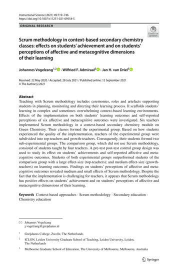
Roosink et al. Journal of NeuroEngineering and Rehabilitation 2015, 12:2http://www.jneuroengrehab.com/content/12/1/2to the individual needs of patients. As such VR has increasingly been explored in the context of rehabilitation.Common applications of VR in rehabilitation include forexample self-displacements or object displacements inrealistic [7] or non-realistic [8,9] virtual (gaming) environments, or the manipulation of virtual body parts, e.g.to replace a missing limb in amputee patients [10-12].Although used extensively in gaming and videoanimation, the use of full-body avatars is still rare inrehabilitation due to the need for accurate movementrepresentations requiring detailed movement samplingand modelling, which can be complex and timeconsuming. One of the few successful examples is astudy by Koritnik et al. who created a full-body“virtual mirror” by recording kinematic data to animate a virtual mirror-image (non-realistic avatar) inreal-time, while healthy adults were stepping in place[13]. Another example is a very recent study by Bartonand colleagues that implemented a virtual mirror foramputee patients. In their study, movements kinematics of the unimpaired leg were combined with themovement timing of the impaired leg to model a realisticavatar with a symmetric gait pattern [14]. In addition,some studies have used full-body video-capture to displaya full-body mirror-image [3,15] or avatar [16] of the subject onto a virtual reality scene.Unfortunately, the VR systems commonly available inrehabilitation have some important limitations. Themodelling of virtual limbs and avatars has generally beenbased on specific movements in a limited number ofmovement planes, whereas rehabilitation may includecomplex movements in multiple movement planes. Inaddition, only a few VR systems allow for a scaling ofmovements (e.g. providing augmented or reduced feedback). Indeed, an altered perception of body movements[17,18] or body size [19,20] could be used to promote orprevent certain movement patterns and could directlyimpact on pain perception [21-23]. For example, previous work has shown that a virtual environment in whichmovements were scaled to attain reduced movementperception increased the range of neck motion in patientswith neck pain as opposed to a virtual environment without scaling [17]. Likewise, a gradual modulation of visualfeedback of step-length during gait (simple bar graphs)systematically modulated step length away from symmetry, even when subjects were explicitly instructed tomaintain a symmetric gait pattern [18]. However, the required level (low, high) and direction (reduction, augmentation) of scaling is likely to depend on the particular bodypart and movement involved as well as on the particulartype of feedback provided.As such, and prior to the development of any intervention protocols, it is important to establish normativedata regarding body perception during active movementPage 2 of 10in VR, for example by assessing the ability to detect different levels and directions of scaled feedback in healthysubjects [18]. To attain this goal we developed a “virtualmirror” that: 1) displays a realistic full-body avatar, 2) responds to full-body movements in all movement planesin real-time, and that 3) allows for the scaling of visualfeedback on movements at any given joint in real-time.The primary objective of this proof-of-concept studywas to assess the ability of healthy adults to detect scaledfeedback on trunk movements using a two-alternativeforced choice paradigm. For each subject, a psychophysical curve was created, and two main variables of interestwere derived, the point of subjective equality (PSE) andthe just noticeable difference (JND). It was expected thathealthy adults would perform consistent with expectations for a two-alternative forced choice paradigm, i.e.that the detection of scaled feedback could be modelledusing a sigmoid curve, and that subjects would displayunbiased perception (no shift in PSE) and high discriminative ability (small JND). Secondary objectives were toassess virtual reality immersion and task adherence(movement kinematics).Technological development of the virtual mirrorThe virtual mirror consists of three main components:1) a motion capture (MOCAP) system, 2) an interactionand rendering system (IRS), and 3) a projection system,see Figure 1. The subject’s movements (rotation andposition) are first acquired using the MOCAP system.The data is then sent to the IRS, which scales the subject’s movements and applies the scaled data to an avatarin real-time. The IRS finally displays the avatar onto aprojection screen. A mirrored projection setup allowsthe subjects to see their avatar as a mirror-image.Motion capture systemThe MOCAP system (Vicon Motion Systems Ltd.,Oxford, UK) is used to acquire the subject’s movements,which are then mapped to an avatar in real-time by theIRS. The system consists of 12 infrared cameras (Bonita10) connected to a computer (Intel Xeon E31270,3.40 GHz; 4 GB RAM; OS: Windows 7, 64 bits; NVIDIAQuadro 2000) running Vicon’s Nexus 1.8.2 acquisitionsoftware. Movements are captured with a sampling frequency of 100 Hz using a set of 41 reflective markers(14 mm) placed on the subject’s entire body. To be able tolocate a marker in 3D space, the MOCAP system must becalibrated. The calibration consists of environment reflection removal, a calibration of the cameras using a wandwith a specific marker configuration, and setting the volume origin.The placement of the markers on the subject’s body isfacilitated by using a motion capture suit, and is determined by a skeleton template file based on Vicon’s
Roosink et al. Journal of NeuroEngineering and Rehabilitation 2015, ge 3 of 10Figure 1 Overview of the different components of the virtual mirror. 1) Motion capture (MOCAP) system including the positioning of 41reflective markers on the subject’s body (A) to create a Vicon skeleton template (B); 2) Interaction and rendering system (IRS) that retrieves andscales the MOCAP data online, maps the modified data onto the avatar and renders the avatar on screen; 3) Projection screen displaying theavatar’s movements as being augmented (left, scaling factor s 1) or reduced (right, scaling factor s 1) as opposed to the subject’s actualmovements (here displayed as a white skeleton).‘HumanRTkm’ model. This model additionally defines ahierarchy of segments (or bones) consisting of 19 segments. A complete list of segments and their hierarchyis presented in Table 1, and a visual representation ispresented in Figure 1 (frame 1). The segments aremapped onto the subject based on another calibrationTable 1 Motion capture: skeleton segments and hierarchyRoot segment Level 1Level 2PelvisHeadThoraxLevel 3Level 4Level 5Clavicle L Humerus L Radius L Hand LClavicle R Humerus R Radius R Hand RL: left, R: right.Femur L Tibia LFoot LToes LFemur R Tibia RFoot RToes Rprocedure. This procedure consists of 1) acquiring a sequence of predefined body movements of the head,shoulders, arms, trunk and legs; 2) labeling the markersof the acquired sequence according to the skeleton template; and 3) calibrating the position and orientation ofthe skeleton joints based on the sequence movements.Once the subject is calibrated, real-time segment positions and orientations are transmitted to the IRSthrough a local network.Interaction and rendering systemThe IRS consists of a computer (Intel Xeon E31270,3.40 GHz; 4 GB RAM; Windows 7, 32 bits; NVIDIAQuadro 2000) running D-Flow (Motek Medical,Amsterdam, The Netherlands). The computer receives theMOCAP data, performs the scaling (see paragraph on
Roosink et al. Journal of NeuroEngineering and Rehabilitation 2015, �Movement scaling’ for details) and maps the resultingdata onto the avatar rig so that the avatar follows the subject’s movements in real-time at a refresh rate of 60 Hz. Arealistic male avatar model was bought (www.TurboSquid.com, Martin T-pose, ID 523309) and rigged for motioncapture using Blender (Blender Foundation, Amsterdam,The Netherlands). The avatar model was then convertedto the OGRE format (Object-Oriented Graphics Rendering Engine, ogre3d.org) to be used in real-time in the IRS.As such, the size and proportions of the avatar vary basedon individual MOCAP data whereas its appearance (e.g.body shape, clothing) remains the same for each subject.In principle, the avatar is placed in an empty scene (greyfloor, black walls). However, a height-adjustable stool thatis present in the laboratory was also modeled in VR andcan additionally be presented and adjusted (i.e. height, positioning in the scene) using an interface programmed inD-flow.Projection systemThe avatar model is projected onto a silver-coatedscreen (projection surface 3.05 m 2.06 m) using a single projector (Hitachi, Tokyo, Japan; CP-WX8255A;1920 1080 High Definition) connected to the IRS computer. To produce the mirror effect, the projector is setin rear-projection mode. Notably, there are no technicallimitations to project the avatar onto other projectiondevices such as a head-mounted display. Additionally,the avatar might be projected in 3D. In the current setup, the avatar can be viewed in full-body size while thesubject remains within an area of about 2.5 by 4 meters.The screen can be approached up to 1 meter. The sizeof the avatar is proportionally scaled with the distance asopposed to the screen. At a distance of about 2 meters,the avatar’s height is approximately 1.5 times smallerthan the subject’s real height.Movement scalingThe movement scaling procedure is summarized inFigure 1. Movement scaling is programmed directly inD-Flow on the IRS using custom scripts. All rotationmanipulations are performed in real-time using quaternions. Starting from the global position and rotationdata of the MOCAP system, the data is first transformedinto the D-Flow coordinate system. Starting from theroot segment (pelvis), the hierarchy of the MOCAP skeleton is used to find the local rotation and position of allother segments. A reference rotation is acquired whilethe subject assumes a static base position. During movement the scaling is applied in the local space of eachsegment on the difference between the reference rotation and the current MOCAP rotation (updated duringmovement) using spherical linear interpolation (SLERP),or quaternion interpolation [24]. The SLERP operationPage 4 of 10returns a rotation interpolated between two rotations q0and q1 according to an interpolation parameter (or scaling factor), s. For parameters s 0 and s 1 SLERP givesq0 and q1, respectively. In our case q0 is the reference rotation and q1 is the current MOCAP rotation. When fora given segment s 1, SLERP returns an interpolatedrotation that is a reduction of the current MOCAP rotation. For s 1 the interpolated rotation is an augmentation of the current MOCAP rotation and follows thesame direction. For s 1 no scaling is applied andSLERP simply returns the current MOCAP rotation.Once the scaled rotation is applied locally on a segment,the positions and rotations of its child segments are updated according to this new scaled rotation. This processis performed upwards in the hierarchy up to the rootsegment (pelvis), resulting in a set of global rotationsand positions that are applied onto the avatar. As such,both rotation amplitudes and velocities are scaled inreal-time (total delay between movements and VR projection ranging between 90 and 120 ms). It is importantto note that the scaling operation is performed locallyon each segment and independently in each axis, so thatin principle the scaling could be applied on any chosensegment depending on the required application.Scaling trunk movementsIn this study, only the trunk, consisting of two segments(pelvis and thorax), was scaled in the sagittal plane (i.e.flexion-extension movements). Scaling factors rangedfrom s 0.667 (corresponding to avatar movementsbeing reduced 1.5 times) to s 1.500 (corresponding toavatar movements being augmented 1.5 times). Therange was determined empirically based on task performance in a two-alternative forced choice paradigmduring pilot-testing in healthy subjects and in patientswith chronic low back pain (for future clinical application). The two extremes (s 0.667 and s 1.500) produced movement scaling that could be clearly identifiedby the subject as being either reduced or augmented,and were used for familiarization and test trials. Twosets of five points equally spaced below and above s 1were used for analyses. As such, on a log scale, eachpoint in the set below 1 had a corresponding inverse inthe set above 1. The final set of scaling factors is listedin Table 2.Proof of concept: perception of scaled trunkmovementsSubjectsThe project was performed in collaboration with theCanadian Armed Forces. Healthy military subjects (agedbetween 18–55 years, men only to comply with the avatar’s gender) were recruited at a regional military base.Exclusion criteria included recurrent low back pain, low
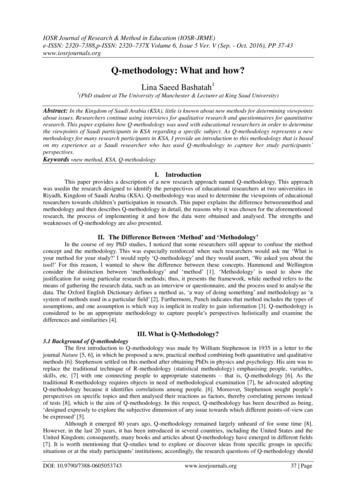
Roosink et al. Journal of NeuroEngineering and Rehabilitation 2015, ble 2 Scaling factors, and number of trialssLog sNumber of trials0.667 0.1763 (test trials)0.749 0.12640.793 0.10150.841 0.07560.891 0.05060.944 1.2610.10151.3360.12641.5000.1763 (test trials)back pain that required medical care or that restrictedwork or recreation during the past 2 years, acute pain(pain score higher than 2/10, 0 no pain, 10 worstpain imaginable) at the time of testing, chronic pain(duration 3 months) during the last 6 months priorto participation, non-corrected visual impairments,repeated fractures, or other medical conditions (inflammatory, neurologic, degenerative, auto-immune,psychiatric) that could interfere with performance during testing. All assessments took place at the CentreInterdisciplinaire de Recherche en Réadaptation et Intégration Sociale of the Institut de réadaptation endéficience physique de Québec. The project was approvedby the local institutional review board (#2013-323). Allsubjects received written and oral information, and signedinformed consent prior to participation.Experimental procedurePreparationDemographic and anthropomorphic data (weight, height,trunk height) were registered, after which the subjectput on a body size-matched (4 sizes available) motioncapture suit (OptiTrack, NaturalPoint, Corvallis, Oregon,USA) on which the markers were placed as describedin the paragraph 'Motion capture system'. After calibrating the subjects for motion capture, they wereplaced in front of the projection screen (distance of2 meters), the lights were dimmed, and the IRS softwarewas activated to display the avatar in front of the subject(mirror mode, s 1 no modulation). A familiarizationperiod including various pre-defined and spontaneousmovements allowed subjects to explore the interactionwith the avatar. Afterwards, the subjects remainedseated facing the screen, but the avatar was medially rotated 90 so that it was displayed from the side (facingPage 5 of 10left) to allow for a better view of trunk flexion-extensionmovements (i.e. side-view mode). A snapshot of the experimental set-up is presented in Figure 2.Subjects were instructed on static base positions forsitting and standing, which required them to keep theirback and neck straight, their head facing the screen,arms falling naturally along the sides of the body, andfeet aligned at shoulder width and pointing forward. Forthe sitting condition, subjects were placed on the stoolthat was adjusted to yield 90 of hip and knee flexion.For the standing condition, the subject was instructed tokeep the knee joint partially flexed in order to maintainbalance during trunk flexion. In the base position, thereference rotation was acquired and the trunk flexionangle was considered to be 0 . Subjects practiced the basics of the trunk flexion task in both positions while observing the simultaneous movements of the avatar onthe screen (side-view, s 1 no modulation). The instructions were to move at a slow pace in one fluentmovement towards a maximum angle of 35 , and thiswas demonstrated by the experimenter. Subjects received feedback on adherence to instructions.Scaling taskThe scaling task was introduced in the sitting position in2 steps. First, the element of moving towards a predefined angle (unknown to the subject) was introduced (4trials). The detection of these predefined angles by theIRS is described in detail under ‘Detecting and controlling flexion angles’. Subjects were required to start bending forward and, upon the appearance of the word “OK”on the screen along with a simultaneous bell-sound,return backwards to the base position. Second, a twoalternative forced choice paradigm was introduced (4 trials). After each trial, subjects had to decide whether themovements of the avatar were greater or smaller thantheir own movements. Subjects did not receive feedbackon performance accuracy. After this brief training period,the experiment was started.The number of experimental trials was weighted perscaling factor to acquire more data for relatively difficulttrials involving small modulations, i.e. trials in which swas close to 1. The scaling factors were then distributedover 3 blocks of 23 trials each. The first 2 trials of eachblock were test trials (unknown to the subject), and werenot further analyzed. The other scaling factors weredistributed pseudo-randomly to ensure that blocks contained a balanced number of relatively easy and relativelydifficult trials. As the tasks had to be performed whilesitting and while standing, the total number of blockswas 6 (3 sitting, 3 standing blocks), and the total numberof trials was 138. Sitting and standing blocks were alternated and the starting block (sitting or standing) wasrandomized across subjects. After each block there was
Roosink et al. Journal of NeuroEngineering and Rehabilitation 2015, ge 6 of 10Figure 2 Snapshot of the experimental procedure during a sitting block. Each block consisted of 23 trials. The scaling factor was differentfor each trial. When subjects reached the required flexion angle (15 , 25 or 35 ), simultaneous visual (OK) and auditory (bell-sound) feedbackwere provided. After returning to the base position, subjects had to decide whether the movements of the avatar were greater or smaller thantheir own movements (two-alternative forced choice paradigm).a short break. After finishing all experimental blocks, theperceived interaction with the virtual mirror (immersion,distraction) was evaluated on a 1–7 scale using a selection of questions from the Presence Questionnaire (seeTable 3 for the complete list of questions) [25]. The totalduration of the experiment (including preparation) wasabout 2 h.Detecting and controlling flexion anglesThree predefined angles (15 , 25 and 35 ) were programmed in the IRS to: 1) have subjects move within asafe range of motion (i.e. to avoid fatigue or pain) and 2)to introduce proprioceptive inter-trial-variability so thatsubjects would have to depend on both visual and proprioceptive feedback to perform the task correctly. Thedetection of flexion angles was based on the sagittalorientation of a vector connecting 2 markers on the backof the subject (C7 and T10). This orientation was considered to be 0 in the base position. When subjectsreached the predefined angle for that trial, the IRSsent out the OK signal (screen) and simultaneous bellsound (audio), indicating to the subject to stop bending forward.The 3 angles were distributed pseudo-randomly acrossthe different blocks. Importantly, the 3 smallest scalingfactors were not combined with a 15 detection angle,and the 3 largest scaling factors were not combined witha 35 detection angle. As such, the resulting avatar’smovements were also restricted to a limited range ofmotion. This avoided extremes in the visual feedbackthat would otherwise allow subjects to base their decision on visual feedback only. The important point froma methodological perspective was that subjects variedtheir flexion angles from trial to trial, and not that theyachieved a specific flexion angle.Outcome parametersFor each individual subject, the responses to the twoalternative forced choice task were averaged over logtransformed scaling factors (see Table 2) and plotted(X log-transformed scaling factor [ 0.126; 0.126]; Y percentage of trials for which the subjects respondedthat the avatar’s movements were greater than their actualmovements [0; 1]). Then a sigmoid curve (Equation 1),with initial value XY0.50 0, with constraints YMAX 1and YMIN 0, and with a variable slope (m), was fitted tothe data (Prism 6 for Windows, Graphpad Software Inc.,
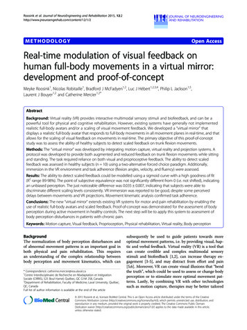
Roosink et al. Journal of NeuroEngineering and Rehabilitation 2015, ble 3 Virtual reality immersion and distraction (basedon the Presence Questionnaire [25])QuestionsImmersionDistractionAV SDHow much were you able to control theavatar (your virtual image)?6.0 0.7How responsive was the avatar to yourmovements?5.8 0.4How quickly did you adjust to the virtualenvironment experience?6.2 1.0How proficient in moving and interactingwith the virtual environment did you feelat the end of the experience?6.2 0.8To what extent did the movements of theavatar seem natural to you?5.1 0.7How well could you examine the detailsof the avatar?5.1 1.1How much delay did you experience betweenyour actions and the response of the system?3.5 2.0How much did the visual display qualityinterfere or distract you from performingassigned tasks or required activities?1.8 1.0How much did the control devices interferewith the performance of assigned tasks orwith other activities?1.3 0.5Page 7 of 10Natik, MA, USA). Data was filtered using a secondorder double pass Butterworth filter (4 Hz). Trunkmovement analyses were performed based on 3markers located on the back of the subject (C7, T10and scapula), and focused on the sagittal plane only.Data analysisScoring for immersion: 1 not able/responsive/etc.; 7 extremely able/responsive/etc. Scoring for distraction: 1 no delay/interference; 7 longdelay/high interference.La Jolla, CA, USA). From each curve, 3 data points wereinterpolated (XY0.25, XY0.50, XY0.75), and used to determine the so-called point of subjective equivalence (PSE,Equation 2) and the just noticeable difference (JND,Equation 3). Theoretically, the chance distribution for atwo-alternative forced choice paradigm predicts a PSEof 0, i.e. there is a 50% chance of responding “greater”or “smaller” when in fact no scaling has been applied. APSE higher than 0 indicates that subjects tend to overestimate their own movements and a PSE lower than 0indicates that subjects tend to underestimate their ownmovements. The higher the slope and the smaller theJND, the better subjects are able to discriminate betweendifferent levels of scaled feedback.Y ¼ YMIN þ ðYMAX ‐YMIN Þ ð1 þ 10ððXY0:50 ‐XÞ mÞ Þð1ÞPSE ¼ XY0:50ð2ÞJND ¼ ðXY0:75 XY0:25 Þ 2ð3ÞTask adherence was assessed by analyzing trunk movements for maximum flexion angles, maximum flexionvelocity and for the fluency of movement around themaximum flexion angle (number of zero-crossings intrunk acceleration between the maximum flexion andmaximum extension velocity) for each of the predefinedflexion angles (15 , 25 , 35 ), using in-house scripts written in Matlab (version R2010b, The Mathworks Inc.,For each of the outcome parameters (XY0.25, PSE, XY0.75,JND, m) the normality of the data distribution (theskewness of the distribution) and presence of outliers(data outside 1.5 times the interquartile range) wasassessed and descriptive statistics were calculated (IBMSPSS for Windows, version 22.0.0.0, USA). Movementdata was analyzed using multivariate tests with withinsubject factor [Angle] (15 , 25 , 35 ). Data is presented intext as mean standard deviation.ResultsA total of 11 healthy subjects participated in the experiment. One subject showed poor task adherence and wasadditionally identified as an outlier based on psychophysical curve metrics and movement data. As such thissubject was excluded from the analyses. The final sampletherefore consisted of 10 male subjects, having a meanage of 28 5 years (range: 22–37), weight of 88 14 kg(range: 62–108), height of 176 10 cm (range: 165–201),and Body Mass Index (BMI) of 28 4 (range: 23–34).Two-alternative forced choice paradigm andpsychophysical curveThe data followed a normal distribution (i.e. skewnessvalues close to 0). Figure 3 presents the data and curvefitting results for a representative subject. In general, thegoodness of fit for these individually fitted curves washigh (R2 range: 0.89 - 0.98). Group averaged interpolatedXY0.25, PSE, and XY0.75 ( SEM) are presented inFigure 4. The 95% confidence interval for the PSEranged from 0.003 to 0.028, indicating that the PSEwas not significantly different from 0. The averageJND was 0.035 0.007 (range 0.026 - 0.042), the average curve slope m was 14.1 2.7 (range 10.1 - 18.6),and the average percentage of correct responses was83% 4% (range 60% - 100%).Virtual reality immersionTable 3 presents the average scores relating to the subjectiv
up, the avatar can be viewed in full-body size while the subject remains within an area of about 2.5 by 4 meters. The screen can be approached up to 1 meter. The size of the avatar is proportionally scaled with the distance as opposed to the screen. At a distance of about 2 meters, the avatar's height is approximately 1.5 times smaller