Transcription
Cho et al. BMC Infectious Diseases(2018) EARCH ARTICLEOpen AccessClinical features and outcomes ofabdominal tuberculosis in southeasternKorea: 12 years of experienceJin-Kyu Cho1†, Young Min Choi2†, Sang Soo Lee2,3,4* , Hye Kyong Park4, Ra Ri Cha2,4, Wan Soo Kim2,4,Jin Joo Kim2,4, Jae Min Lee2,3,4, Hong Jun Kim2,3, Chang Yoon Ha2,3, Hyun Jin Kim2,3,4, Tae Hyo Kim2,3,Woon Tae Jung2,3 and Ok Jae Lee2,3AbstractBackground: Abdominal tuberculosis (TB) is an uncommon form of infection with Mycobacterium tuberculosis inKorea. In this study, we aimed to highlight the clinical features, diagnostic methods, and outcomes of abdominal TBover 12 years in Southeastern Korea.Methods: A total of 139 patients diagnosed as having abdominal TB who received anti-TB medication from January2005 to June 2016 were reviewed. Among them, 69 patients (49.6%) had luminal TB, 28 (20.1%) had peritoneal TB, 7(5.0%) had nodal TB, 23 (16.5%) had visceral TB, and 12 (8.6%) had mixed TB.Results: The most frequent symptoms were abdominal pain (34.5%) and abdominal distension (21.0%). Diagnosisof abdominal TB was confirmed using microbiologic and/or histologic methods in 76 patients (confirmed diagnosis), while the remaining 63 patients were diagnosed based on clinical presentation and radiologic imaging (clinicaldiagnosis). According to diagnostic method, frequency of clinical diagnosis was highest in patients withluminal (50.7%) or peritoneal (64.3%) TB, while frequency of microscopic diagnosis was highest in patientswith visceral TB (68.2%), and frequency of histologic diagnosis was highest in patients with nodal TB (85.2%).Interestingly, most patients, except those with nodal TB, showed a good response to anti-TB agents, with 84.2%showing a complete response. The mortality rate was only 1.4% in the present study.Conclusions: Most patients responded very well to anti-TB therapy, and surgery was required in only a minority ofcases of suspected abdominal TB.Keywords: Tuberculosis, Abdomen, Extra-pulmonary, Luminal, PeritonealBackgroundAbdominal tuberculosis (TB) is defined as infectionof the gastrointestinal tract, peritoneum, abdominalsolid organs, and/or abdominal lymphatics withMycobacterium tuberculosis [1]. Abdominal TB constitutes approximately 12% of extrapulmonary TB casesand 1 to 3% of total TB cases [1, 2]. Abdominal TB is one* Correspondence: 3939lee@naver.com†Jin-Kyu Cho and Young Min Choi contributed equally to this work.2Department of Internal Medicine, Gyeongsang National University School ofMedicine and Gyeongsang National University Hospital, 15, Jinju-daero 816beon-gil, Jinju-si, Gyeongnam 52727, Republic of Korea3Institute of Health Sciences, Gyeongsang National University, Jinju, Republicof KoreaFull list of author information is available at the end of the articleof the most common forms of extrapulmonary TB [3].Abdominal TB is relatively rare, but it is recognized thatabdominal TB is increasing in both developing and developed countries [4–8]. Abdominal TB accounts for approximately 4% of all TB cases in Korea [9]. Diagnosis ofabdominal TB is often overlooked and delayed due to lackof specific symptoms and no specific diagnostic test. Ahigh index of suspicion is necessary for early diagnosis ofabdominal TB; however, it remains a considerable diagnostic dilemma and can mimic many other diseases, suchas Crohn’s disease, abdominal lymphoma, and malignancyof the abdominal organs.Abdominal TB can usually be classified into 4 forms:luminal, peritoneal, nodal, and visceral involving the The Author(s). 2018 Open Access This article is distributed under the terms of the Creative Commons Attribution 4.0International License (http://creativecommons.org/licenses/by/4.0/), which permits unrestricted use, distribution, andreproduction in any medium, provided you give appropriate credit to the original author(s) and the source, provide a link tothe Creative Commons license, and indicate if changes were made. The Creative Commons Public Domain Dedication o/1.0/) applies to the data made available in this article, unless otherwise stated.
Cho et al. BMC Infectious Diseases(2018) 18:699intra-abdominal solid organs [10]. The most commonforms are luminal (ileocecal area) and peritoneal [11].The modes of infection of abdominal TB include swallowing infected sputum, ingestion of bacilli from infected milk products or meat, hematogenous spreadfrom a lung focus, spread via lymphatics from infectedlymph nodes, and contiguous spread from adjacentorgans [12]. The clinical presentation of abdominal TBdepends on the site of infection. Abdominal pain, diarrhea, bleeding from the luminal tract, intestinal obstruction, fever, and weight loss are frequent features ofintestinal TB; ascites and abdominal distension arecommon manifestations of peritoneal TB [8]. Diagnosisof abdominal TB may also vary depending on the site ofinfection [13]. Colonoscopy is useful in patients suspected of having intestinal TB, while laparoscopy andbiopsy are more useful in peritoneal TB; although asciticfluid analysis is more accessible, its acid-fast bacilli(AFB) culture has a low sensitivity.The aim of the present study was to evaluate theclinical features, diagnostic methods, and outcomes ofabdominal TB, including luminal, peritoneal, nodal, visceral, and mixed TB.MethodsStudy populationsBetween January 2005 and June 2016, a total of 139consecutive adult patients aged 18 years diagnosed ashaving abdominal TB began treatment with anti-TBdrugs at Gyeongsang National University Hospital,located on the southeast coast of Korea. Age, sex, bodymass index, alcohol consumption, history of TB, historyof malignancy, TB infection site, date of anti-TB drugprescription, regimen of anti-TB drugs, laboratory data,underlying disease, clinical features, diagnostic method,and clinical outcome were reviewed. The present studywas approved by the Institutional Review Board of theGyeongsang National University Hospital.Page 2 of 8diagnosis. Confirmed diagnosis was defined as microbiologic or histologic evidence of M. tuberculosis [14].Patients were classified into 5 groups according to siteof TB infection: (1) luminal, (2) peritoneal, (3) nodal, (4)visceral, and (5) mixed. Luminal TB was further dividedinto esophageal, gastric, duodenal, jejunal, ileocecal, andcolorectal. Peritoneal TB was divided into 3 types [10,15]: (1) the wet ascitic type was associated with largeamounts of free or loculated ascites; (2) the fixed fibrotictype was associated with involvement of the omentumand mesentery and was characterized by presence ofbowel loop entanglement; and (3) the dry plastic typewas characterized by peritoneal and mesenteric thickening with caseous nodules and presence of adhesions. Toavoid confusion, we classified peritonal TB into twotypes, the wet and dry type (fixed fibrotic and dry plastictype). Nodal TB was divided into mesenteric, portahepatitis, celiac axis, peripancreatic, and combined. Visceral TB was divided into hepatic, splenic, genitourinary,adrenal, and combined.Clinical outcomesAbdominal TB outcomes were classified as follows[16]: (1) complete response: resolution of symptoms,disappearance of AFB on smear or culture, disappearance of tuberculous granulomas, and disappearance orhealing of active tuberculous lesions on relook colonoscopy; (2) partial response: resolution of symptomsand partial disappearance of tuberculous lesions atend of treatment; (3) no response: persistence ofsymptoms, persistence of AFB on smear or culture,persistence of tuberculous granulomas, and persistence of active tuberculous lesions on relook colonoscopy at end of treatment; (4) lost to follow up:treatment interrupted for 2 consecutive months; (5)death: death from any cause during treatment; (6)recurrence: colonoscopic or radiologic documentationof recurrent lesions after a complete response hadbeen achieved; and (7) transfer out: transferred toanother reporting and recording unit.Definitions and classificationAbdominal TB was defined as infection of the luminaltract, peritoneum, intra-abdominal lymph nodes, and/orintra-abdominal solid-organs with M. tuberculosis. Diagnosis of abdominal TB was based on: (1) positive AFBsmear or culture from ascites, urine, or biopsy specimen(microbiologic diagnosis); (2) demonstration of caseatinggranulomas on biopsy specimen (histologic diagnosis);(3) typical presentation and good response to anti-TBagents (clinical diagnosis); or (4) high index of suspicionin susceptible patients and good response to anti-TBagents (clinical diagnosis). When peritoneal TB couldnot be diagnosed by biopsy, the high ascitic adenosinedeaminase ( 33 IU/L) criteria was used for clinicalStatistical analysisAll analyses were performed using PASW version 18(SPSS Inc., Chicago, IL, USA). Continuous variables wereexpressed as median (interquartile range). Intergroupdifferences in quantitative data were measured using theMann-Whitney U test, while the Fisher exact test wasused for qualitative data. All analyses were 2-sided and a Pvalue of 0.05 was considered statistically significant.ResultsPatient characteristicsA total of 139 patients (72 male, 51.8%) with a medianage of 47.0 years were diagnosed has having abdominal
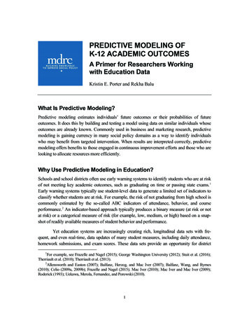
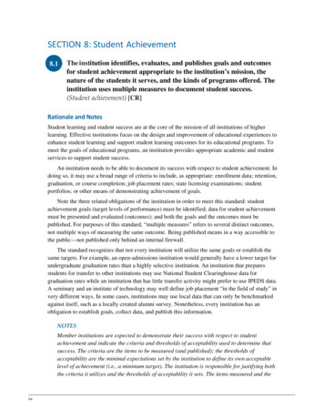
Cho et al. BMC Infectious Diseases(2018) 18:699TB. Among them, 69 patients (49.6%) were diagnosedwith luminal TB, 28 (20.1%) with peritoneal TB, 7(5.0%) with nodal TB, 23 (16.5%) with visceral TB,and 12 (8.6%) with mixed TB (Fig. 1). The ileocecum(67.1%) was the most frequently involved site in patients with luminal TB. Wet ascitic type (75.7%) wasthe most common type in patients with peritonealTB. Genitourinary tract (69.0%) and porta hepatitis(35.7%) were the most frequently involved sites inpatients with visceral and nodal TB, respectively(Table 1). The demographic characteristics of the patients are presented in Table 2. There were no significant differences in sex, body mass index, chronichepatitis B, HIV infection, alcohol consumption, livercirrhosis, end-stage renal disease, history of malignancy, history of TB, or anemia at diagnosis amongthe 5 study groups. The mixed TB group was significantly younger (median age, 32.0 years) than the other4 groups. The rate of history of TB was significantlyhigher in the visceral TB group (21.7%) than in theperitoneal TB group (0%).Abdominal pain was the most common symptom(34.5%; Table 3). Incidence of abdominal distension wassignificantly higher in the peritoneal TB group than inthe other 4 groups. However, there were no significantdifferences in the frequencies of other symptoms amongthe 5 study groups.Fig. 1 Sites of abdominal tuberculosis (TB)Page 3 of 8Diagnostic methodsIn the 139 patients with abdominal TB, diagnosis wasconfirmed microbiologically in 51 patients (37.0%) andhistologically in 59 patients (42.8%). Confirmed diagnosis was achieved in 76 patients (54.7%), while theremaining 63 patients (45.3%) were diagnosed clinically(Table 4). The frequency of microbiologic diagnosis(AFB smear or culture) was significantly higher in thevisceral TB group (68.2%) than in the luminal (33.3%)and peritoneal (25.0%) TB groups. The frequency ofhistologic diagnosis (caseating granulomas on biopsyspecimen) was significantly higher in the nodal TBgroup (85.7%) than in the luminal (39.1%) and peritoneal (28.6%) TB groups. The frequency of clinicaldiagnosis was significantly higher in the peritoneal TBgroup (64.3%) than in the nodal (14.3%) and visceral(13.6%) TB groups.Surgery was performed in 21 patients (15.1%) andprovided a good diagnostic yield in 90.4% (19/21).Among them, 10 underwent surgery for diagnosis ofsuspected abdominal TB, and 11 for management of numerous complications, such as intussusception, abscess,perforation, obstruction, and hemorrhage. The frequencyof surgery was higher in the nodal (42.9%) and visceral(43.5%) TB groups than in the luminal (2.9%) andperitoneal (14.3%) TB groups. Percutaneous biopsy wasperformed in 16 patients (11.5%); endoscopy or
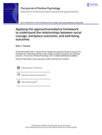
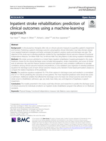
Cho et al. BMC Infectious Diseases(2018) 18:699Page 4 of 8Table 1 Site of involvement in abdominal tuberculosis (n 139)SiteN (%)Luminal TBEsophageal3 (3.9%)Stomach0 (0%)Duodenal3 (3.9%)Jejunal7 (9.2%)Ileocecal51 (67.1%)Colorectal12 (15.8%)Peritoneal TBWet ascitic type28 (75.7%)Dry ascitic type9 (24.3%)Nodal TBMesenteric2 (14.3%)Porta hepatis5 (35.7%)Along the celiac axis1 (7.1%)Peripancreatic2 (14.3%)Retroperitoneal1 (7.1%)Combined3 (21.4%)Visceral TBHepatic3 (10.3%)Splenic4 (13.8%)Genitourinary20 (69.0%)Adrenal1 (3.4%)Combined1 (3.4%)Mixed TBLuminal and peritoneal4 (33.3%)Luminal and nodal1 (8.3%)Luminal and peritoneal, and nodal2 (16.7%)Peritoneal and visceral2 (16.7%)Nodal and visceral2 (16.7%)Peritoneal and visceral, and nodal1 (8.3%)TB: tuberculosisData are presented as the median (interquartile range) for continuous dataand percentages for categorical datacolonoscopy was performed in 70 patients (50.4%). Asciticdiagnosis through paracentesis was achieved in 33 patients(23.7%). Among them, a positive yield for AFB culture wasobserved in only 8 patients (24.2%). Diagnostic yield forconfirmed diagnosis from surgery, percutaneous biopsy,endoscopy/colonoscopy, and paracentesis was 90.4, 81.3,48.6, and 24.2%, respectively.Treatment outcomesOf the 139 patients who underwent anti-TB treatment,117 (84.2%) showed a complete response, 1 (0.7%) showeda partial response, 2 (1.4%) showed no response, 12 (8.6%)were lost to follow-up, 3 (2.2%) had recurrence, and 3(2.2%) transferred out (Table 5). The frequency ofcomplete response was significantly lower in the nodal TBgroup than in the other 4 study groups. Only 2 patients(1.4%) died during anti-TB therapy. One patient waspositive for HIV and multidrug-resistant abdominal TBinvolving the liver, spleen, and genitourinary tract. Themajor cause of death in this patient was concomitant TBmeningitis. Another non-HIV-positive patient died due tointestinal obstruction with sepsis.Twenty-one patients (15.2%) had adverse effects dueto anti-TB agents, and 9 (6.5%) developed drug-inducedliver injury. The median duration of anti-TB treatmentwas 194 days; 131 patients were treated with first-lineanti-TB agents, while 8 received second-line anti-TBagents. Figure 2 shows an overall decrease of approximately 60% in number of TB cases in southeasternKorea in the period 2005–2008 to 2013–2016.DiscussionIn the current study, we described the distribution of abdominal TB at a tertiary hospital in southeastern Korea.The most frequent site of abdominal TB was the luminaltract (49.6%) followed by the peritoneum (20.1%), solidviscera (16.5%), mixed organs (8.6%), and lymph nodes(5.0%). The most common presentation was abdominalpain (34.5%), whereas abdominal distension was aunique presentation in patients with peritoneal TB.According to diagnostic method, the frequency of clinical diagnosis was highest in the luminal and peritonealTB groups, while the frequency of microscopic diagnosiswas highest in the visceral TB group, and the frequencyof histologic diagnosis was highest in the nodal TBgroup. Interestingly, 117 patients (84.2%) showed acomplete response and the mortality rate was only 1.4%.Abdominal TB poses a considerable diagnosticchallenge due to the lack of specific symptoms and pathognomonic findings. Moreover, no single diagnosticmethod is sufficient for diagnosis. Based on our findings,colonoscopy and paracentesis may be useful in cases ofluminal and peritoneal TB, where mucosal or peritoneallesions are accessible. However, colonoscopy andperitoneal fluid analysis have a low diagnostic yield forconfirmed diagnosis of abdominal TB (49.2 and 22.2%,respectively). Therefore, the majority of luminal andperitoneal TB cases were diagnosed based on clinicalresponse to anti-TB agents and radiologic findings.Among these, laparotomy was performed in only 2 and4 patients in the luminal and peritoneal TB groups, respectively. Peritoneal fluid analysis is the most usefulnonoperative diagnostic method for peritoneal TB. Highascitic adenosine deaminase activity level ( 33 IU/L)and low serum ascitic albumin gradient ( 1.1) have asensitivity of 97% and specificity of 100% [17]. Therefore,laparoscopy could be used to rule out other
Cho et al. BMC Infectious Diseases(2018) 18:699Page 5 of 8Table 2 Demographic characteristics of 139 patients with abdominal tuberculosisCharacteristicAll patients(n 139)Luminal TB(n 69)Peritoneal TB(n 28)Nodal TB(n 7)Visceral TB(n 23)Mixed TB(n 12)Age47.0 (32.0–58.0)48.0 (38.5–58.5)52.5 (27.3–63.0)53.0 (27.0–57.0)46.0 (33.0–56.0)32.0 (25.5–45.8) d,Male sex72 (51.8%)36 (52.2%)18 (64.3%)3 (42.9%)9 (39.1%)6 (50%)BMI (m/kg2)21.80 (19.56–24.53)21.3 (19.6–24.1)21.6 (19.3–24.5)23.7 (20.1–27.8)20.1 (22.8–25.3)20.8 (18.0–22.8)Chronic hepatitis B10 (7.2%)5 (7.2%)2 (7.1%)0 (0%)2 (8.7%)1 (8.3%)Chronic hepatitis C3 (2.2%)1 (1.4%)0 (0%)2 (28.6%) b, e, h0 (0%)0 (0%)HIV-infection2 (1.4%)0 (0%)0 (0%)0 (0%)1 (4.3%)1 (8.3%)Alcohol 40 g/day16 (11.5%)6 (8.7%)6 (21.4%)0 (0%)2 (8.7%)2 (16.7%)Cirrhosis3 (2.2%)2 (2.9%)0 (0%)0 (0%)0 (0%)1 (8.3%)Diabetes9 (6.5%)3 (4.3%)3 (10.7%)1 (14.3%)2 (8.7%)0 (0%)ESRD or CAPD8 (5.8%)3 (4.3%)3 (10.7%)1 (14.3%)1 (4.3%)0 (0%)History of malignancy7 (5.0%)3 (4.3%)2 (7.1%)0 (0%)2 (8.7%)0 (0%)History of TB13 (9.4%)7 (10.1%)0 (0%)1 (14.3%)5 (21.7%) f0 (0%)Anemia at diagnosis46 (33.1%)21 (30.4%)11 (39.3%)3 (42.9%)5 (21.7%)6 (50.0%)Leukocytosis at diagnosis30 (21.6%)16 (23.2%)3 (10.7%)3 (42.9%)3 (13.0%)5 (41.7%) gg, jHIV: human immunodeficiency virus; ESRD: end-stage renal disease; CAPD: continuous ambulatory peritoneal dialysiData are presented as the median(interquartile range) for continuous data and percentages for categorical dataDefined as hemoglobin of 12 g/dlDefined as a white blood cell count of 10,000/mm3a: P 0.05 Luminal TB vs. Peritoneal TB, b: P 0.05 Luminal TB vs. Nodal TB, c: P 0.05 Luminal TB vs. Visceral TB, d: P 0.05 Luminal TB vs. Mixed TB, e: P 0.05Peritoneal TB vs. Nodal TB, f: P 0.05 Peritoneal TB vs. Visceral TB, g: P 0.05 Peritoneal TB vs. Mixed TB, h: P 0.05 Nodal TB vs. Visceral TB, i: P 0.05 Nodal TB vs.Mixed TB, j: P 0.05 Visceral TB vs. Mixed TBintra-abdominal malignancies and to minimize any possible diagnostic delay in these groups. On the other hand,diagnosis of nodal TB was confirmed from surgical procedures or percutaneous biopsy in 6 patients (confirmeddiagnosis), while 1 patient was diagnosed based on clinicalresponse to anti-TB agents and computed tomographyimaging (clinical diagnosis). Diagnosis of visceral TB wasconfirmed from surgical procedures or percutaneousbiopsy in 13 patients, and from urine AFB in 7 patients(confirmed diagnosis), while 3 patients were diagnosedbased on clinical response to anti-TB agents and computed tomography imaging (clinical diagnosis). AlthoughTable 3 Clinical features in 139 patients with abdominal tuberculosisCharacteristicAll patients (n 139)Luminal TB (n 69)Peritoneal TB (n 28)Nodal TB (n 7)Visceral TB (n 23)Mixed TB (n 12)Abdominal pain48 (34.5%)23 (33.3%)7 (25.0%)3 (42.9%)9 (39.1%)6 (50.0%)Fever16 (11.5%)5 (7.2%)6 (21.4%)0 (0%)3 (13.0%)2 (16.7%)Anorexia9 (6.5%)6 (8.7%)1 (3.6%)1 (14.3%)0 (0%)1 (8.3%)Body weight loss5 (3.6%)4 (5.8%)1 (3.6%)0 (0%)0 (0%)0 (0%)Abdominal distension29 (21.0%)2 (2.9%)22 (78.6%) a, e,0 (0%)0 (0%)5 (41.7%) d, jf, gBloody stool11 (7.9%)10 (14.5%)0 (0%)0 (0%)1 (4.3%)0 (0%)Dyspnea2 (1.4%)0 (0%)1 (3.6%)0 (0%)1 (4.3%)0 (0%)Diarrhea6 (4.3%)5 (7.2%)0 (0%)0 (0%)0 (0%)1 (8.3%)Hematuria1 (0.7%)0 (0%)0 (0%)0 (0%)1 (4.3%)0 (0%)Dysuria2 (1.4%)0 (0%)0 (0%)0 (0%)2 (8.7%)0 (0%)Palpable mass3 (2.2%)1 (1.4%)0 (0%)1 (14.3%)1 (4.3%)0 (0%)Scrotal swelling1 (0.7%)0 (0%)0 (0%)0 (0%)1 (4.3%)0 (0%)Jaundice1 (0.7%)0 (0%)0 (0%)1 (14.3%)0 (0%)0 (0%)No symptom31 (22.3%)23 (33.3%) a,1 (3.6%)2 (28.6%)5 (21.7%)0 (0%)dData are presented as the median (interquartile range) for continuous data and percentages for categorical dataa: P 0.05 Luminal TB vs. Peritoneal TB, b: P 0.05 Luminal TB vs. Nodal TB, c: P 0.05 Luminal TB vs. Visceral TB, d: P 0.05 Luminal TB vs. Mixed TB, e: P 0.05Peritoneal TB vs. Nodal TB, f: P 0.05 Peritoneal TB vs. Visceral TB, g: P 0.05 Peritoneal TB vs. Mixed TB, h: P 0.05 Nodal TB vs. Visceral TB, i: P 0.05 Nodal TB vs.Mixed TB, j: P 0.05 Visceral TB vs. Mixed TB
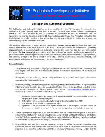
Cho et al. BMC Infectious Diseases(2018) 18:699Page 6 of 8Table 4 Diagnostic method in 139 patients with abdominal tuberculosisCharacteristicAll patients(n 139)Luminal TB(n 69)Peritoneal TB(n 28)Nodal TB(n 7)Visceral TB(n 23)Mixed TB(n 12)Microbiologic diagnosis51 (37.0%)23 (33.3%)7 (25.0%)2 (28.6%)15 (68.2%) c, f4 (33.3%)12 (54.5%)6 (50.0%)3 (13.6%)6 (50.0%) j10 (43.5%) c, f2 (16.7%)Histological diagnosis59 (42.8%)27 (39.1%)8 (28.6%)Clinical diagnosis63 (45.3%)35 (50.7%) c18 (64.3%) e,Operation21 (15.1%)2 (2.9%)4 (14.3%)3 (42.9%)*Confirmed diagnosis by operation19 (90.4%)2 (100%)4 (100%)3 (100%)9 (90%)1 (50%)6 (21.4%)3 (42.9%)3 (13.0%)4 (33.3%)4 (66.7%)2 (66.7%)3 (100%)4 (100%)0 (0%)0 (0%)0 (0%)4 (33.3%) g, j0 (0%)0 (0%)1 (25%)006 (50.0%) g, j0 (0%)0 (0%)2 (33.3%) da, b, c, dPercutaneous biopsy group16 (11.5%)0 (0%)*Confirmed diagnosis by percutaneousbiopsy13 (81.3%)0/0Endoscopy or colonoscopy70 (50.4%)65 (94.2%) a, b, c,*Confirmed diagnosis by endoscopyor colonoscopy34 (48.6%)32 (49.2%)0 (0%)Paracentesis33027 (96.4%) a,*Confirmed diagnosis by paracentesis8 (24.2%)0 (0%)6 (21.4%) a,fd6 (85.7%)b, efe, f, g1 (14.3%)bData are presented as the median (interquartile range) for continuous data and percentages for categorical data*A Confirmed diagnosis was defined as microbiologic or histological diagnosisa: P 0.05 Luminal TB vs. Peritoneal TB, b: P 0.05 Luminal TB vs. Nodal TB, c: P 0.05 Luminal TB vs. Visceral TB, d: P 0.05 Luminal TB vs. Mixed TB, e: P 0.05Peritoneal TB vs. Nodal TB, f: P 0.05 Peritoneal TB vs. Visceral TB, g: P 0.05 Peritoneal TB vs. Mixed TB, h: P 0.05 Nodal TB vs. Visceral TB, i: P 0.05 Nodal TB vs.Mixed TB, j: P 0.05 Visceral TB vs. Mixed TBnodal and visceral TB are mainly treatable medically,surgery is still often required for suspected abdominal TBand management of complications, such as infection,perforation, and hemorrhage.A 6-month course of anti-TB therapy for luminal TBis recommended in treatment guidelines [18, 19]. Twoprevious prospective, randomized studies confirmed ahigh cure rate of 90% after both 6 and 9 months ofstandard anti-TB therapy [6, 16]. In addition, someretrospective studies have shown that anti-TB agents areusually highly effective and associated with low mortality(0–6%) in abdominal TB [7, 8, 13, 20, 21]. However,abdominal TB has a high mortality rate (6–20%), andthe majority of patients had acute complications andTable 5 Treatment outcomes in 139 patients with abdominal tuberculosisCharacteristicAll patients(n 139)Luminal TB(n 69)Peritoneal TB(n 28)Nodal TB(n 7)Visceral TB(n 23)Mixed TB(n 12)Duration oftreatment, days194.0 (176.0–258.0)189.0 (175.0–219.0)200.0 (165.0–258.0)258.0 (16.0–285.0)284.0 (178.0–284.0)245.0 (177.5–349.0)Side effect21 (15.2%)11 (15.9%)3 (11.1%)0 (0%)4 (17.4%)3 (25.0%)Drug-inducedliver injury9 (6.5%)5 (8.7%)2 (7.1%)0 (0%)0 (0%)1 (8.3%)Nephrectomy4 (2.9%)0 (0%)0 (0%)0 (0%)4 (17.4%) c,First-line anti-TBdrug131 (94.2%)66 (95.7%)27 (96.4%)7 (100%)20 (87.0%)11 (91.7%)Second-line anti-TBdrug8 (5.8%)3 (4.3%)1 (3.6%)0 (0%)3 (13.0%)1 (8.3%)Complete response117 (84.2%)62 (89.9%)22 (78.6%)3 (42.9%) b, e,20 (87.0%)10 (83.3%)Partial response1 (0.7%)0 (0%)0 (0%)1 (14.3%)0 (0%)0 (0%)No response2 (1.4%)1 (1.4%)0 (0%)0 (0%)1 (4.3%)0 (0%)Lost to follow up12 (8.6%)4 (5.8%)5 (17.9%)0 (0%)1 (4.3%)2 (16.7%)Recurrence3 (2.2%)1 (1.4%)1 (3.6%)1 (14.3%)0 (0%)0 (0%)Transfer out3 (2.2%)1 (1.4%)0 (0%)2 (28.6%)0 (0%)0 (0%)Death2 (1.4%)1 (1.4%)0 (0%)0 (0%)1 (4.3%)0 (0%)h, if0 (0%)Data are presented as the median (interquartile range) for continuous data and percentages for categorical dataa: P 0.05 Luminal TB vs. Peritoneal TB, b: P 0.05 Luminal TB vs. Nodal TB, c: P 0.05 Luminal TB vs. Visceral TB, d: P 0.05 Luminal TB vs. Mixed TB, e: P 0.05Peritoneal TB vs. Nodal TB, f: P 0.05 Peritoneal TB vs. Visceral TB, g: P 0.05 Peritoneal TB vs. Mixed TB, h: P 0.05 Nodal TB vs. Visceral TB, i: P 0.05 Nodal TB vs.Mixed TB, j: P 0.05 Visceral TB vs. Mixed TB
Cho et al. BMC Infectious Diseases(2018) 18:699Page 7 of 8Fig. 2 Cases of abdominal tuberculosis (TB), 2005–2008, 2009–2012, and 2013–2016, respectively. In 2010, incidence of abdominal TB tendedto decreaserequired emergency exploratory laparotomy in severalother studies [22–25]. In our study population, mostpatients with abdominal TB showed a good response toanti-TB agents and had a good prognosis. Moreover,only 3 cases relapsed and required additional treatment,and the mortality rate was only 1.4%, suggesting that ifdiagnosed early, abdominal TB can be treated successfully with anti-TB agents, unlike high-mortality studygroups. However, nodal TB is difficult to treat based onthe low rate of complete response (42.9%), and surgeryis often still required for suspected diagnosis.Drug-induced liver injury during anti-TB treatment isthe most common reason leading to discontinuation oftherapy [26]. In addition, hepatitis B and C virus aremajor risk factors for liver injury [27, 28]. We noteddrug-induced liver injury in 9 patients (6.5%), including2 with hepatitis B and 1 with hepatitis C. In 8 patients,all drugs were stopped and a step-wise rechallenge wasinitiated after complete resolution of hepatotoxicity; 1patient died due to intestinal obstruction.The limitations of this study include its single-center,retrospective nature, relatively small sample size, especially in the nodal TB group, and incomplete diagnosticconfirmation in all patients. In addition, precise information on alcohol consumption, underlying disease, anddrug compliance were unavailable from medical recordreview. Our study has a low frequency of mixed TBcompared to previous studies [14]. Because of the retrospective nature of this study, computed tomography wasnot performed in all patients with ascites. This is themajor limitation of this study. Thus, further prospectivestudies on abdominal TB including more patients arerequired.ConclusionAbdominal TB can be of various forms, includingluminal, peritoneal, nodal, and visceral. A high index ofclinical suspicion is required to make a diagnosis of abdominal TB due to the nonspecific clinical symptomsand radiologic features. Early diagnosis with prompttreatment is essential for a promising prognosis. Mostcases respond well to medical therapy, and surgery isrequired in only a minority of cases.AbbreviationsAFB: acid-fast bacilli; TB: tuberculosisAcknowledgmentsNone.FundingThere was no financial support for this study.Availability of data and materialsThe datasets generated and/or analyzed during the current study are notpublicly available due to ethical and confidentiality reasons but are availablefrom the corresponding author on reasonable request under theGyeongsang National University Hospital Ethics Committee’s approval. Thedata that support the findings of this study are available on request to thecorrespondence author. (Sang Soo Lee, Email:3939lee@naver.com).Authors’ contributionsConception and design: SSL, Data collection: HKP, Young Min CRC, WSK, JJK,JML, HongJK, CYH, HyunJK, THK, WTJ, and OJL, Data analysis andinterpretation: JKC, YMC and SSL, Manuscript writing: JKC and SSL; Finalapproval of manuscript: All authors.
Cho et al. BMC Infectious Diseases(2018) 18:699Ethics approval and consent to participateThe project was approved by the Institutional Review Board of GyeongsangNational University Hospital. Informed consent was waived given that all ofthe personal data obtained were anonymized before analysis.Consent for publicationNot applicable.Competing interestsThe authors declare that they have no competing interests.Publisher’s NoteSpringer Nature remains neutral with regard to jurisdictional claims inpublished maps and institutional affiliations.Author details1Department of Surgery, Gyeongsang National University Hospital, 15,Jinju-daero 816 beon-gil, Jinju-si 52727, Gyeongnam, Republic of Korea.2Department of Internal Medicine, Gyeongsang National University School ofMedicine and Gyeongsang National University Hospital, 15, Jinju-daero 816beon-gil, Jinju-si, Gyeongnam 52727, Republic of Korea. 3Institute of HealthSciences, Gyeongsang National University, Jinju, Republic of Korea.4Department of Internal Medicine, Gyeongsang National UniversityChangwon Hospital, Changwon, Republic of Korea.Received: 22 March 2018 Accepted: 18 December 2018References1. Sheer TA, Coyle WJ. Gastrointestinal tuberculosis. Curr Gastroenterol Rep.2003;5(4):273–8.2. Farer LS, Lowell AM, Meador MP. Extrapulmonary tuberculosis in the UnitedStates. Am J Epidemiol. 1979;109(2):205–17.3. Donoghue HD, Holton J. Intestinal tuberculosis. Curr Opin Infect Dis. 2009;22(5):490–6.4. Lingenfelser T, Zak J, Marks IN, Steyn E, Halkett J, Price SK. Abdominaltuberculosis: still a potentially lethal disease. Am J Gastroenterol. 1993;88(5):744–50.5. Marshall JB. Tuberculosis of the gastrointestinal tract and peritoneum. Am JGastroenterol. 1993;88(7):989–99.6. Park SH, Yang SK, Yang DH, Kim KJ, Yoon SM, Choe JW, et al. Prospectiverandomized trial of six-month versus nine-month therapy for intestinaltuberculosis. Antimicrob Agents Chemother. 2009;53(10):4167–71.7. Ramesh J, Banait GS, Ormerod LP. Abdominal tuberculosis in a district generalhospital: a retrospective review of 86 cases. QJM. 2008;101(3):189–95.8. Mamo JP, Brij SO, Enoch DA. Abdominal tuberculosis: a retrospective reviewof cases presenting to a UK district hospital. QJM. 2013;106(4):347–54.9. Lee CM, Lee SS, Lee JM, Cho HC, Kim WS, Kim HJ, et al. Early monitoring fordetection of antituberculous drug-induced hep
Gyeongsang National University Hospital. Definitions and classification Abdominal TB was defined as infection of the luminal tract, peritoneum, intra-abdominal lymph nodes, and/or intra-abdominal solid-organs with M. tuberculosis. Diag-nosis of abdominal TB was based on: (1) positive AFB smear or culture from ascites, urine, or biopsy specimen