Transcription
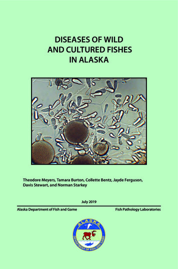
DISEASES OF WILDAND CULTURED FISHESIN ALASKATheodore Meyers, Tamara Burton, Collette Bentz, Jayde Ferguson,Davis Stewart, and Norman StarkeyJuly 2019Alaska Department of Fish and GameFish Pathology Laboratories
The Alaska Department of Fish and Gameprinted this publication at a cost of 9.83 inAnchorage, Alaska, USA.
About This Field GuideThis field guide is a product of the Ichthyophonus Diagnostics, Educational andOutreach Program which was initiated and funded by the Yukon River Panel’sRestoration and Enhancement fund and facilitated by the Yukon River DrainageFisheries Association in conjunction with the Alaska Department of Fish andGame. The original impetus driving the production of this guide was from aconcern that Yukon River fishers were discarding Canadian-origin Chinooksalmon believed to be infected by Ichthyophonus. It was decided to develop aneducational program that included the creation of a field guide containingphotographs and descriptions of frequently encountered parasites within YukonRiver fish.This field guide is to serve as a brief illustrated reference that lists many of thecommon (and not so common) parasitic, infectious, and noninfectious diseasesof wild and cultured fishes encountered in Alaska. The content is directedtowards lay users, as well as fish culturists at aquaculture facilities and fieldbiologists and is not a comprehensive treatise nor should it be considered ascientific document. Interested users of this guide are directed to the listed fishdisease references for additional information.Information contained within this field guide is published from the laboratoryrecords of the Alaska Department of Fish and Game, Fish Pathology Sectionthat has regulatory oversight of finfish health in the State of Alaska. This thirdprinting includes several new entries, some new photographs and updatedinformation on previous diseases and parasites. This version may bedownloaded as a PDF from the ADF&G website at the following web disease/pdfs/fish disease book.pdf3
Text written and provided by: Theodore Meyers, Tamara Burton, Collette Bentz, andJayde Ferguson, Alaska Department of Fish and Game, Fish Pathology Laboratories,333 Raspberry Road, Anchorage, Alaska 99518; P.O. Box 115526 (physical 3333Glacier Highway), Juneau, Alaska 99811-5526.Manuscript Photographs: Alaska Department of Fish and Game, Fish PathologyLaboratory photo archives except where indicated.Publication design by Southfork Graphic Services.Cover Photograph: Ichthyophonus growing in laboratory culture. 2019 Alaska Department of Fish and Game, third printingThe original printing of this publication was produced with funding and support fromthe Yukon River Panel and its members and the Yukon River Drainage FisheriesAssociation. Current funding is provided by the ADF&G, Commercial Fisheries andSport Fish Divisions.The Alaska Department of Fish and Game (ADF&G) administers all programs andactivities free from discrimination based on race, color, national origin, age, sex,religion, marital status, pregnancy, parenthood, or disability. The departmentadministers all programs and activities in compliance with Title VI of the Civil RightsAct of 1964, Section 504 of the Rehabilitation Act of 1973, Title II of the Americanswith Disabilities Act of 1990, the Age Discrimination Act of 1975, and Title IX of theEducation Amendments of 1972. If you believe you have been discriminated againstin any program, activity, or facility please write ADF&G ADA Coordinator, P.O. Box115526, Juneau, AK 99811-5526; U.S. Fish and Wildlife Service, 4401 N. FairfaxDrive, MS 2042, Arlington, VA 22203; or Office of Equal Opportunity, U.S.Department of the Interior, 1849 C Street NW MS 5230, Washington DC 20240.For information on alternative formats and questions on this publication, pleasecontact the department’s ADA Coordinator at (VOICE) 907-465-6077,(Statewide Telecommunication Device for the Deaf) 1-800-478-3648,(Juneau TDD) 907-465-3646, or (FAX) 907-465-6078.
Table of ContentsVirusesKudoa . 62Aquareovirus .2Myxobolus neurotropus . 64Erythrocytic Inclusion Body Syndrome (EIBS) .4Myxobolus squamalis . 66Erythrocytic Necrosis Virus (VENV) .6Tetracapsuloides bryosalmonae (PKD) . 68Infectious Hematopoietic Necrosis Virus (IHNV) .8North American Viral HemorrhagicSepticemia Virus (NA-VHSV).10Pacifc Salmon Paramyxovirus .12Piscine Orthoreovirus (PRV).14BacteriaBacterial Coldwater Disease (BCWD).16Helminths (Worms)Acanthocephalans (spiny headed worms).70Anisakid Larvae (nematode) . 72Philometra (nematode).74Philonema (nematode) .76Black Spot Disease (trematode) .78Encysted Digenean Metacercariae (trematode) . 80Bacterial Gill Disease .18Larval Diplostomulum of the Eye (trematode) . 82Bacterial Kidney Disease (BKD) . 20Gyrodactylus and Dactylogyrus (monogene) . 84Enteric Redmouth Disease (ERM) . 22Piscicola (annelid). 86Furunculosis.24Diphyllobothrium (cestode) . 88Fusobacteria-like Agent . 26Schistocephalus (cestode). 90Marine Tenacibaculosis. 28Triaenophorus (cestode) .92Motile Aeromonas and Pseudomonas Septicemia . 30Mycobacteriosis of Fish.32Vibriosis. 34FungiPhaeohyphomycosis of Safron Cod . 36Phoma herbarum . 38ProtozoaEpistylis (Heteropolaria) . 40Hexamita.42Ichthyobodiasis (Costiasis) . 44Ichthyophonus. 46Saprolegniasis – Cotton Wool Disease. 48Trichodiniasis. 50Trichophrya (Capriniana) . 52White Spot Disease . 54X-Cell Tumors . 56CnidariaCeratonova (Ceratomyxa) shasta. 58Henneguya . 60ArthropodsExternal Parasitic Copepods. 94Salmincola (copepod) . 96Sarcotaces (copepod) . 98Non-infectious DiseasesBloat (Water Belly). 100Blue Sac Disease of Fry. 102Coagulated Yolk Disease (White Spot Disease). 104Drop-out Disease . 106Gas Bubble Disease (GBD). 108Mushy Halibut Syndrome .110Neoplasia (Tumors).112Organ and Tissue Anomalies .116Pigment Aberrations in Fish.118Sunburn (Back-Peel). 120ReferenceGlossary of Terms . 122Fish Disease References .127
VIRUSESAquareovirusassociated with epizootic fish mortality producing severe hemorrhaging infingerlings and yearlings resulting in upto 80% mortality.I. Causative Agent and DiseaseAquareovirus is a genus in the virusfamily Reoviridae. These icosahedral(60-80 nm) 11 segmented doublestranded RNA viruses (over 50) havebeen isolated from a variety of marineand freshwater aquatic animals worldwide including finfish, and bivalve molluscs. Genetic analyses have identified 7different genotypes or species (A-G) ofaquareoviruses. Most of these virusesproduce self-limiting infections of lowpathogenicity and are not associatedwith extensive disease or mortality.Exceptions include isolates from 7 fishspecies that have been associated withfish mortality, most notably the grasscarp aquareovirus (G). The viral agentsare most often isolated from asymptomatic adult carrier fish during routinescreening examinations.IV. TransmissionTransmission is horizontal via wateror from fish to fish. Isolates from bivalvemollusks likely represent virus that hasbeen shed into the water column from afish host and then bioaccumulated intoshellfish tissues by filter feeding.V. DiagnosisDetection of Aquareovirus is byisolation of the virus in cultures ofsusceptible fish cell lines inoculatedwith infected tissue. The virus causes aunique cytopathic effect (CPE) characterized by focal areas of cellular fusion(syncytia) and cytoplasmic destructioncreating a vacuolated or foamy appearance. The exception is grass carp speciesG that produces a diffuse CPE. Presumptive identifications are made based onthe typical CPE and are confirmed byserology, electron microscopy or polymerase chain reaction (PCR).II. Host SpeciesIn the Pacific Northwest states ofWashington, Oregon and California,adult Chinook salmon appear to be themost frequent species infected withaquareovirus A or B. The virus has alsobeen isolated from adult coho and chumsalmon and steelhead. Rainbow trouthave been experimentally infected withthe virus resulting in mild hepatitis withno overt disease or mortality. In Alaska,aquareoviruses have been isolatedfrom Chinook salmon (species B) andgeoduck clams (species A).VI. Prognosis for HostThe prognosis for the fish host isgood in the majority of cases where thevirus is not a primary pathogen. Thereare no corrective therapies for viralinfections in fish except avoidance.VII. Human Health SignifcanceThere are no human health concernsassociated with aquareoviruses.III. Clinical SignsFish naturally infected with aquareoviruses generally do not exhibit clinicalsigns of disease. Experimental infectionscan produce focal necrotic lesions in thelivers of rainbow trout, chum salmon andbluegill fry. Other pathogenic exceptionsinclude the grass carp species G that is2
VIRUSES Large rounded plaques of syncytial cell CPE (arrow) of Aquareovirus inbluegill fry cells.Double capsid morphology of Aquareovirus particles(arrow) in negative stain; transmission electronmicroscopy, X 91,000.3
VIRUSESErythrocytic Inclusion Body Syndrome (EIBS)V. DiagnosisIsolation and replication of thevirus in available fish cell lines hasbeen unsuccessful. Thus, diagnosis isby observation of the small pale blueinclusion bodies in the cytoplasm ofinfected erythrocytes with confirmationby transmission electron microscopy(TEM). The virus is found free in thecytoplasm or more commonly occurs inmembrane bound cytoplasmic inclusionbodies within erythrocytes.I. Causative Agent and DiseaseErythrocytic inclusion body syndrome (EIBS) is caused by an unclassified icosahedral virus (70-80 nm) thatinfects erythrocytes of several salmonidfishes in fresh and seawater. Typically,EIBS presents with single or multiplepale, basophilic inclusions (0.4-1.6um) in the cytoplasm of erythrocytesin stained peripheral blood smears.Affected fish may be asymptomatic,but more often have varying degreesof anemia and secondary bacterial andfungal infections. In severe cases ofuncomplicated anemia, cumulative fishmortality over 20% has been reportedwith hematocrits less than 20%.VI. Prognosis for HostSevere fish losses caused directlyby EIBS are rare. However, fish becomeweakened from the anemia and mortality from other associated environmentalstressors or secondary pathogens can besignificant. The disease generally is selflimiting with recovery and immunity insurvivors.II. Host SpeciesEIBS has been found in Chinook,coho and Atlantic salmon in the Pacific Northwest, Japan, Norway and theBritish Isles. Other salmonid speciesshowing variable susceptibilities by experimental injection with infected bloodhomogenates include cutthroat trout,masou salmon and chum salmon.VII. Human Health SignifcanceThere are no human health concernswith the EIBS virus.NOTE: In Alaska, only one case of EIBShas been reported in 2004 affectingjuvenile Chinook salmon in seawater netpens. Subsequent studies haveshown that EIBS in Japanese farmedcoho salmon may be caused by a strainof Piscine Orthoreovirus (PRV-2).Molecular studies have determined thatPRV is present in Alaskan coho andChinook salmon. See PRV chapter formore detail.III. Clinical SignsFish are lethargic, anorexic andanemic with chronic mortality oftenassociated with secondary infectionsby other pathogens. Five stages of EIBShave been described: preinclusion, inclusion body formation, cell lysis with lowhematocrits, recovery with increasinghematocrits and full recovery.IV. TransmissionThe disease can be transmittedhorizontally while surviving fish generally recover and develop an acquiredimmunity against reinfection that istransferable by passive immunization.4
VIRUSES Erythrocytes of Chinook salmon with small basophilic cytoplasmicinclusion bodies (arrow) typical of EIBS, X 1000.EIBS inclusion body (not membrane-bound) composed ofvirus particles in the cytoplasm of an erythrocyte; transmissionelectron microscopy, X 56,400.5
VIRUSESErythrocytic Necrosis Virus (VENV)IV. TransmissionTransmission of this virus is likelyhorizontal from fish to fish based on thefew experimental studies using waterborne exposure. Adult carrier fish ofsusceptible species are likely reservoirsof the virus that is transmitted to juvenile fish. Anadromous fish likely becomeinfected during their marine phase oflife. There is some suggestion that thevirus is vector-borne and one instanceof infection in juvenile salmonids infreshwater suggesting vertical transmission from adult anadromous parent fish.I. Causative Agent and DiseaseErythrocytic necrosis or viralerythrocytic necrosis (VEN) is causedby several similar iridoviruses havingdouble-stranded DNA and a hexagonalshape ranging in size from 130-330 nm.The viruses infect erythrocytes causinga hemolytic disease often resulting inanemia and secondary infections byother pathogens including VHSV TypeIVaII. Host SpeciesThere are likely several differentstrains of the virus worldwide in themarine environment infecting a largevariety of more than 20 anadromous andmarine fish species. In Alaska, VENVhas been detected in Pacific herringfrom several locations but has not yetbeen observed in salmonids. Resultsfrom experimental infections and occurrence of epizootics in young-of-the-yearPacific herring indicate that juveniles aremore susceptible than older fish.V. DiagnosisDiagnosis is made with blood smearsshowing characteristic eosinophilicinclusion bodies (1-4 um) present in thecytoplasm of erythrocytes when stainedwith Giemsa or Wright stains. Impression smears of hematopoietic headkidney can be substituted for blood. Thevirus is confirmed by the observationof iridovirus particles associated withinclusion bodies using electron microscopy (TEM). VEN viruses have not beensuccessfully cultured in available fishcell lines, however an unvalidated PCRis available for the virus in Pacific herring.III. Clinical SignsAdult herring generally show noclinical signs of disease. In juvenilePacific herring, fish are anemic exhibiting nearly white gills and pale visceralorgans. Liver color may be green dueto breakdown of blood hemoglobinreleasing excess biliverdin. Hemotocritsmay be as low as 2 to 10%, erythrocytesare fragile causing hemolysis of bloodsamples, and immature erythrocytespredominate in peripheral blood. Highmortality with dead fish on the shorelineaccompanied by extensive congregationsof predator birds may occur in areaswhere juvenile herring are weakened bythe disease.VI. Prognosis for HostThe virus in Alaskan juvenile Pacificherring caused one of the first naturalepizootics reportedly associated withmass fish mortality. Chronic to subacutemortality in juvenile Pacific herring canalso occur, especially when stressed.VII. Human Health SignifcanceNo human health concerns are associated with VEN virus.6
VIRUSES Anemic Pacifc herring with very pale gills and green livers fromexcessive biliverdin commonly seen with VENErythrocytes of Pacifc herring with large eosinophiliccytoplasmic inclusion bodies (arrow), some surrounded by pinklattices composed of virus particles; Dif Quik, X 400. TEM of infected erythrocyte showing largevirus particles (arrow) comprising the latticesurrounding inclusion bodies in stained smears,X 15,600.7
VIRUSESInfectious HematopoieticNecrosis Virus (IHNV)III. Clinical SignsInfected fish may exhibit: lethargy,whirling behavior, cranial swelling, abdominal swelling, exophthalmia, anemiaand darkened body coloration; hemorrhaging of musculature and base of fins;fecal casts; pre-emergence in sac-fry;pale liver, spleen and kidney; stomach/intestine filled with milky or wateryfluid with petechial hemorrhaging ofmesenteries or visceral tissues. Gills arepale, moderately hyperplastic and bloodsmears often contain necrobiotic bodies.I. Causative Agent and DiseaseInfectious hematopoietic necrosis virus (IHNV) is a bullet-shapednovirhabdovirus that is enzootic to theNorth American Pacific Northwest butwas inadvertently established in the USSnake River Valley in Idaho and in several countries of Asia and Europe. Thethree genetic clades of IHNV (U,M,L)can infect several salmonid species andhave had severe economic impacts onintensively cultured salmon and trout.IHNV in Alaska (U clade) has beenlimited primarily to sockeye salmon andrarely Chinook and chum salmon wheninfected sockeye are present in their water supplies. Culture of sockeye salmonin Alaska by avoidance of IHNV hasbeen successful through the rigorous useof the ADF&G sockeye salmon culturepolicy. The disease, infectious hematopoietic necrosis (IHN), is an acute, systemic infection causing necrosis of thekidney tissues and other visceral organsresulting in extensive mortality in hatchery reared sockeye salmon juveniles aswell as in wild stocks of out-migratingsockeye salmon smolts.IV. TransmissionHorizontal transmission through water via feces or sex products or carcassdegradation is the most common routeof infection. Virus occurs commonly inovarian fluids and on the surface of eggs.Rarely, vertical transmission can occurwithin eggs (internal) and possibly withadhesion of virus particles to sperm during fertilization. Incubation and courseof the disease can be strongly influencedby water temperature as reported in theLower 48. Optimum temperature is 1012 C but IHN losses have been reportedabove 15 C. Mortalities occur within4-6 days post-exposure peaking at 8-14days. In Alaska, the disease can causeup to 100% mortality in sockeye salmonat water temperatures as low as 1-2 Cwhere exponential mortality may takelonger to occur. No natural reservoirs ofIHNV have been confirmed other thanthose susceptible fish species that arecarriers of the virus. However, transientdetections of IHNV have been reportedin organic sediments, invertebrates, andsome forage species of marine fish whenassociated with ongoing or recent IHNVepizootics in susceptible salmonid speciesII. Host SpeciesFish species susceptible to infectionand disease by IHNV include: sockeye,Chinook, chum, amago, yamame andAtlantic salmon; cutthroat trout and rainbow/steelhead trout. Brook and browntrout are experimentally susceptible toinfection and mortality while lake troutare intermediate in susceptibility. Arcticchar and grayling are resistant whilecoho salmon are also resistant but cancarry the virus when in the presence ofother susceptible virus-infected fish species. Mortality is highest in young fishand resistance to infection and diseaseincreases with age.8
VIRUSESV. DiagnosisSusceptible fish cell cultures are inoculated with kidney and spleen tissues(whole fry if small) or ovarian fluidsfrom fish suspected of having IHNV.Presumptive diagnosis results from diffuse or plaqued lysis of inoculated cellmonolayers (cytopathic effect). Virus isdefinitively identified by PCR.In Alaskan hatcheries, all infected lots offish are destroyed. The occurrence of thedisease is avoided through preventativemeasures including a virus-free watersupply, rigorous disinfection, isolationof egg and fish lots and containment ofdiseased fish. There is an effective DNAvaccine used in Canada that is also licensed in the US but has been restrictedcommercially due to unlikely safetyconcerns regarding GMO products.VI. Prognosis for HostPrognosis for infected fish is poor.Survivors of epizootics and subclinicalinfections become lifelong carriers ofthe virus. There is no known therapy forfish that have been infected with IHNV.VII. Human Health SignifcanceThere are no human health concernsassociated with IHN virus.Hemorrhaging at the base of the fns issometimes observed in IHN disease. Exaggerated (top, middle) cephalic bumpson sockeye salmon fry commonly occur withIHN disease.Scoliosis in sockeye salmon smolt survivingIHN. IHN virus particles (arrow) budding from cellmembrane of an EPC cultured fsh cell; TEM,X 34,000.Necrotic macrophages or kidney cells(necrobiotic bodies-arrows) with debris inperipheral blood, X 1000.9
VIRUSESNorth American Viral HemorrhagicSepticemia Virus (NA-VHSV)100% of exposed juvenile fish with lowerchronic mortality occurring in adults.Infected juvenile herring develop hemorrhages of the skin around the mouth andisthmus and/or at the base of fins whileoccasional hemorrhages occur in adultfish along the flanks that may progressto ulcers. Fin erosion and lethargicswimming behavior may also be present.Experimentally infected juvenile rainbow trout exhibited darkened body colorand hemorrhaging at the base of fins andvent associated with low mortality.I. Causative Agent and DiseaseNorth American viral hemorrhagicsepticemia virus (NA-VHSV) Type IVais a bullet-shaped RNA novirhabdovirus.It is molecularly distinct from the similarType IVb in the Great Lakes, USA thatis pathogenic for a large number ofnonsalmonid species. It is also differentfrom Type IVc in marine/freshwater species of Atlantic Canada and most VHSVstrains in Asia (IVa occurs in Japan/Korea) and Europe (Types I, II, III) thatare pathogenic for some marine speciesand rainbow trout.IV. TransmissionTransmission of VHSV is horizontalthrough ambient seawater from fish tofish and likely by ingestion of infectedfish. Individual infected juvenile Pacificherring can shed up to 106.5 plaque forming units (PFU) of virus per ml. Primaryvirus infection is through the epidermisand possibly gill tissues followed bysystemic infection (viremia). BecauseVHSV in the Pacific Northwest is indigenous to Pacific herring and other foragespecies utilized by salmon, these preyare a likely source of VHSV periodically detected in adult coho and Chinooksalmon in Washington State.II. Host SpeciesNA-VHSV Type IVa infects manymarine fish species in the northernPacific Ocean including anadromouscoho and Chinook salmon. In Alaska,the virus is reported from Pacificherring, Pacific cod, Pacific hake andwalleye pollock. Two isolates from pinksalmon have been the only occurrencesof VHSV from a free ranging Alaskansalmonid. The virus is enzootic inpopulations of Northern Pacific herringand sardines causing epizootic mortality. Experimental studies indicate thatjuvenile Alaskan Chinook, coho, pinkand sockeye salmon are refractory to thevirus by waterborne exposure.V. DiagnosisCultures of susceptible fish celllines are inoculated with kidney, spleen,liver, ovarian fluids or epidermal lesionsfrom suspect fish. Presumptive diagnosis is made when characteristic cytopathic effect (CPE) or lysis occurs in cellmonolayers from virus infection. Virusidentification is confirmed by PCR.III. Clinical SignsDetection of NA-VHSV from anadromous salmonids in Washington andOregon has generally been at very lowlevels and prevalences and not associated with clinical disease. In Pacific cod,secondary VHSV infection can be detected at low levels in skin erosions andulcers caused by other primary pathogens. Septicemia with skin hemorrhagesmay also occur. In Pacific herring, thevirus can be acutely lethal for up toVI. Prognosis for HostSusceptible juvenile herring sustainup to 100% mortality which may notoccur in adult fish or is lower and more10
VIRUSESvariable but generally lower in other forage species.chronic. Herring that survive virus infection develop apparent immunity to reinfection. Noteworthy, is that low levelsof VHSV can occasionally be detected ina small percentage of apparently healthyherring from most populations. Clinicaldisease and mortality from the virus isVII. Human Health SignifcanceThere are no human health concernsassociated with NA-VHS virus.Skin hemorrhaging in infected Pacifc herring Left: Pacifc herring with typical VHS hemorrhage; Right: Skin hemorrhaging in infectedPacifc cod (photo: NMFS staf)Electron micrograph of VHSV particles (arrow) in a cultured EPCfsh cell, X 56,50011
VIRUSESPacific Salmon ParamyxovirusI. Causative Agent and DiseasePacific salmon paramyxovirus(PSPV) is a large, irregular shaped,enveloped, single-stranded RNA virusabout 125-250 nm in diameter belongingin the family Paramyxoviridae and thegenus Aquaparamyxovirus. The virus isof low virulence and not associated withdisease or mortality. The viral agent isgenerally isolated from asymptomaticcarrier fish during routine viral screening.This virus has the unique characteristicamong fish viruses of hemagglutinatingerythrocytes from fish, some mammals(human, rabbit, horse, guinea pig andswine), and birds. Hemagglutinationallows viral placement in the Paramyxoviridae and confirmation of a paramyxovirus along with other proceduresincluding ultrastructural morphologyby electron microscopy, fluorescent antibody testing (FAT), PCR and sequencing.II. Host SpeciesThe most common host in NorthAmerica is adult Chinook salmon fromAlaska, Oregon and Washington. Unconfirmed isolates have been reportedfrom other salmonids. In Norway, Atlantic salmon paramyxovirus (ASPV)has been isolated from seawater rearedAtlantic salmon.VI. Prognosis for HostThe prognosis for the host is goodregarding the non-pathogenic nature ofthe North American isolates of PSPV.The role of Norwegian ASPV in causingPGI is questionable since other agentshave been present confounding thetrue etiology of fish mortality. In thiscase perhaps corrective therapy wouldinclude optimizing the environment andavoidance. Further study is warranted.III. Clinical SignsNo clinical signs of disease areassociated with fish infected by PSPV.The ASPV paramyxovirus in Norway isreportedly associated with the diseasesyndrome, proliferative gill inflammation (PGI).VII. Human Health SignifcanceThere are no human health concernsassociated with paramyxoviruses in fish.IV. TransmissionThe mode of transmission is horizontal by water or fish to fish. A marinereservoir for the virus is suspected.V. DiagnosisDetection of paramyxovirus is byisolation in cultures of susceptible fishcell lines inoculated with infected tissue.The virus causes a cytopathic effect(CPE) characterized by retracted androunded cells after an extensive incubation period. Presumptive identificationsare made by observing the typical CPE.12
VIRUSESChinook salmon, Ninilchik River, AK. Falsely colorized ultrastructureof PSPV in cultured infected CHSE-214 cell with three virus particlesin diferent stages of budding from cell membrane (arrow centerparticle); blue (B) spiked envelope; purple (P) nucleocapsidwithin particles from pool of subsurface nucleocapsid material in thecytoplasm. Inset: Same image not colorized. (From Meyers and Batts2016).20 umCytopathic efect of PSPV from Chinook salmon, Ninilchik River, AK in CHSE-214 cells at14 C demonstrating clustered rounding and refractility of cells (Right) at 9 days postinoculation of previously passaged material. Normal cell culture (Left). Phase contrast(From Meyers and Batts 2016).13
VIRUSESPiscine Orthoreovirus (PRV)I. Causative Agent and DiseasePiscine orthoreovirus (PRV), alsoknown as Atlantic salmon reovirus,was identified in 2010 by next generation sequencing of tissues from farmedAtlantic salmon in Norway dying fromthe disease “heart and skeletal muscleinflammation” (HSMI). The virus hasdouble-stranded RNA with 10 segmentsand is 72 nm in diameter. There arethree strains of the virus (PRV1, 2, 3)possibly influencing disease outcomein diff
Department of the Interior, 1849 C Street NW MS 5230, Washington DC 20240. For information on alternative formats and questions on this publication, please . contact the department's ADA Coordinator at (VOICE) 907-465-6077, (Statewide Telecommunication Device for the Deaf) 1-800-478-3648, (Juneau TDD) 907-465-3646, or (FAX) 907-465-6078.


![INDEX [randycherry ]](/img/21/x-20-20tv-20fakebook-20-20hal-20leonard.jpg)