Transcription
Interventional RadiologyIn-Training Test Questionsfor Diagnostic Radiology ResidentsMarch, 2013Sponsored by:Commission on EducationCommittee on Residency Training in Diagnostic Radiology 2013 by American College of Radiology. All rights reserved.1891 Preston White Drive -- Reston, VA 20191-4326 -- 703/648-8900 -- www.acr.org
1. Concerning acute gastrointestinal hemorrhage, which statement is TRUE?A. Radionuclide scanning should not be performed.B. Bright red blood per rectum excludes an upper gastrointestinal bleed.C. The angiographic diagnosis is based upon the visualization of contrast extravasation into thebowel lumen.D. Bleeding from Mallory-Weiss tears may be diagnosed upon injection of either the superior orinferior mesenteric arteries.Rationales:A. Incorrect. Radionuclide scanning is more sensitive than arteriography in detecting gastrointestinalhemorrhage and can be helpful in localizing the bleed.B. Incorrect. About 10% of patients with brisk upper gastrointestinal hemorrhage, bleeding proximal to theligament of Treitz, will have bright red blood per rectum.C. Correct. The hallmark of gastrointestinal hemorrhage is extravasation of contrast material into the bowel.D. Incorrect. Mallory-Weiss tears occur at the gastroesophageal junction, not in the distribution of either thesuperior or inferior mesenteric arteries.Citations:Kaufman JA, Lee MJ. Vascular and Interventional Radiology: The Requisites. St. Louis, Mo: Mosby; 2004.Valji K. Vascular and Interventional Radiology. Philadelphia, Pa: W.B. Saunders; 1999.2. Concerning inferior vena cava filters, which statement is TRUE?A.B.C.D.Removable filters are not available.The ideal location for filter placement is at the iliac vein confluence.Current filters require surgical cut down for placement.Current filters can be placed from femoral or jugular venous approach.Rationales:A. Incorrect. Removable filters are now commercially available.B. Incorrect. The ideal location is just below the renal veins.C. Incorrect. Most devices are placed percutaneously.D. Correct. Current devices can be placed via a transfemoral or transjugular access.Citations:Kinney TB. Update on inferior vena cava filters. J Vasc Interv Radiol. 2003;14:425-440.
3.A 67-year-old man presents with acute onset of back pain. You are shown a thoracic aortogram. What isthe MOST likely diagnosis?A.B.C.D.Intraluminal thrombusTraumatic lacerationDissecting hematomaMycotic aneurysmRationales:A. Incorrect.B. Incorrect.C. Correct. Aortic dissection is the separation of the intima from the adventia by blood within the medial layer ofthe artery.D. Incorrect.Citations:Kaufman JA, Lee MJ. Vascular and Interventional Radiology: The Requisites. St. Louis, Mo: Mosby; 2004.
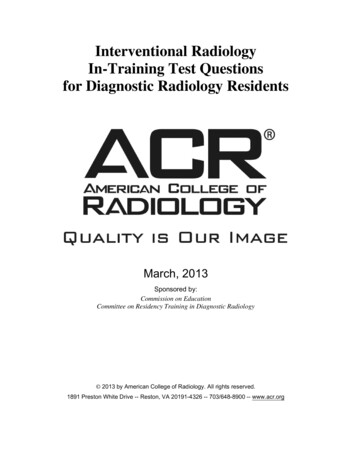
4. A 52-year-old construction worker had bluish discoloration and numbness of the fifth finger of his righthand. You are shown an arteriogram (Figure 4) of the right hand and wrist. The proximal arteries wereintact. What is the MOST likely diagnosis?A.B.C.D.Paget-Schroetter syndromeGiant cell arteritisSclerodermaHypothenar hammer syndromeRationales:A. Incorrect. All 4 possible answers are associated with occlusions of upper extremity blood vessels. However,Paget-Schroetter is a syndrome of venous occlusion at the thoracic outlet.B. Incorrect. Giant cell arteritis is associated with long strictures of the subclavian and axillary arteries.C. Incorrect. Scleroderma does cause small vessel occlusions of the arteries of the hand and wrist and should beseriously considered in the differential diagnosis, but the patient is a male construction worker, and it is theulnar artery that is occluded.D. Correct. Finger ischemia resulting from repetitive trauma to the ulnar artery, often the result of occupationalexposure, is hypothenar hammer syndrome.Citations:Taylor LM. Hypothenar hammer syndrome. J Vasc Surg. 2003;37:697.Valji K. Vascular and Interventional Radiology. Philadelphia, Pa: W.B. Saunders; 1999.Vedantham S, Gould J. Case Review Vascular and Interventional Imaging. St. Louis, Mo: Mosby; 2004.
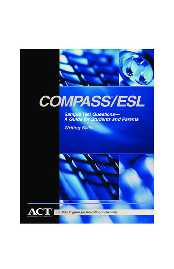
5. You are shown a single image from a percutaneous transhepatic cholangiogram of a jaundiced patient(Figure 2). What is the MOST LIKELY diagnosis?A.B.C.D.Gallbladder carcinomaSclerosing cholangitisPancreatic carcinomaCholedochoceleRationales:A. Incorrect. Gallbladder carcinoma causes obstruction at the level of the proximal common hepatic duct orhigher. Because gallbladder carcinoma usually arises in the setting of chronic cholecystitis you will not expect tosee a distended gallbladder.B. Incorrect. Sclerosing cholangitis typically causes diffuse, multifocal strictures often with a beaded appearancein the intra and extrahepatic ducts.C. Correct. The “rat tailed” appearance of the extrinsic obstruction and the location of the stricture at the levelof the head of the pancreas make this diagnosis the favorite. The “Courvoisier’s gallbladder” is also typical.D. Incorrect. A choledochocele appears as a focal cystic collection of contrast at the level of theampulla.
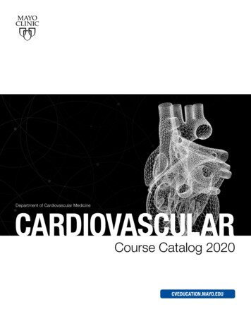
6. You are shown a single image from a non-contrast-enhanced CT scan of the chest (Figure 3). Which ofthe following statements is true?A.B.C.D.An excisional biopsy should be the next diagnostic step.A percutaneous needle biopsy should be the next diagnostic step.Radiofrequency ablation is the standard therapy for this disorder.Coil embolization is a standard therapy for this disorder.Rationales:The correct answer is D. The findings are diagnostic for a pulmonary arteriovenous malformation. The nodule isthe saccular communication between the feeding pulmonary artery and the draining pulmonary vein. It isincorrect to think this is a solid tumor, perhaps a small lung cancer. The diagnosis could be confirmed with a CTangiogram, but certainly it would be incorrect to perform a needle biopsy of a vascular lesion. Standard therapyis coil embolization. It would be too invasive to excise this lesion, particularly as they are so often multiple.Radiofrequency ablation at this point is an experimental technique for malignant lung lesions not amenable tosurgery. Not recommended here.Citations:SCVIR Syllabus Series: Thoracic and Visceral Vascular Interventions, Chapter 5. Valgi p 278, Kaufman p211.Fernando HC, Hoyas AD, Litle V, Belani CP, Luketich JD. Radiofrequency ablation: identification of the idealpatient. Clin Lung Cancer. 2004;6:149-53.
7. The patient presented with sudden onset of facial and upper extremity swelling. You are shown biplanarangiographic images (Figures 4A and 4B). What action should be taken?A.B.C.D.Emergency surgeryCatheter directed thrombolysisSystemic chemotherapyAngioplasty and stent placementRationales:The correct answer is A. The clinical history confirmed by the angiogram is diagnostic of superior vena cavasyndrome. Acute superior vena cava syndrome is considered a medical emergency. The treatment options forsuperior vena cava syndrome are endovascular therapy and external beam irradiation. The response toangioplasty and stent placement is more rapid than to radiotherapy. In this case there is no clot to lyse.Citations:Kaufman JA, Lee MJ. Vascular and Interventional Radiology. The Requisites. Mosby 2004.Vendatham S, Gould JE. Case Review Vascular & Interventional Imaging. Mosby 2004.
8. You are shown duplex Doppler ultrasound images of the right greater saphenous vein during a Valsalvamaneuver (Figure 5). What is the MOST LIKELY diagnosis?A.B.C.D.Superficial thrombophlebitisVenous insufficiencyArteriovenous fistulaPhlegmasia cerulea dolensRationales:The correct answer is B. Superficial thrombophlebitis and phlegmasia can be easily eliminated as correctanswers because there is flow through a patent vein and no clot is demonstrated. An arteriovenous fistula willshow a pulsatile arterial wave form. The retrograde flow through the greater saphenous vein on Valsalvademonstrates incompetence of the venous valves. Venous insufficiency is the correct answer.Citation: Zwiebel WJ, Pellerito JS. Introduction to Vascular Ultrasonography. Elsevier Saunders 2005.
9. You are shown an image from an abdominal aortogram. Which one of the following BEST describesthe patient?A.B.C.D.25-year-old woman with recently discovered hypertension45-year-old man with acute renal failure70-year-old man with poorly controlled hypertension80-year-old woman being evaluated for an aortic stent graftRationales:A. Incorrect. This is an elderly patient with atheromatous disease. In a young woman, a more common cause forrenal vascular hypertension would be fibromuscular dysplasia, the most characteristic appearance of which is astring-of-bead appearance along the course of the renal artery.B. Incorrect. Even for a 45 year-old, this is advanced atherosclerotic disease and renal failure is unusual withunilateral renal artery stenosis. Not the best choice.C. Correct. The age is right for the amount of disease in the aorta. The renal artery stenosis could easily explainthe difficult to control hypertension.D. Incorrect. Aortic stent grafts are typically used as an alternative to surgery for abdominal aortic aneurysms.Citations:Kaufman JA, Lee MJ. Vascular and Interventional Radiology. The Requisites. Mosby. 2004.
10. Drug-eluting stents have been used in the treatment of coronary artery disease. What is the purpose ofthe drugs?A.B.C.D.To retard post-procedure elastic recoil.To retard post-procedure platelet aggregation.To retard the development neointimal hyperplasia.To retard the progression of atherosclerotic occlusive disease.Rationales:The correct answer is C. The advantage of a stent over angioplasty alone is it prevents elastic recoil. The purposeof the eluted drugs is to retard neointimal hyperplasia as a cause for in-stent stenosis. These are not antiplateletmedications or drugs to inhibit progressive atherosclerosis. Although drug eluting stents have revolutionized thetreatment of coronary artery disease, it has been difficult to show that they are an improvement over barestents for the treatment of superficial femoral artery disease.Citations:Oliva VL, Soulez G. Sirolimus-eluting stents versus the superficial femoral artery: second round. J Vasc IntervRadiol 2005; 16:313-315.
Vascular and Interventional Radiology: The Requisites. St. Louis, Mo: Mosby; 2004. Valji K. Vascular and Interventional Radiology. Philadelphia, Pa: W.B. Saunders; 1999. 2. Concerning inferior vena cava filters, which statement is TRUE? A. Removable filters are not available. B. The ideal location for filter placement is at the iliac vein .