Transcription
(2022) 22:776He et al. BMC en AccessRESEARCHThe value of HPV genotypes combinedwith clinical indicators in the classificationof cervical squamous cell carcinomaand adenocarcinomaZhimin He1,2, Rongsheng Chen3, Shangying Hu4, Yajiao Zhang5, Yang Liu1,2,3, Chengwei Li1,2, Fajin Lv1,2,3,6* andZhibo Xiao3*AbstractBackground: To investigate the differences in HPV genotypes and clinical indicators between cervical squamouscell carcinoma and adenocarcinoma and to identify independent predictors for differentiating cervical squamous cellcarcinoma and adenocarcinoma.Methods: A total of 319 patients with cervical cancer, including 238 patients with squamous cell carcinoma and 81patients with adenocarcinoma, were retrospectively analysed. The clinical characteristics and laboratory indicators,including HPV genotypes, SCCAg, CA125, CA19-9, CYFRA 21–1 and parity, were analysed by univariate and multivariate analyses, and a classification model for cervical squamous cell carcinoma and adenocarcinoma was established.The model was validated in 96 patients with cervical cancer.Results: There were significant differences in SCCAg, CA125, CA19-9, CYFRA 21–1, HPV genotypes and clinical symptoms between cervical squamous cell carcinoma and adenocarcinoma (P 0.05). Logistic regression analysis showedthat SCCAg and HPV genotypes (high risk) were independent predictors for differentiating cervical squamous cellcarcinoma from adenocarcinoma. The AUC value of the established classification model was 0.854 (95% CI: 0.804–0.904). The accuracy, sensitivity and specificity of the model were 0.846, 0.691 and 0.899, respectively. The classificationaccuracy was 0.823 when the model was verified.Conclusion: The histological type of cervical cancer patients with persistent infection of high-risk HPV subtypes andlow serum SCCAg levels was more prone to being adenocarcinoma. When the above independent predictors occur,the occurrence and development of cervical adenocarcinoma should be anticipated, and early active interventiontreatment should be used to improve the prognosis and survival of patients.Keywords: Cervical cancer, Adenocarcinoma, Squamous cell carcinoma, Human papilloma virus, Tumour marker*Correspondence: fajinlv@163.com; 202530@cqmu.edu.cn1State Key Laboratory of Ultrasound in Medicine and Engineering,College of Biomedical Engineering, Chongqing Medical University,Chongqing 400016, China3Department of Radiology, The First Affiliated Hospital of ChongqingMedical University, Chongqing 400016, PR ChinaFull list of author information is available at the end of the articleCervical cancer is the most common malignant tumourof the female reproductive system, and it ranks fourthin both the number of new cases of cancer and the proportion of cancer deaths among women in the world. In2020, there were approximately 604,000 new cases and342,000 deaths worldwide, so cervical cancer poses a serious threat to the lives and health of women worldwide The Author(s) 2022. Open Access This article is licensed under a Creative Commons Attribution 4.0 International License, whichpermits use, sharing, adaptation, distribution and reproduction in any medium or format, as long as you give appropriate credit to theoriginal author(s) and the source, provide a link to the Creative Commons licence, and indicate if changes were made. The images orother third party material in this article are included in the article’s Creative Commons licence, unless indicated otherwise in a credit lineto the material. If material is not included in the article’s Creative Commons licence and your intended use is not permitted by statutoryregulation or exceeds the permitted use, you will need to obtain permission directly from the copyright holder. To view a copy of thislicence, visit http:// creat iveco mmons. org/ licen ses/ by/4. 0/. The Creative Commons Public Domain Dedication waiver (http:// creat iveco mmons. org/ publi cdoma in/ zero/1. 0/) applies to the data made available in this article, unless otherwise stated in a credit line to the data.
He et al. BMC Cancer(2022) 22:776[1]. Squamous cell carcinoma (SCC) is the most common histological type of cervical cancer, accounting for70% 75% of cases, followed by adenocarcinoma (AC),accounting for 10% 25% of cases [2]. A series of studieshave shown that compared with concurrent squamouscell carcinoma, adenocarcinoma is more aggressive andless sensitive to radiotherapy and chemotherapy, with ahigher metastasis rate, poorer prognosis and lower survival rate [3–9]. Therefore, many scholars believe that different clinical treatment strategies should be developedaccording to the different characteristics of cervical squamous cell carcinoma and adenocarcinoma, which is ofgreat significance for determining accurate and personalized treatment plans [10–12]. Excitingly, new treatmentstrategies for cervical adenocarcinoma have been exploratory studies and applications. Noriyuki Okonogi et al.[13, 14] found that carbon-ion radiotherapy (CIRT) orconcurrent cisplatin and CIRT showed promising resultsin the treatment of cervical adenocarcinoma, which maybe a promising therapeutic strategy for cervical adenocarcinoma. The different epidemiology and prognosis ofcervical squamous cell carcinoma and adenocarcinoma,as well as the individualized treatment options beingexplored, will inevitably lead to the need for new differential diagnosis methods for cervical squamous cell carcinoma and adenocarcinoma. Therefore, the differentialdiagnosis of cervical squamous cell carcinoma and adenocarcinoma is closely related to the treatment and prognosis of patients.In 2021, the second edition of the Screening and Treatment Guidelines for Cervical Precancerous Lesionsreleased by the World Health Organization (WHO)clearly recommended human papillomavirus (HPV)DNA testing as the preferred screening method for cervical cancer [15]. Persistent HPV infection is the maincause of cervical cancer, and there are many genotypes[16]. When HPV genotypes are 60%-70% nucleotidehomologous, they cluster in the same species, and themost common HPV species, alpha 7 (HPV 18, 39, 45, 59,68 and 70) and alpha 9 (HPV 16, 31, 33, 35, 52, 58 and67), account for 80% of all cervical cancer cases [17].HPV species are associated with the survival prognosis ofcervical cancer patients. Cervical cancer patients who areHPV negative or only infected with HPV alpha 7 have aworse prognosis and higher risk; cervical cancer patientswith coinfection of HPV alpha 7 and HPV alpha 9 are atmedium risk; and cervical cancer patients infected withonly HPV alpha 9 or other HPV genotypes are at lowerrisk [18]. Therefore, at present, risk subtypes based onthe prognosis of HPV species are mostly used for the efficacy evaluation and prognosis prediction of radiotherapyand chemotherapy in patients with cervical cancer [18,19], but their application in the differential diagnosis ofPage 2 of 10cervical squamous cell carcinoma and adenocarcinomahas not been reported.At present, clinical indicators such as tumour markersare often used in relevant studies on the identification ofcervical squamous cell carcinoma and adenocarcinoma.Such indicators are easy to obtain clinically and play animportant role in the differential diagnosis and prognosis prediction of tumours [20–25]. Lehtovirta P, Borras G,Liu Y et al. studied the differences in squamous cell carcinoma antigen (SCCAg) and carbohydrate antigen 125(CA125), CA125 and carbohydrate antigen 19–9 (CA199), and SCCAg and CA19-9 levels between cervicalsquamous cell carcinoma and adenocarcinoma, and theresults showed that the levels of CA125 and CA19-9 werehigher in adenocarcinoma, while SCCAg had a higherlevel in squamous cell carcinoma [26–28]. However, theindicators used in these studies were not comprehensive, and there were intersections, but whether they areindependent predictors of the differential diagnosis ofcervical squamous cell carcinoma and adenocarcinomahas not been clarified. At the same time, it is not clearenough whether clinical indicators such as pregnancy,birth, body mass index (BMI), menopause, smoking history, clinical symptoms, and routine inflammatory indicators have any value in differentiating cervical squamouscell carcinoma from adenocarcinoma. Therefore, thisstudy combined HPV genotypes with clinical indicatorsto provide a reference and basis for the noninvasive differential diagnosis of cervical squamous cell carcinomaand adenocarcinoma.Materials and methodsStudy patientsThe inclusion criteria of this study were as follows: (1)the surgical method was radical hysterectomy and pelvic lymph node dissection, and cervical cancer wasconfirmed by postoperative pathology combined withimmunohistochemistry; (2) preoperative chemotherapy,radiotherapy, coning or other treatments were not performed; and (3) the preoperative clinicopathological datawere complete. The exclusion criteria were as follows:(1) other malignant tumours or major diseases; and (2)rare histological types of cervical cancer, such as adenosquamous carcinoma, clear cell carcinoma, or small cellcarcinoma.In this study, a total of 415 cases of cervical canceradmitted to the Department of Gynecology of the FirstAffiliated Hospital of Chongqing Medical University fromJanuary 2018 to September 2021 were enrolled accordingto the inclusion and exclusion criteria. The 319 cases (238cases of squamous cell carcinoma and 81 cases of adenocarcinoma) from January 2018 to December 2020 wereused as the primary cohort for retrospective analysis and
He et al. BMC Cancer(2022) 22:776Page 3 of 10establishing a clinical classification model; 96 cases (76cases of squamous cell carcinoma and 20 cases of adenocarcinoma) from January 2021 to September 2021 wereused as the validation cohort to verify the model effect.The demographics information of the study populationare shown in Table 1. The study was approved by theMedical Ethics Committee of the First Affiliated Hospital of Chongqing Medical University (No.2021–395), thestudy being conducted according to the guidelines of theDeclaration of Helsinki, and the informed consent of thesubjects was exempted.Clinical indicators and HPV genotypesGeneral clinical features and laboratory indicatorsincluded age, clinical symptoms, smoking history, parity,gravidity, menopausal status, BMI, red blood cell count(reference value range 3.8–5.1, 10 12/L), white bloodcell count (reference value range 3.5–9.5, 10 9/L),platelets (reference value range 101–320, 10 9/L), neutrophil percentage (reference value range 40–75, %),lymphocyte percentage (reference value range 20–50,%), SCCAg (reference value range 0–2.7, ng/ml),CA125 (reference value range 0–35, U/ml), CA19-9(reference value range 0–27, U/ml), CYFRA 21–1 (reference value range 0–3.3, ng/ml), carcinoembryonicantigen (CEA, reference value range 0.2–10, ng/ml),and HPV genotypes.The above indicators were collected after the patientswere admitted to hospital and before radical hysterectomy. Patients’ blood routine data (RBC, WBC, platelets, neutrophil percentage, lymphocyte percentage) andtumour marker data (SCCAg, CA125, CA19-9, CYFRATable 1 The demographic information of the study populationClinical IndicatorsPrimary cohort (n 319)Validation cohort (n (25.4%)20(20.8%)Subtypes0.361Menopausal .0%)Asymptomatic or Other69(21.7%)25(26.0%)Contact Bleeding166(52.0%)43(44.8%)Irregular Vaginal Clinical Symptoms0.444Smoking trophil hocyte 2.9)0.005*CYFRA 1.9(1.3,3.2)0.789Low Risk233(73.1%)78(81.2%)Medium Risk16(5.0%)4(4.2%)High Risk70(21.9%)14(14.6%)HPV Subtypes*0.253A p value of 0.05 was considered to indicate significant difference
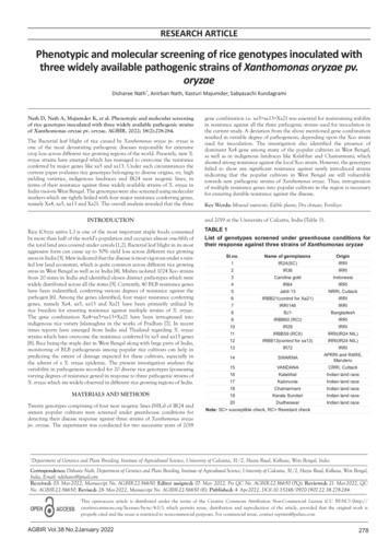
He et al. BMC Cancer(2022) 22:77621–1, CEA) were obtained by blood samples drawn fromveins. The patient’s HPV-DNA test was carried out usingcervical secretions and exfoliated cells of the cervix. Thespecific method was as follows: a disposable cervicalsampler special cervical brush was placed in the cervicalopening, rotated 5 times clockwise, put into 2 ml cell special preservation solution, fully rinsed, and then brokenalong the crease of the brush handle, leaving the brushhead for examination.The clinical symptoms were assessed as follows: contact bleeding, irregular vaginal bleeding, asymptomaticor other. For the HPV risk subtypes, high risk refers toHPV negative or only HPV alpha 7 positive; medium riskrefers to both HPV alpha 7 and HPV alpha 9 being positive; and low risk refers to positive only for HPV alpha 9or other HPV genotypes [18, 19].Statistical analysisStatistical analysis was performed using SPSS statistical software version 22.0. The measurement data conforming to a normal distribution are presented as themean standard deviation (SD), and the comparisonbetween the two groups was performed by two independent sample t tests. If not normally distributed, themeasurement data are expressed as the median (interquartile range), and significant differences between twogroups were analysed using the Mann–Whitney U test.The qualitative data are represented as n (%) and werecompared using the chi-square (χ2) test or Fisher’s exacttest. Spearman correlation analysis was used for correlation analysis. Independent predictors of cervical squamous carcinoma and adenocarcinoma were analysed bybinary logistic regression. The Hosmer–Lemeshow testwas used to analyse the goodness-of-fit of the model.Receiver operating characteristic (ROC) curves and calibration curves were drawn to evaluate the prediction efficiency of the model. P 0.05 was considered statisticallysignificant.ResultsDistribution of clinical indicators and HPV subtypesin squamous cell carcinoma and adenocarcinomaThe levels of CA125 and CA19-9 in the cervical adenocarcinoma group were higher than those in the squamous cell carcinoma group, while the level of CYFRA21–1 was lower, and the differences were statisticallyPage 4 of 10significant (P 0.05). The SCCAg level was significantlyhigher in cervical squamous cell carcinoma than in adenocarcinoma, and the difference was statistically significant (P 0.001). The main clinical symptoms of thecervical squamous cell carcinoma group were contactbleeding (54.2%), followed by irregular vaginal bleeding(27.7%). The clinical symptoms of the adenocarcinomagroup were mainly contact bleeding (45.7%), followed byasymptomatic or other symptoms (32.1%), and there wasa significant difference between the two groups (P 0.05).Regarding the HPV risk subtypes, the high-risk subtype(61.7%) was more common in the adenocarcinoma group,while the low-risk subtype (86.1%) was more commonin the squamous cell carcinoma group, and there was asignificant difference between the two groups (P 0.05)(Fig. 1). HPV-negative patients accounted for 22.2% ofpatients with cervical adenocarcinoma and only 3.8% ofpatients with squamous cell carcinoma. Except for theabove indicators, there was no significant difference inother indicators between the two groups (P 0.05). Thedistribution of clinical indicators and HPV risk subtypesbetween squamous cell carcinoma and adenocarcinomais shown in Table 2.Independent Predictor Analysis of Squamous CellCarcinoma and AdenocarcinomaBinary logistic regression analysis was performed usingthe significantly different indicators between cervicalsquamous cell carcinoma and adenocarcinoma combinedwith clinical experience indicators as independent variables and the classification of the two as dependent variables. It was found that SCCAg (P 0.009, OR 0.671,95% CI 0.497–0.905) and HPV risk subtypes (P 0.001,OR 19.722, 95% CI 9.252–42.040) were independentpredictors for distinguishing cervical squamous cell carcinoma from adenocarcinoma. For each unit increase inthe SCCAg level, the risk of the cervical cancer subtypebeing adenocarcinoma decreased by 0.671 times. Patientsinfected with high-risk HPV subtypes were 19.722 timesmore likely to develop adenocarcinoma of cervical cancer than those infected with low-risk HPV subtypes. Thedetails are shown in Table 3.Evaluation of model performanceAccording to the multivariate analysis of HPV risk subtypes and clinical indicators, a clinical classification(See figure on next page.)Fig. 1 Split violin plot of the differences between cervical squamous cell carcinoma and adenocarcinoma in A SCCAg (Mann–Whitney Utest; P 0.001), B CA125 (Mann–Whitney U test; P 0.031), C CA199 (Mann–Whitney U test; P 0.030), D CYFRA 21–1 (Mann–Whitney U test;P 0.023). The solid line represents the median, the dashed line represents the interquartile range, and the violin shape is the probability densityestimated based on nuclear density in the split violin plot; Stacked histogram of the differences between cervical squamous cell carcinoma andadenocarcinoma in E clinical symptoms (chi-square test; P 0.030), F HPV subtypes (chi-square test; P 0.001)
He et al. BMC Cancer(2022) 22:776Fig. 1 (See legend on previous page.)Page 5 of 10
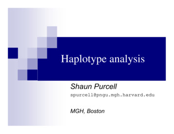
He et al. BMC Cancer(2022) 22:776Page 6 of 10Table 2 Comparison of clinical indicators and HPV subtypesbetween SCC and ACClinical IndicatorsSCC (n 238)AC (n LT209(173.00,241.57) 208(179.16,242.50) 0.959SCCAg1.5(0.9,2.9)1(0.7,1.6) trophil Percentage 58.1(53.00,63.50)57.8(51.85,64.35)0.913Lymphocyte Percentage 31.45(27.10,36.43)31(25.20,35.70)0.349CYFRA 124(52.1%)33(40.7%)Menopausal Status0.077Smoking )0.030*Clinical SymptomsAsymptomatic orOtherContact BleedingIrregular %)66(27.7%)18(22.2%) 0.001*HPV Subtypes*Low Risk205(86.1%)28(34.6%)Medium Risk13(5.5%)3(3.7%)High Risk20(8.4%)50(61.7%)A p value of 0.05 was considered to indicate significant differencemodel mainly based on SCCAg and HPV risk subtypeswas established. The nonparametric Hosmer–Lemeshow test results (P 0.963) indicated that the model hada high goodness of fit. A ROC curve was drawn for theclassification model of cervical squamous cell carcinomaand adenocarcinoma. The area under the curve (AUC)was 0.854 (95% CI: 0.804–0.904, P 0.001), and the modelaccuracy rate was 0.846. The prediction probability underthe maximum Youden index (0.59) was taken as the cutoff value (cut-off 0.277), and the sensitivity and specificity of the model were 0.691 and 0.899, respectively,as shown in Fig. 2. The calibration curve graph showedthat the calibration curve was close to the ideal 45 curve,which indicated that the model had good calibrationcapabilities, as shown in Fig. 3. The data of cervical cancer patients from January 2021 to September 2021 wereused for model validation, and the classification accuracy was 0.823, indicating that the model was stable andreproducible.DiscussionIn current study, the age and distribution proportion ofhistological subtypes of the study population were consistent with previous studies and known knowledge [2].In our study, the most common clinical symptom ofcervical squamous cell carcinoma and adenocarcinomawas contact bleeding, while asymptomatic or otherrare symptoms mostly occurred in adenocarcinoma,which was completely consistent with previous literature reports [8]. At the same time, the results showedthat there were no significant differences between cervical squamous cell carcinoma and adenocarcinoma inage, gravidity, parity, BMI, menopause, smoking history,or routine inflammatory indicators (RBC, WBC, platelets, neutrophil percentage, and lymphocyte percentage)Table 3 Independent predictor analysis of SCC and ACVariableBS.EWaldPOR95% CI for pausal 0.2460.304–1.439Clinical SymptomsContact Bleeding-0.4130.3961.0870.2970.661Irregular Vaginal 163.5390.0601.0300.999–1.063CYFRA 0030.0100.0890.7651.0030.983–1.02459.676 0.001*HPV*Medium Risk0.8950.7241.5260.2172.4450.592–10.119High Risk2.9820.38659.613 0.001*19.7229.252–42.040A p value of 0.05 was considered to indicate significant difference
He et al. BMC Cancer(2022) 22:776Page 7 of 10Fig. 2 Receiver operating characteristic (ROC) curve analysis showed the effect of SCCAg combined with HPV subtypes on the classification ofcervical squamous cell carcinoma and adenocarcinoma. The area under the curve (AUC) was 0.854 (95% CI: 0.804–0.904, P 0.001)Fig. 3 Calibration curve of the established model. It depicts the agreement between the model-predicted classification outcomes and the actualobserved classification outcomes. The diagonal dotted line represents a perfect prediction by an ideal model. The red solid line represents theperformance of the model, of which a closer fit to the diagonal dotted line represents a better prediction(P 0.05). Among them, there was no significant difference in parity or BMI between cervical squamous cellcarcinoma and adenocarcinoma, which was consistent with previous research results [29, 30]. However,previous studies suggested that smoking was significantly associated with the risk of cervical squamous cellcarcinoma (RR 1.50) but not with the risk of adenocarcinoma (RR 0.86) [30]. We considered that the samplesize of the smoking group in this study was too small tocompare the difference in smoking history between cervical squamous cell carcinoma and adenocarcinoma.In addition, there was no significant difference between
He et al. BMC Cancer(2022) 22:776squamous cell carcinoma and adenocarcinoma in theseclinical and conventional inflammatory indicators. Weconsidered that cervical squamous cell carcinoma andadenocarcinoma are both malignant tumour subtypes ofepithelial origin, and the application of conventional clinical and inflammatory indicators in the classification oftumour subtypes is limited and cannot be used for theirdifferentiation [28, 31].In the study of tumour markers, we jointly studiedSCCAg, CA125, CA19-9, CYFRA 21–1 and CEA. Theresults showed that the levels of CA125 and CA19-9 incervical adenocarcinoma were higher than those in squamous cell carcinoma, while the level of SCCAg in cervicalsquamous cell carcinoma was higher than that in adenocarcinoma; the difference was statistically significant(P 0.05), which was consistent with previous researchresults [26–28]. There was no significant difference inCEA between them (P 0.084), which was consistentwith previous research results [27]. Meanwhile, the levelof CYFRA 21–1 in cervical squamous cell carcinomawas higher than that in adenocarcinoma, and the difference was statistically significant (P 0.05). This resultmay be because CYFRA 21–1 is a product of cytokeratin19, which is mainly distributed in squamous and monolayer epithelial cells [32]. Multivariate analysis furtherconfirmed that SCCAg was an independent predictorof cervical squamous cell carcinoma and adenocarcinoma (P 0.009, OR 0.671, 95% CI 0.497–0.905).SCCAg is a subcomponent of TA-4 extracted from cervical squamous cell carcinoma, and its serum level can beused as one of the auxiliary indicators for the diagnosis,efficacy evaluation and prognosis prediction of cervicalsquamous cell carcinoma [23–25, 33–35]. Therefore, webelieve that serum SCCAg levels can play a good role inthe differentiation of cervical squamous cell carcinomaand adenocarcinoma.In the correlation study of HPV risk subtypes and theidentification of cervical squamous cell carcinoma andadenocarcinoma, the results of this study showed thatthe HPV-negative rate of cervical adenocarcinoma wasapproximately 22.2%, while that of cervical squamouscell carcinoma was approximately 3.8%. This is consistentwith the results in previous studies that approximately20%-30% of patients with cervical adenocarcinoma wereHPV negative, while only approximately 5% of patientswith cervical squamous cell carcinoma were HPV negative [36–38]. Moreover, cervical cancer patients persistently infected with high-risk HPV subtypes tended tohave adenocarcinoma, while cervical cancer patients persistently infected with low-risk HPV subtypes tended tohave squamous cell carcinoma (P 0.001). That is, cervical adenocarcinoma is more likely to show no HPV infection or only HPV alpha 7 (HPV 18, 39, 45, 59, 68 and 70)Page 8 of 10positivity, whereas cervical squamous cell carcinoma ismore likely to show only HPV alpha 9 (HPV 16, 31, 33,35, 52, 58 and 67) positivity or positivity of other HPVgenotypes. This result may be because HPV 18, as themost common genotype of HPV alpha 7, is most associated with cervical adenocarcinoma, while HPV 16, as themost common genotype of HPV alpha 9, is closely relatedto cervical squamous cell carcinoma [39, 40]. In furthermultivariate analysis, we found that the HPV risk subtype was also an independent predictor for differentiatingcervical squamous cell carcinoma from adenocarcinoma.Compared with patients infected with low-risk HPVsubtypes, patients infected with high-risk HPV subtypeswere approximately 19 times more likely to develop adenocarcinoma (P 0.001, OR 19.722, 95% CI 9.252–42.040). Patients with high-risk cervical cancer whoare HPV negative or only infected with HPV alpha 7have a worse clinical prognosis [18, 19, 41]. Our resultsshowed that the histological type of these cervical cancerpatients was more prone to being adenocarcinoma. Thisis consistent with the clinical characteristics of adenocarcinoma, with more aggressiveness, insensitivity toradiotherapy and chemotherapy, a higher metastasis rate,poorer prognosis and a lower survival rate comparedwith the same period of squamous cell carcinoma [3–9].Therefore, we believe that HPV risk subtypes have goodclassification ability between cervical squamous cell carcinoma and adenocarcinoma.At the same time, this study established a clinical classification model for the differential diagnosis of cervicalsquamous cell carcinoma and adenocarcinoma based onSCCAg and HPV risk subtypes, and the ROC curve wasdrawn. The AUC of the model was 0.854 (95% CI: 0.804–0.904, P 0.001), and the model accuracy was 0.846. Theprediction probability under the maximum Youden indexwas taken as the cut-off value, and the sensitivity andspecificity of the model were 0.691 and 0.899, respectively. Nonparametric Hosmer–Lemeshow test results(P 0.963) indicated that the model had a high goodness of fit, suggesting that the classification model hadgood differentiation and calibration abilities and coulddistinguish cervical squamous cell carcinoma and adenocarcinoma well. In addition, the data of cervical cancerpatients from January 2021 to September 2021 were usedfor model validation, and the classification accuracy was0.823, indicating the good stability and reproducibility ofthe model.Of course, this study also has some limitations. (1)As a retrospective study, this study has a certain selection bias. For example, the sample size of the smokinggroup was small, which makes it difficult to evaluate the difference in smoking history between cervical squamous cell carcinoma and adenocarcinoma.
He et al. BMC Cancer(2022) 22:776(2) Imaging features were not included in this study,and we only explored the differences in clinical features and laboratory parameters between cervicalsquamous cell carcinoma and adenocarcinoma. In thefuture, we will combine imaging features for furtherresearch.In conclusion, HPV risk subtypes and SCCAg areindependent predictors for differentiating cervicalsquamous cell carcinoma from adenocarcinoma andcan play a good role in classification. The histologicaltype of cervical cancer patients with persistent infection of high-risk HPV subtypes and low serum SCCAglevels is more prone to being adenocarcinoma, whilethe histological type of cervical cancer patients withpersistent infection of low-risk HPV subtypes and highserum SCCAg levels tends to be squamous cell carcinoma. Clinical attention should be given to the occurrence and development of cervical adenocarcinoma,and early intervention should be given to improve theprognosis and survival of patients.AbbreviationsAC: Adenocarcinoma; SCC: Squamous cell carcinoma; HPV: Human papillomavirus; SCCAg: Squamous cell carcinoma antigen; CA125: Carbohydrateantigen 125; CA19-9: Carbohydrate antigen 19–9; BMI: Body mass index; CEA:Carcinoembryonic antigen; RBC: Red blood cell count; WBC: White blood cellcount; PLT: Platelets; OR: Odds ratio; ROC: Receiver operating characteristic;AUC : The area under the curve.AcknowledgementsNot applicable.Authors’ contributionsZMH, FJL and ZBX conceived and designed the study. ZMH, RSC and CWLcollected patient data. ZMH, SYH and YJZ analysed and interpreted the patientdata. ZMH was a major contributor in writing the manuscript. YL, FJL and ZBXreviewed the manuscript. All authors read and approved
The value of HPV genotypes combined . Table 3 Independent predictor analysis of SCC and AC * A p value of 0.05 was considered to indicate signicant dierence . The study was approved by the Medical Ethics Committee of the First Ali-ated Hospital of Chongqing Medical University (No.2021-395), the study .