Transcription
ARTICLE IN PRESSVision Improvement in Retinal Degeneration Patientsby Implantation of Retina Together with RetinalPigment EpitheliumNORMAN D. RADTKE, ROBERT B. ARAMANT, HEYWOOD M. PETRY, PARKE T. GREEN,DIANE J. PIDWELL, AND MAGDALENE J. SEILER PURPOSE:To demonstrate efficacy and safety of theimplantation of neural retinal progenitor cell layers(sheets) with its retinal pigment epithelium (RPE) inretinitis pigmentosa (RP) and dry age-related maculardegeneration (AMD) patients with 20/200 or worsevision in the surgery eye. DESIGN: Interventional nonrandomized clinical trial. METHODS: Ten patients (six RP, four AMD) receivedretinal implants in one eye and were followed in a phase IItrial conducted in a clinical practice setting. Early Treatment Diabetic Retinopathy Study (EDTRS) was the primary outcome measure. All implant recipients and nine of10 tissue donors were deoxyribonucleic acids typed. RESULTS: Seven patients (three RP, four AMD) showedimproved EDTRS visual acuity (VA) scores. Three ofthese patients (one RP, two AMD) showed improvement inboth eyes to the same extent. Vision in one RP patientremained the same, while vision in two RP patients decreased. One RP patient has maintained an improvement invision from 20/800 to 20/200 ETDRS for more than fiveyears; at the six-year examination, it was still maintained at20/320 while the nonsurgery eye had deteriorated to handmotion vision. This patient also showed a 22.72% increasein light sensitivity at five years compared to microperimetryresults at two years; the other patients showed no improvedsensitivity. Although no match was found between donorsand recipients, no rejection of the implanted tissue wasobserved clinically. CONCLUSIONS: Seven (70%) of 10 patients showedimproved VA. This outcome provides clinical evidence ofthe safety and beneficial effect of retinal implants andcorroborates results in animal models of retinal degeneration. (Am J Ophthalmol 2008;xx:xxx. 2008 byElsevier Inc. All rights reserved.)See accompanying Editorial on page xxx.Accepted for publication Apr 7, 2008.From the Retina Vitreous Resource Center (N.D.R.), AnatomicalSciences and Neurobiology (R.B.A.), Ophthalmology andVisual Sciences(R.B.A., M.J.S.), and Psychological and Brain Sciences (H.M.P.), University of Louisville, Louisville, Kentucky; Doheny Retina Institute(R.B.A., M.J.S.), Department of Ophthalmology, University of SouthernCalifornia, Los Angeles, California; Anatomy and Neurobiology (R.B.A.,M.J.S.), UC Irvine, Irvine, California; Nidek Inc, Fremont, California(P.T.G.); Jewish Hospital, Louisville, Kentucky (D.J.P.); and Transplantation Center, The Cleveland Clinic, Cleveland, Ohio (D.J.P.).Inquiries to Norman D. Radtke, Retina Vitreous Resource Center, 240Audubon Medical Plaza, Louisville, KY 40217; e-mail: nradtke@rvrc.com0002-9394/08/ 34.00doi:10.1016/j.ajo.2008.04.009 2008 BYRETINITIS PIGMENTOSA (RP) DENOTES A GROUP OFinherited disorders that involve progressive loss ofphotoreceptors or retinal pigment epithelium (RPE)1and progressive visual impairments. Age-related maculardegeneration (AMD) is the leading cause of blindness inthe population older than 55 in the United States2 andWestern Europe. Geographic atrophy (GA) of the RPEis a form of advanced AMD that is associated withcentral vision loss,2 progresses gradually, and does notregress over time.3In each of these degenerative disorders, if the damagedphotoreceptors can be replaced with new cells that connect with the remaining host retina,4,5 it may be possibleto restore visual function. Human fetal donor tissue transplanted to athymic nude rats can develop retinal layers incontact with a monolayer of cotransplanted RPE.6 Transplants of fetal retinal layers have also been shown torestore visual function in the brain in several models ofretinal degeneration7,8 and to make synaptic connectionswith the neural circuitry of the host retina.9 After injecting freshly harvested retinal progenitor cells in the rho / mouse, increased ganglion cell responses and pupillaryreflexes have been interpreted as visual improvements,10similar to previous results by other groups.11,12 However,the reliability of ganglion cell recordings and pupillometryfor establishing transplant effects has been questioned.13,14Limited clinical trials of retinal implantation have beenunderway since 1998. Between January 1, 1998 and January 1, 2001, phase I of a clinical trial was performed todemonstrate the safety of implanting freshly harvestedlayers of fetal retina together with its RPE. Results showedthe safety of the procedure.15The Food and Drug Administration (FDA) then allowedthe surgery to be performed in patients with 20/200 vision orworse in the surgery eye. A phase II trial was conductedbetween 2002 and 2005 with a group of 10 patients (six RPand four AMD). This article presents the results.METHODS STUDY DESIGN:The study was performed in a clinicalpractice setting. Subjects had 1) decreased central visualacuity (VA) of 20/200 or worse by Early TreatmentELSEVIER INC. ALLRIGHTS RESERVED.1
ARTICLE IN PRESSweek. Since visual stimulation during development isimportant for the formation of synaptic connectivity (review19), these same patients were also asked to watchexperimental visual stimulation videos on a volunteerbasis. The videos consisted of moving dots and stripes withdifferent colors and were developed by a member of thisresearch team, who discussed their design with a lowvision specialist, a psychophysics specialist, and severalpatients.Diabetic Retinopathy Study (ETDRS) vision testing for atleast one year before the procedure in the surgery eye, 2) adiagnosis of RP or AMD, and 3) better vision in thenonsurgery eye than the surgery eye. There was no controlgroup. Success was defined as preservation or improvementof vision after one year as measured by EDTRS.Subjects older than 21 years of age were recruited fromDr Radtke’s private practice or via the practice’s website.All applicants met strict inclusion/exclusion criteria. Qualified subjects were given detailed information about thestudy and informed consent was obtained. The WesternInstitutional Review Board (IRB), FDA, or NortonHealthcare Research Office did not require a data andsafety monitoring committee for this study, so one was notappointed. The Investigational New Drug Application(IND) number was very stringent and subject to close FDAscrutiny. IMPLANTATION METHOD AND SURGICAL PROCEDURE: The dissection of fetal retina together with its RPE IMMUNOLOGY TESTS: All retinal implant recipientsand nine of the 10 donors were deoxyribonucleic acids(DNA) typed for major histocompatibility complex(MHC) antigens HLA-A, B, C, DR, and DQ at JewishHospital (Louisville, Kentucky, USA). Recipients weretyped after signing the consent form, well in advance ofthe transplant surgery. One donor could not be typedbecause of poor tissue quality. The donor DNA was extractedusing a DNA tissue extraction kit (QIAGEN, Valencia,California, USA) and typed for HLA antigens by polymerasechain reaction amplification using sequence-specific primers(Pel Freeze, Brown Deer, Wisconsin, USA).At each recipient follow-up visit, a blood sample wastested for donor-specific antibodies. Anti-HLA antibodieswere detected using two techniques with sensitive flowcytometric procedures: a pool of normal T cells and HLAantigen– coated beads (One Lambda, Canoga Park, California, USA).has been described previously.6,15 Eyeballs were dissectedfree from surrounding tissues and incubated in dispase(Collaborative Biomedical Products, Bedford, Massachusetts, USA) for 10 to 30 minutes. The final dissection wasperformed in the surgery room.A standard three-port 20-gauge vitrectomy was performed, excising the posterior hyaloid face in all cases.Immediately before implantation, 2-mm2 to 5-mm2 piecesof fetal retina with its RPE were cut (average 3.8 mm2)under a dissection microscope and loaded into a custommade implantation instrument with a flat plastic nozzle tipat a 130-degree angle.15,16 The implantation instrumentmaintained the orientation of the donor tissue. After aretinotomy superior nasal to the fovea, the surgeon inserted the loaded nozzle tip into the subretinal space in themacular area and then released the nozzle, which placedprepared sheets of retina/RPE into the target area. Theretinotomy was sealed by laser. Surgery success was definedas good placement of the tissue under the fovea withcompletely flat recipient retina and good sealing of theretinotomy superior nasal to the fovea. Immediately afterimplantation, the medium in which the donor tissue hadbeen prepared was tested for sterility and endotoxins.No immunosuppressant drugs were given. Six patients(Patients 1, 6, 7, 8, 9, and 10) received oxygen postoperatively to reduce surgical trauma.17,18 Patient 1 was on aventilator postoperatively and received oxygen for 10 days;the other patients received nasal cannula oxygen for one STUDY EVALUATION METHODS: All patients were followed for one year and offered annual follow-up examinations. Four patients elected to participate in these optionalannual exams (Patient 3 for two years, Patients 4 and 5 forthree years, and Patient 1 for six years). All patientsreceived a clinical examination, funduscopy, and fluorescein angiography (FA); the nonsurgery eye was also examined postoperatively to rule out sympathetic ophthalmia ateach examination time point (three times preoperatively;and one week and one, three, six, nine, and 12 monthspostoperatively). A complete refraction was performed oneach patient before VA was measured by EDTRS. Allpatients also had their lens removed and had an intraocular lens implant in the posterior chamber with the capsuleopened preoperatively.Early Treatment Diabetic Retinopathy Study vision wastested three times prior to surgery and at least seven timespostoperatively using a previously described protocol,20with an ESV-3000 Illuminator cabinet (Vector Vision,Greenville, Ohio, USA; test luminance 85 cd/m2). Testingwas performed at 4 m; measurements at 1 m were performed when fewer than 15 letters were read at 4 m. Thetotal ETDRS letters correctly identified were counted untilone line (5 letters) was completely missed. The line abovewas then converted to Snellen acuity by measuring thedistance of the patient from the chart, whether 4 m or 1 m.This value was divided into 20 and multiplied by the meter DONOR TISSUE: Donor tissues were derived from human fetal eyes, age range 10 to 15 weeks gestation, isolatedfrom dead macerated fetuses after elective abortions. Donors were not compensated or approached to donate tissueuntil after the decision was made to terminate the pregnancy. All donors signed an informed consent form priorto donation.2AMERICAN JOURNALOFOPHTHALMOLOGYMONTH 2008
ARTICLE IN PRESSFIGURE 1. Study Patient 8 with retinitis pigmentosa (RP). The left column shows preoperative fundus images and the rightcolumn shows postoperative images. Position of the transplant is indicated by dotted rectangle. (Top left) Preoperative fundus. (Topright) Fundus at one year postoperative. (Top, small image insert) Fundus at one month postoperative with the position of thepigmented transplant indicated (arrow). (Middle left) Early fluorescein angiograph (FA), preoperative. (Middle right) Early FA, oneyear postoperative. (Bottom left) Late FA, preoperative. (Bottom right) Late FA, one year postoperative. The fundus images andFA document the presence of choriocapillaris underneath the transplant with no leakage. This implies evidence of no clinicalrejection. Pre-op ⴝ preoperative; Post-op ⴝ postoperative.number next to each line on the left side of the chart. Forexample, if the patient reads the 40-m line and is 4 maway, the vision is 20/200 by Snellen. ETDRS tests wereperformed in the clinical practice of Dr Radtke and at anindependent test site (Patients 1, 3, 4, and 5 were testedonly in Dr Radtke’s practice). Testers were masked to thesurgery eye. If there was a variation between test sites, theresults from the independent site were reported.Secondary evaluation methods used at the beginning ofthe study were scanning laser ophthalmoscopy (SLO) andVOL. XX, NO. Xmultifocal electroretinography (mfERG). As the studyprogressed and new imaging technology became available,optical coherence tomography (OCT) and microperimetry(MP1) replaced the SLO and mfERG methods. MP1 testswere performed in the clinical practice of Dr Radtke. OCTtests were done in Louisville, Kentucky, at the clinicalpractices of Drs Radtke and Kumar. SLO and OCT testswere also performed at the University of Medicine andDentistry of New Jersey (UMDNJ), Newark, New Jersey,and mfERG tests at The Smith-Kettlewell Eye ResearchRETINAL IMPLANTATIONINHUMANS3
ARTICLE IN PRESSFIGURE 2. Study Patient 7 with age-related macular degeneration. The left column shows preoperative fundus images and the rightcolumn shows postoperative images. Position of the transplant is indicated by dotted rectangle. (Top left) Preoperative fundus. (Topright) Fundus at one year postoperative. (Top, small image insert) Fundus at one month postoperative with the position of thepigmented transplant indicated (arrow). (Middle left) Early FA, preoperative. (Middle right) Early FA, one year postoperative.(Bottom left) Late FA, preoperative. (Bottom right) Late FA, one year postoperative. The fundus images and FA document thepresence of choriocapillaris underneath the transplant with no leakage. This implies evidence of no clinical rejection.dent test sites for SLO and mfERG tests. These twoevaluation methods were discontinued as full retina MP1testing proved more accurate.As stated above, OCT and MP1 tests were introducedlater in the study. OCT tests were performed preoperatively and postoperatively on two RP patients and twoAMD patients, and postoperatively on one AMD patient.MP1 tests with fixation and sensitivity (MP1; NidekAdvanced Vision Information System (NAVIS); NidekTechnologies, Vigonza, Italy) were performed on threepatients preoperatively (one RP and two AMD) and fivepatients postoperatively (two RP and three AMD). MP1continuously registers fixation behavior by automated real-Institute, San Francisco, California. Again, testers weremasked to the identity of the surgery eye.Scanning laser ophthalmoscopy tests with a small, dimstimulus were performed on three RP patients preoperatively and one AMD and two RP patients postoperatively.The reference cross was fixed by manual tracking at clearlydefined vascular landmarks.21 Photopic mfERGs were recorded (VERIS Science software; EDI Inc, San Mateo,California, USA) as previously described.22 These recordings were performed three times preoperatively on fourpatients (two RP and two AMD) and five times postoperatively on three patients (two RP and one AMD). Severalsubjects were unable to travel the distance to the indepen4AMERICAN JOURNALOFOPHTHALMOLOGYMONTH 2008
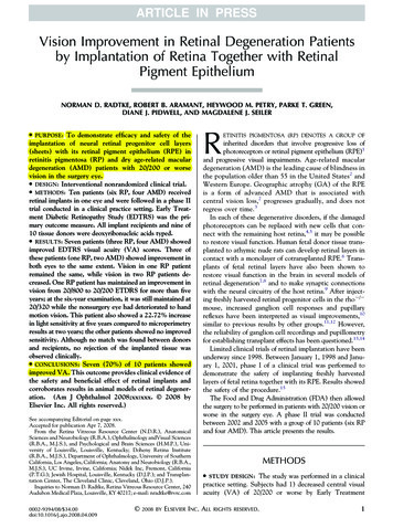
ARTICLE IN PRESSFIGURE 3. Study Patient 8 with RP. This optical coherencetomography of implant at one month (Top) and one yearpostoperatively (Bottom) shows the area (arrows) with thickened retina indicating implant. The retinal pigment epitheliumand choroid are highly reflective (red).time fundus tracking and alignment21 and digitally registers the infrared photograph from the current to theprevious test, allowing use of the same pattern and preciseplacement of the pattern’s previous location.RESULTS IMPLANT SURVIVAL:No rejection of the implantedtissue was observed clinically. No leakage was observed onFAs, suggesting that the implanted RPE cells could havereestablished a new blood-retinal barrier. Figures 1 and 2show examples of RP Patient 8 and AMD Patient 7,respectively. The pigmentation of the transplant was lostin eight of 10 cases at three months postoperatively. Figure3 shows OCTs of Patient 8 at one month and one yearpostoperatively. IMMUNOLOGY TESTS:All typed donor-recipient pairshad at least one antigen mismatch at each of the A, B, andDR loci (Table 1). These mismatches indicate that thegrafts had the potential to be recognized as foreign by therecipient’s immune system.Recipients were tested for anti-HLA antibody preimplant and postimplant. Four female recipients (Patients 1and 3, RP; Patients 10 and 6, AMD) had demonstrableanti-HLA antibody preimplant either from exposurethrough pregnancy or via blood transfusion. Several ofthese antibodies were specific for HLA antigens present onthe retinal grafts but no new donor-specific antibodydeveloped throughout the follow-up period. Patient 2 hada negative antibody screen pretransplant and developed anantibody-like reactivity at 10 months postimplant that wasentirely absent in the subsequent sample at 15 monthspostimplant. The anti-HLA specificity of this reactivityVOL. XX, NO. Xcould not be defined because of nonspecific reactivity ofthis patient’s serum with the latex beads used in the assay.In Patients 1 and 10, the antibody titers increased at nine and12 months postimplant, respectively. However, the HLAspecificity did not change, indicating that no de novoanti– donor-specific antibody developed. In Patient 1, theantibody titer returned to preimplant levels in a samplecollected three months after the apparent increase in titer.For Patient 10, the increase in titer was detected in thelast sample collected and no subsequent samples wereavailable to follow the titer. In this case, as was true forall patients with reactivity in the antibody screeningassays, the specificity of the antibodies present did notchange during the increase in titer. In all cases, theincrease was not restricted to the antibody specific tothe donor antigen but all specificities increased simultaneously. This pattern of activation may be seen incases of infection, vaccination, or generalized immuneactivation and is not typical of a donor-specific response, although this cannot be entirely excluded.Patient 6 had weak antibody but the titer never increased over the two-year time period tested. With theexception of Patient 2, no de novo donor-specificantibody was detected postimplant. Anti-HLA antibodydetected preimplant persisted postimplant in all cases.With the exception of Patient 2, whose anti-HLAspecificity could not be defined, there were no instanceswhere a patient developed de novo antibody that couldbe demonstrated to be specific for an HLA antigenpresent in their donor. Interestingly, all five patientswith preimplant anti-HLA antibody had visual improvement (Tables 2 and 3). EARLY TREATMENT DIABETIC RETINOPATHY STUDYRESULTS: Tables 2 and 3 present the results of ETDRStesting. All subjects were evaluated three times preoperativelyand at least seven times postoperatively. Four patients electedto participate in optional annual follow-up exams (Patient 1for six years; Patients 4 and 5 for three years, Patient 3 for twoyears). Clinical evidence of visual improvement by ETDRSwas seen in seven of 10 patients (three RP, four AMD). InPatients 1 and 3 (RP) and 5, 6, and 7 (AMD), the vision inthe surgery eye improved, whereas the vision in the nonsurgery eye did not change significantly or worsened. Patients 2(RP) and 10 (AMD) showed vision improvement in botheyes to the same extent. Patients 4 and 9 (RP) showed adecrease in vision, and Patient 8 (RP) remained the same.Patient 1 (RP) has maintained a previously reported23 visionimprovement of 20/800 to 20/200 ETDRS for five years; atthe six-year examination it was still maintained at 20/320while the nonsurgery eye had deteriorated to hand motionvision (see Table 2). OTHER RESULTS:No changes were observed in themfERG tests. The SLO tests showed an improvement in VAin Patient 1 between nine months and 27 months postoper-RETINAL IMPLANTATIONINHUMANS5
ARTICLE IN PRESSTABLE 1. Recipient and Donor Human Lymphocyte Antigen Tissue entPatient11 donor22 donor33 donor44 donor55 donor66 donor77 donor88 donor99 donor1010 donorAB2, 31, 3024, 3311, 2, 231, 211, 23, 242, 1, 21, 301, 1, 261, 32, 321, 32, 302, 321, 2914, 2713, 1413, 1444, 4944, 578, 35, 44, 8127, 508, 458, 528, 5139, 577, 577, 447, 5751, 577, 607, 44CBw4/64, 64, 64, 64, 4, 6, 6, 4, 64, 66, 4, 64, 64, 64, 64, 64, 64, 6, 4, 6No type available6, 86, 87, 164, 67, 15, 4, 186, 7, 167, 167, 166, 76, 75, 76, 77, 147, 107, 16DRDR51/52/53DQ1, 1317, 71, 77, 137, 17, 714, 7, 111, 717, 1517, 184, 87, 167, 158, 117, 1511, 154, 157, 15DR52DR52, 53DR53, DR52, 53DR53, DR52, 53DR52, DR52, 53DR53, DR52, 2, 51DR53, 51DR53, 515, 62, 2, 52, 62, 2, 5, 2, 62, 52, 62, 44, 85, 96, 94, 76, 96, 76, 82, 6Bold italic indicates a recipient/donor match. As the data show, donors and recipients are mismatched in most human lymphocyte antigens(HLA). No tissue for HLA testing was available for the LC donor.TABLE 2. Results of Study Patients with Retinitis PigmentosaVision of Surgery Eye (ETDRS)Patient IDand Ageat 6 Months1 Year20/800 OS20/40020/1604.0 (02/02)2.00.9LP OSHMHM6.0 (02/03)5.05.0HM OD20/64020/4005.0 (03/03)3.52.020/640 OD20/64020/8003.5 (10/03)3.54.0LP ODLPLP6.0 (06/05)6.06.020/320 OSLPLP1.8 (12/05)6.06.0Average change in surgeryeye SEMVision of Nonsurgery Eye (ETDRS)Last Result ofPoststudySurveillanceChange @ 1Year (decimal)Pos 3.1Pos 1.0Pos 3.0Neg 0.5NC 0Neg 4.20.4 1.16 years (02/08)20/320 1.81 year (04/04)HM 5.02 years (06/05)CF @ 3 feet3 years (03/07)20/800 4.01 year (10/06)LP 6.01 year (3/07)LP 6.0PreoperativeVision to Date20/400 ODHM2.0 (02/02)5.0 (02/08)LP ODHM6.0 (02/03)5.0 (04/04)20/400 OS20/6402.0 (03/03)3.5 (07/04)20/800 OS20/8004.0 (10/03)4.0 (03/07)20/250 OS20/2001.6 (06/05)1.0 (10/06)20/160 OD20/1600.9 (12/05)0.9 (03/07)Average change innonsurgery eye SEMChange @ 1Year (decimal)NC 0.2Pos 1.0Neg 1.5NC 0Pos 0.6NC 00.05 0.35CF count fingers; ETDRS Early Treatment Diabetic Retinopathy Study; HM hand motion; LP light perception; NC no change;Neg vision deterioration; OD right eye; OS left eye; Pos vision improvement; SEM standard error of mean.For calculation of change in decimal vision values, HM was set as 5.0 and LP was set as 6.0. There is no significant difference in the averagechange between surgery eye and nonsurgery eye, but the standard deviations are significantly different.atively.23 MP1 tests showed no improved sensitivity with theexception of Patient 1 (see Figure 4). Her MP1 results showeda defined area of cluster points just nasal and adjacent to theimplant over which the average sensitivity was calculated to6AMERICAN JOURNALhave increased from 1.6 decibels [dB] (year three postoperatively) to 2.7 dB or 22.72% (year five postoperatively). Thisresult correlates well with her improved ETDRS VA scoreand subjective function.OFOPHTHALMOLOGYMONTH 2008
ARTICLE IN PRESSTABLE 3. Results of Study Patients with Age-Related Macular DegenerationVision of Surgery Eye (ETDRS)Patient IDand Ageat Implant5886*767711076PostoperativePreoperative6 Months1 Year20/640 OD20/24020/4003.6 (02/04)1.42.020/400 OS20/24020/2002.0 (08/05)1.41.020/400 OS20/34020/1602.0 (08/05)1.80.920/400 OS20/32020/2602.0 (03/06)1.81.5Average change in surgeryeye SEMChange @ 1Year (decimal)Pos 1.6Pos 1.0Pos 1.1Pos 0.51.05 0.22Last Result ofPoststudySurveillance3 years (04/07)20/400 2.01 year (10/06)20/200 1.01 year (10/06)20/160 0.91 year (04/07)20/260 1.5Vision of Nonsurgery Eye (ETDRS)PreoperativeVision to Date20/160 OS20/2000.9 (01/03)1.0 (04/07)20/200 OD20/2001.0 (08/05)1.0 (10/06)20/60 OD20/2000.5 (08/05)1.0 (10/06)20/240 OD20/1601.4 (03/06)0.9 (04/07)Average change innonsurgery eye SEMChange @ 1Year (decimal)NC 0.1NC 0Neg 0.5Pos 0.5 0.02 0.2ETDRS Early Treatment Diabetic Retinopathy Study; NC no change; Neg vision deterioration; OD right eye; OS left eye;Pos vision improvement; SEM standard error of mean.Difference between change in surgery eye and change in nonsurgery eye appears significant (P .05); however, too few values forstatistical comparisons.*Patient 6’s preoperative vision was very variable, between 20/260 and 20/640 in the surgery eye (mean, 2.27 0.23) and between 20/200and 20/640 in the nonsurgery eye (mean, 1.89 0.21) (average of 11 different measurements). The other patients did not show suchvariability. The values shown in the table are from the last test before surgery in August 2005.FIGURE 4. Study Patient 1 with RP. Results of microperimetry MP1 testing show retina at three years postimplant (Left) and fiveyears postimplant (Right). A polygon in yellow and green highlights the area of interest and the average sensitivity within that areais calculated. Improvement in sensitivity is evident in the area of the polygon adjacent to the graft area. In the left image, taken inNovember 2004, the mean sensitivity in the polygon area is 1.6 decibels (dB); in the right image, taken in March 2007, the meansensitivity is 2.7 dB. The 22.72% increase in sensitivity of 1.1 dB, from 1.6 dB to 2.7 dB, corresponded well with improved visualacuity by Early Treatment Diabetic Retinopathy Study and the patient’s subjective assessment.DISCUSSIONNO EFFECTIVE TREATMENT EXISTS FOR THE RECOVERY OFvisual loss from RP and AMD. Oral vitamin A therapyhas slowed the rate of electroretinogram loss in RP butVOL. XX, NO. Xcannot recover lost vision.24 Gene and pharmacologictherapies are also being studied. A clinical trial of genetherapy in Leber congenital amaurosis24 is underway,and Neurotech has started phase II clinical trials forciliary neurotrophic factor (CNTF) for RP.25 MultipleRETINAL IMPLANTATIONINHUMANS7
ARTICLE IN PRESScenters26 –28 are actively pursuing development and useof an artificial retina.This study demonstrates visual improvement as measuredby ETDRS in seven of 10 patients. In our study, RPE absenceor presence at one year in the transplant area, genetic typing,gender, or age of patients did not correlate with improvementin vision. Multiple variables such as size of implant, time fromharvest to implantation, duration of surgery, fetus age, ormode of inheritance (autosomal dominant [AD], autosomalrecessive [AR], gender-linked, or sporadic) were not significant in predicting visual outcomes. Without treatment, thelikely outcome was that the vision of all subjects in the studywould have deteriorated further.No surgical complications occurred. The custom-madeimplantation instrument maintains proper orientation ofthe fragile donor tissue and allows the surgeon to gentlyplace the tissue flat, without roll-up, and without any pushor injection pressure and minimal additional fluid. Thiseliminates the requirement for a subretinal fluid bleb andthus minimizes trauma to the implant and host retina.16At more than five years postoperatively, no graft encapsulation, tissue destruction, or macular edema indicatingrejection was seen clinically or with FA on any subjects.However, without histologic data, the presence of moresubtle graft rejection cannot be excluded. Previous reportsof human retinal implantation have varied in the incidence of rejection depending on whether the implantinvolved patches of cultured RPE cells29 or dissociatedcells and whether the patient had exudative or nonexudative manifestations of AMD.Human Lymphocyte Antigen typing of recipients anddonors confirmed that none of the implants were HLAmatched. In solid organ transplant, HLA mismatches at theclass II DR locus are particularly conducive to immunerecognition and rejection episodes.30 In all cases here, therewas at least one DR antigen mismatch, indicating that DRmatching did not contribute to graft survival. Several patientsthat had previously been sensitized to HLA antigens demonstrated increased titers of the antibody they were previouslysensitized to. However, transient increases in antibody titerwithout any new antibody specificity can develop after manymanipulations that have pro-inflammatory effects, such assurgeries or vaccinations.31 Since no patients developed newantibody specificity directed toward their retinal donor, thereis evidence that the patients’ immune systems were notrecognizing the donor antigens and responding to them.Frequently, if patients are re-exposed to an antigen they havebeen sensitized to previously, their titer of antibody willincrease and will persist.32 We did not see that kind ofresponse in any of our patients that demonstrated previoussensitization. With the exception of one patient (Patient 2)whose anti-HLA specificity could not be defined, there wasno de novo donor-specific HLA antibody detected in any ofour patients.It is well documented that intraocular spaces are, to alarge extent, immunoprivileged sites.33 Lack of apparent8AMERICAN JOURNALcell-mediated rejection of the retinal tissue implanted tothe subretinal space likely derives from this immunologicprotection.33 Additionally, the absence of detectable graftdamage from donor-specific antibodies that were present insome patients before implantation would indicate that theblood-brain barrier is prohibiting antibody infiltration intothe subretinal space. Placement of allogeneic tissue intothe anterior chamber of the eye results in a protectiveresponse called anterior chamber–associated immune deviation (ACAID).33 Similar immune deviation could beoccurring in retinal implant recipients, which could explain graft survival despite the presence of HLA-mismatched graft antigens. If regulatory T cells are developingin response to retinal implants, they are not inhibitingmemory B-cell antibody production as all antibody specificities, donor directed and non– donor specific, persistedin all sensitized patients studied here.32In eight of 10 cases, the transplanted RPE cells lost theirpigmentation at three to six months postoperatively, whichcould indicate graft rejection. However, the phenomenon ofpigment
retinitis pigmentosa (RP) and dry age-related macular degeneration (AMD) patients with 20/200 or worse . versity of Louisville, Louisville, Kentucky; Doheny Retina Institute (R.B.A., M.J.S.), Department of Ophthalmology, University of Southern . Audubon Medical Plaza, Louisville, KY 40217; e-mail: nradtke@rvrc.com