Transcription
Living Image 4.4 Software Release NotesNew Features and ImprovementsContents1.Purpose .22.New Features.22.1.Cherenkov Imaging Mode .22.2.Updates for IVIS Lumina Series III System .32.2.1. Illumination Module .32.2.2. Lumina III Filters .32.3.Expanded Probe Database in the Imaging Wizard .42.4.FLIT algorithm improvements .62.5.3D Data Visualization Improvements .62.6.Longitudinal Study window .62.7.Tool Palette reorganization and simplification .72.8.DyCE Trans-illumination imaging.72.9.Acquisition improvements .72.10. Browser displays saturated images .72.11. Perform Smoothing and Binning on a sequence .82.12. Volume Slice Viewer access shortcuts.82.13. 3D ROI enhancements .92.14. Support for Mac OS X 10.8 .92.15. Support for high-speed I/O board with NI-DAQmx driver .103.Improvements .104.Known Issues in Living Image 4.4 .115.System Requirements.126.Video Card Requirements.13
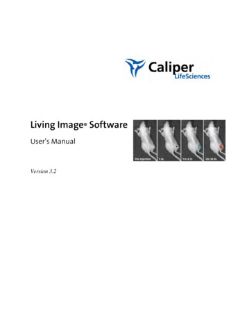
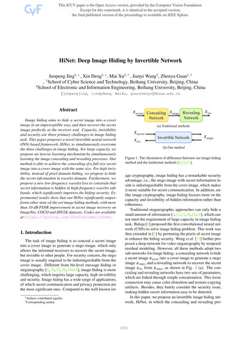
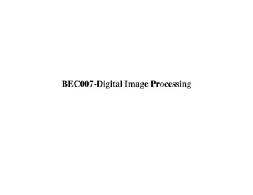
Living Image 4.4 Software Release NotesNew Features and Improvements1. PurposeThis document provides a brief overview of new features and improvements in Living Image 4.4. Thisrelease serves as an update for Living Image 4.3.1 version on IVIS Lumina, Kinetic, Lumina XR, Spectrumand Spectrum CT instruments. Major features include the introduction of the Cherenkov imaging modein the Wizard, integration of the entire PerkinElmer catalogue of fluorescent agents, support for IVISLumina Series III, FLIT algorithm improvements, and tool palette simplification. Living Image 4.4 releasealso provides improvements to the existing software features and bug fixes.2. New Features2.1. Cherenkov Imaging ModeThe Living Image 4.4 Imaging Wizard now offers a Cherenkov Imaging Mode which guides the userthrough optimal camera parameter settings for high signal-to-noise when imaging the Cherenkovlight emitted from radionuclide-injected animal subjects. One can easily set up a bio-distribution scanof radiopharmaceuticals using the Cherenkov DyCE mode. The user can choose from a selection ofradionuclides at acquisition time, so that the data may be decay-corrected if necessary at analysistime, such as for kinetic curves from DyCE analysis. (Note: The Cherenkov emission spectrum is thesame for all radionuclides. One can choose Spectral Unmixing in Cherenkov mode to unmixradionuclides from other light-emitting probes of very different spectra, but one cannot spectrallyunmix to distinguish Cherenkov signal between radionuclides).Figure 1: Imaging Wizard workflow for Cherenkov mode
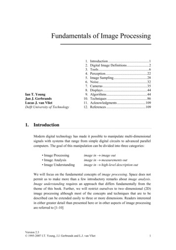
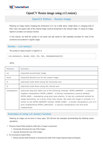
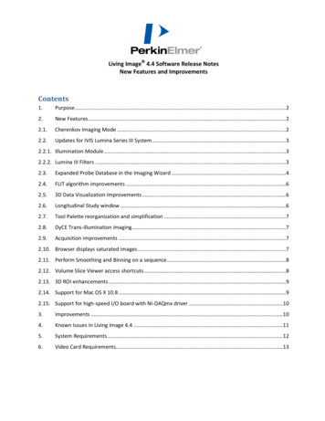
Living Image 4.4 Software Release NotesNew Features and Improvements2.2. Updates for IVIS Lumina Series III System2.2.1. Illumination ModuleThe IVIS Lumina III imaging system contains a new 150W extended range tungsten halogen lamp.This new illumination module gives extended excitation power into the near infraredwavelengths. Figure 2 shows the power increase for the IVIS Lumina III systems at wavelengthsgreater than 700nm. This power increase will yield higher signal-to-noise ratios for fluorescentprobes that are specifically designed to be excited at NIR wavelengths where the autofluorescence from animal tissue is low. The new illumination module is also specifically designedto handle the extra heat caused by the transmission of additional IR light which includes usingspecially designed high temperature fiber optic cables.Figure 2: Comparison of the spectral output for the Lumina III and the Lumina II illumination modules2.2.2. Lumina III FiltersThe Lumina III has a newly designed 22 position excitation filter wheel which is filled with 19narrow band excitation filters and one block for bioluminescent imaging. The excitation filtershave a 20nm band pass and span the wavelength region from 410-790nm. The exciters weredesigned specifically to do spectral unmixing on the excitation side (Table 1). The emission filterswere also redesigned to span the wavelength region of 500-865nm using only one filter wheelassembly thereby eliminating the need for the user to change emission filter wheels. The new setof seven emission filters have a 40nm band pass and hit all major emission peaks for the newlyexpanded probe database (see section 2.3).
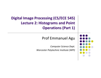
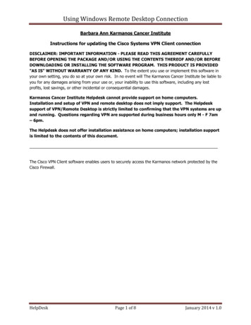
Living Image 4.4 Software Release NotesNew Features and ImprovementsLumina LTExcitation FiltersCenter 7103074530Emission FiltersCenter na IIIExcitation FiltersCenter 78020Emission FiltersCenter lBandwidth52040570406204067040710407904084540Table 1: Description of the fluorescent excitation and emission filters that are currently in the Lumina Series III instruments.Note: the Lumina LT has a different set of excitation and emission filters.2.3. Expanded Probe Database in the Imaging WizardLiving Image now offers an expanded probe database used by the Imaging Wizard. Table 2 is analphabetical list of the 99 fluorescent reporters that are currently in the software. The database isorganized in a tree-like structure in order to simplify the selection of the desired probe (see Figure 3).Each probe is categorized as an agent, dye, protein or quantum dot. If the user is unsure where thereprobe is located there is also an alphabetical listing. If the probe being used is not located in thedatabase, the user can input the peak excitation and emission wavelengths into the software in orderto determine filter selection. Frequently used probes will be saved in the “Recently Used” list.
Living Image 4.4 Software Release NotesNew Features and Improvements1.2-DG 75026. BoneProbe 68051. Integri Sens e 64576. QDot 8052.Al exa Fl uor 48827. Ca tB 680 FAST52. Integri Sens e 68077. Reni nSens e 680 FAST3.Al exa Fl uor 51428. Ca tB 750 FAST53. Integri Sens e 75078. RJ-2-DG-7504.Al exa Fl uor 53229. Ca tK 680 FAST54. i RFP79. Roda mi ne Red5.Al exa Fl uor 54630. COX 255. mCherry80. Superha nce 6806.Al exa Fl uor 55531. Cy356. MMPSens e 645 FAST81. tdToma to7.Al exa Fl uor 56832. Cy557. MMPSens e 68082. Texa s Red8.Al exa Fl uor 59533. Cy5.558. MMPSens e 750 FAST83. TLectinSens e 6809.Al exa Fl uor 61034. Cy759. mPl um84. Tra ns feri Sens e 75010. Al exa Fl uor 63335. DADO60. Neutrophi l El a s tas e 680 FAST 85. TurboFP60211. Al exa Fl uor 64736. Di D61. Os teoSens e 680 EX86. TurboFP63512. Al exa Fl uor 66037. Di R62. Os teoSens e 750 EX87. TurboGFP13. Al exa Fl uor 68038. Ds Red (RFP)63. Os teoSens e 80088. TurboRFP14. Al exa Fl uor 70039. Doxorubi ci n64. PKH2689. TurboYFP15. Al exa Fl uor 75040. eGFP65. ProSens e 68090. Vi vo Ta g 80016. Ami noSPARK 68041. FITC66. ProSens e 750 EX91. Vi voTa g 64517. Ami noSPARK 75042. Fol a teRSens e 68067. ProSens e 750 FAST92. Vi voTa g 68018. Angi oSens e 680 EX43. Ga s troSens e 75068. PSA 750 FAST93. Vi voTa g 75019. Angi oSens e 750 EX44. Genha nce 68069. Qdot 52594. Vi voTa g-S 68020. Angi oSPARK 68045. Genha nce 75070. Qdot 54595. Vi voTra ck 680 (dye)21. Angi oSPARK 75046. GFP71. QDot 56596. XLCF 68022. Annexi n Vi vo 75047. HER2Sens e 64572. QDot 58597. XLCF 75023. Ba cteri a l Detection 75048. Hypoxi Sens e 68073. QDot 60598. XLCF 77024. Ba cteri Sens e 64549. ICG74. QDot 65599. YFP25. Bombes i nRSens e 68050. Integi n 75075. QDot 705Table 2: List of the expanded reporter library that has been incorporated into the Living Image 4.4 softwareFigure 3: Screenshot of the Imaging Wizard showing the organization of the “tree-like” structure for the expanded probe database.
Living Image 4.4 Software Release NotesNew Features and Improvements2.4. FLIT algorithm improvementsArtifacts caused by excitation beam coupling to tissue observed in FLIT reconstructions analyzed byLI4.3.1 and prior versions have been greatly reduced. The reduction of these artifacts allows forgreatly improved interpretation of 3D fluorescence tomographic reconstructions.2.5. 3D Data Visualization ImprovementsFor CT volume, Living Image 4.4 offers improved contrastbetween tissues, higher dynamic range of the scene, andmore realistic lighting on the tissue interfaces. For DLIT andFLIT 3D source data, voxels are now rendered with highervisual contrast, better depth rendering and improved spatialregistration accuracy with CT volume data. Renderingsettings for source voxels are optimized for provided colorpalettes and “Jet” is introduced as the new default colorpalette.2.6. Longitudinal Study windowThe 3D voxel display minimum and maximum color scalevalues can be manually set for improved flexibility in datavisualization. The maximum color scale value is limited to be 100 max(vi), where i 1,.,N for N total voxels of intensity vi.The minimum color scale value is limited to be 0.
Living Image 4.4 Software Release NotesNew Features and Improvements2.7. Tool Palette reorganization and simplificationThe Tool Palette has been reorganized for improved ease of use.The Image Information palette has been retired and its toolsincorporated into the Image Adjust section, which now alsocontains Filtering tools. Additionally, when the Tool Palette isdocked in the main application window, it automatically adjusts todisplay selected tools in full view.2.8. DyCE Trans-illumination imagingDyCE image acquisition and analysis is now available for transillumination fluorescence imaging with single-point illumination,allowing for increased sensitivity of dynamic fluorescence imagingto deep sources. Fluorescence DyCE data acquired with transillumination excitation will be analyzed with DyCE in NTF Efficiencyunits by default. Users will have the choice to analyze the data inRadiant Efficiency units as well.2.9. Acquisition improvementsLiving Image 4.4 software includes the following enhancements tothe data acquisition workflow: Ability to close and save a sequence dataset while another sequence acquisition is in progressTrans-illumination Setup window restricts point selection to rectangle (applies to IVISSpectrum and Spectrum CT)“Mask Grid Points to Subject” option is applied by default in Trans-illumination SetupWindow (applies to IVIS Spectrum and Spectrum CT)A warning caption appears on image thumbnail if loading trans-illumination image datacontaining saturated pixels (applies to IVIS Spectrum and Spectrum CT)Imaging chamber door can be opened earlier during a CT scan while CT image reconstructionis in progress (applies to IVIS Spectrum CT)DyCE sequence setup limits max number of images to 200DyCE time interval begins at the start of first luminescent exposure after the photograph2.10. Browser displays saturated imagesThe Living Image Browser has been improved to indicate images with saturated pixels. A red iconwill alert the user if optical data sequence contains any images with saturated pixels, thoseimages are not recommended for analysis.
Living Image 4.4 Software Release NotesNew Features and Improvements2.11. Perform Smoothing and Binning on a sequenceSmoothing and Binning image operations can now be applied to the entire sequence, as opposed topreviously applicable to individual image only. The tools are available in the Image Adjust tool palettefor sequence and single image modes. Smoothing computes the average signal of a specified numberof pixels (for example, 5 x 5) and replaces the original signal with the average signal. Binning specifiesthe number of pixels in the image data that are grouped together to form a larger pixel.2.12. Volume Slice Viewer access shortcutsWithin the 3D View tab, simply clicking on a 2D plane will now automatically switch the tool paletteto the ‘Slice’ tab to enable quicker adjustment of the grayscale ranges. Double-clicking the Coronal,Sagittal, or Transaxial window pane now automatically launches the Volume Slice Viewer. Then,switch to single slice view or choose to render thick slice, adjust slice thickness, scroll, and zoom in onindividual slice features.
Living Image 4.4 Software Release NotesNew Features and Improvements2.13. 3D ROI enhancementsTo simplify manipulation of an ROI in 3D space, Living Image 4.4 includes the following enhancementsto the 3D ROI tool: ROI position – Instead of defaulting the center of the image, new 3D ROIs are now placed atthe intersection of slice planes.ROI Size - Larger default size for 3D ROI.ROI handles - Larger ROI handles for easier manipulation.ROI translational adjustment - Double-click the ROI in the perspective view to open 3D ROIProperties dialog box for finer adjustments .ROI angular adjustment - Angle parameters in the ROI Properties dialog box provide anotherway to rotate the 3D ROI in the x, y, or z plane.Measurement units for CT images - The units for thresholding a Spectrum CT image are nowin HU, enabling easier comparison between animal and across time points.2.14. Support for Mac OS X 10.8Living Image 4.4 analysis module is compatible with the Mac OS X 10.8 (code-named “MountainLion”). The Living Image 4.4 Installation and Licensing Guide describes the necessary steps to installand license Living Image application on the Mac OS X 10.8.
Living Image 4.4 Software Release NotesNew Features and Improvements2.15. Support for high-speed I/O board with NI-DAQmx driverLiving Image 4.4 provides transparent support for the National Instruments 6535-series high-speedI/O board for use on new or upgraded IVIS Spectrum systems. Systems running with this new boardrequire the installation of both the NI-DAQmx software library (version 9.6.1 or higher) and theMicrosoft .NET Framework 4.0. Installation of NI-DAQmx does not require the removal of the olderNIDAQ libraries.The Living Image software will automatically determine if the new software driver is present andwhich I/O driver to use. The new board must be installed in a PCI Express (PCIx) slot. The software willautomatically determine its location in the computer’s backplane.3. ImprovementsThe following table is a summary of the -5279LI-5263LI-5262LI-5215LI-5616SummaryNew “Large Animal” imaging mode on Lumina XRSupport for IVIS Spectrum LT instrumentLimit Filter Pair mode in the Imaging Wizard to only one pairAllow binning/smoothing operation for spectral unmixing resultsSupport for Lumina III with removable emission filter wheelsPre-defined filter selection table for Spectral Unmixing scan on the IVIS SpectrumPrompt user earlier to acquire new CT reference imagesDecay-correct data for Cerenkov data in Spectral Unmixing/DyCEAllow analysis of NTF Efficiency DyCE imagesWarn user against 3D reconstruction acquisition when FOV D is selectedSupport manual co-registration with Bruker MRTool Palette scrolling improvement'Info' tab is available in 3D View windowDisplay unit and min/max for the color bar in Unmixing tabBrightness controls for "Image Cube" in Spectral UnmixingImprovements to volume masking in surface topographyImproved usability in of histogram toolDisplay Em Gain in the Browser for kinetic dataAbility to switch between auto and manual exposure in the DyCE Sequence SetuptableRecognize DyCE dataset if partially loadedNew volume color table 'Jet' for 3D reconstruction sourcesImprove accessibility to X-ray MIP imageNew default display settings in surface & CT for 3D dataset acquired on Spectrum CTConvert traditional NI-DAQ I/O to NI-DAQmx I/O
Living Image 4.4 Software Release NotesNew Features and Improvements4. Known Issues in Living Image 4.44.1 LI-5089 High Resolution CT scan can sometimes cause application crash orcomputer freezeIn the case of a Living Image crash or computer freeze at the time of high resolution CT scan,please restart the computer. If the problem persists, power cycle the IVIS Spectrum CT.4.2 LI-5206 Tool Palette disappears or 3D tools missing in the tool paletteThis issue may affect Spectrum CT acquisition computer after acquisition of a 3D sequence. If thetool palette is missing or loads an incomplete set of tools, please save, close and reload thedataset. Docking the tool palette will alleviate these symptoms. To dock the tool palette in themain window, drag the palette to the right of left size of the window and release, or choose thedefault option in User Preferences dialog.4.3 LI-5771 Quantum/ Spectrum co-registration with carbon bedRegistration of a Quantum uCT image with the structured light surface from an IVIS Spectrum isfacilitated by a hardware bed with a custom designed fiducial. In some cases, unexpecteddeflection in the hardware bed makes it algorithmically challenging to detect the fiducial. Until acomplete software solution is implemented, a modified hardware bed will be available uponrequest.4.4 3D settings on computers with dual graphic cardsIf your computer (mostly laptops) is equipped with dual graphic cards, please follow the nextfigure to default the high-performance graphics card for the Living Image software. Otherwise,Living Image 3D viewer, especially with the 3D Multi-Modality tool, may not function correctlywhen running on low-end integrated graphics hardware. Here is an example of a laptop withboth Intel integrated graphics and NVIDIA graphics. Open the NVIDIA control panel and manage3D settings. In the program settings, add Living Image executable (livingimage.exe) to customizeand then select the preferred graphics processor to “High-performance NVIDIA” processor.
Living Image 4.4 Software Release NotesNew Features and Improvements5. System RequirementsPC:Windows 7 32-bit / Windows XP * 32-bit2GHz Core 2 Duo or higher processor recommended4GB RAM* Please note that future revisions of Living Image will discontinue support of Windows XP on the analysismoduleWindows 7 64-bit2GHz Quad Core (i5, i7) processors8GB RAM recommended for IVIS Spectrum CT data analysisMac:Mac OS X 10.6 to 10.82GHz Core 2 Duo or higher processor recommended4GB RAM or higher recommended for IVIS Spectrum CT data analysis* for analysis module only; for 3D Multi-Modality support Mac OS X 10.6 to 10.7 is required onMac computers equipped with ATI Radeon video card.
Living Image 4.4 Software Release NotesNew Features and ImprovementsNote: Mac PowerPC platform is not supported in Living Image 4.4Please note that support for Mac OS X 10.5 has been discontinued in Living Image 4.46. Video Card Requirements3D Multi-Modality tools require that the graphics processing unit (GPU) meet the minimumspecifications shown below. If the appropriate license is not installed or the GPU does not meet thesespecifications, the 3D Multi-Modality tools will not appear in the tool palette.Minimum GPU specificationsSpecificationOpenGL Version Requirement*DescriptionOpenGL 2.0 and aboveOpenGL Extension Requirement*GL-EXT-Texture3DGraphics Card Memory:Recommended: 1GB (Dedicated)Consumer Graphics Cards (Desktop/ Supported:Mobile, Windows/Mac)– NVIDIA GeForce 8 Series and above (8, 9, 100, 200, 300 and 400series)– ATI Radeon HD 4000 Series and above (4000 and 5000 series)Recommended:– Desktop - NVIDIA GeForce GT 240 and above– Mobile - NVIDIA GeForce GT 230M and aboveWorkstation Graphics Cards (Desktop/Mobile, Windows/Mac)Supported:– NVIDIA Quadro NVS Series and Above (NVS & FX series)– ATI FireGL V5600 and Above (FireGL, FirePro & CrossFire series)Recommended:– Desktop - Quadro FX 1800 and above– Mobile - Quadro FX 880M and above*If these specifications are not met, the 3D Multi-Modality tools will not appear in the tool palette.
The IVIS Lumina III imaging system contains a new 150W extended range tungsten halogen lamp. This new illumination module gives extended excitation power into the near infrared wavelengths. Figure 2 shows the power increase for the IVIS Lumina III systems at wavelengths . Living Image 4.4 software includes the following enhancements to