Transcription
(2022) 15:253Garcia‑Torres et al. Parasites & arasites & VectorsOpen AccessRESEARCHReview and statistical analysis of clinicalmanagement of feline leishmaniosis causedby Leishmania infantumMaria Garcia‑Torres1, María Cristina López1, Séverine Tasker2,3, Michael Rex Lappin4, Carles Blasi‑Brugué1 andXavier Roura1*AbstractBackground: There is limited information about feline leishmaniosis (FeL) management in clinical practice. Leishmania infantum is the species of Leishmania most frequently reported in both dogs and cats in countries of the Mediter‑ranean region (henceforth ‘Mediterranean countries’), Central and South America, and Iran. This study was conductedto provide veterinary clinicians with an updated overview of evidence-based information on leishmaniosis in cats.Methods: A review was performed using PubMed, Science Direct, Google Scholar and Web of Science. Case reportsof FeL caused by L. infantum were sought for the period 1912 to 1 June 2021.Results: Sixty-three case reports are included in this review. Fifty-nine out of the 63 cats were from Europe, mostlyfrom Mediterranean countries (88.9%). Most of them were domestic short-haired cats (90%) with a mean age of7.9 years, and had access to the outdoors (77.3%). Sixty-six percent of the cats had comorbidities, of which felineimmunodeficiency virus infection was the most frequent (37.7%). Dermatological lesions (69.8%) was the mostfrequent clinical sign, and hyperproteinemia (46.3%) the most frequent clinicopathological abnormality. Serology wasthe most performed diagnostic method (76.2%) and was positive for 93.7% of cats. Medical treatment was appliedin 71.4% of cats, and allopurinol was the most used drug (74.4%). Survival time was greater for treated cats (520 days;71.4% of cats) than non-treated cats (210 days; 25.4%).Conclusions: The majority of the cats had comorbidities, of which feline immunodeficiency virus was the mostfrequent. Dermatological lesions were frequently reported, and systemic clinical signs and clinicopathological abnor‑malities were also common. Serology may be useful for the diagnosis of FeL in clinical practice, and a positive titerof 1/40 may be a useful cut-off for sick cats. The reported treatments and dosages varied, but there was a good clini‑cal response and longer survival in most of the cats treated with allopurinol monotherapy.Keywords: Cats, Leishmaniasis, Serology, Allopurinol, Practitioners*Correspondence: Xavier.Roura@uab.cat1Hospital Clínic Veterinari, Universitat Autònoma de Barcelona, Barcelona,SpainFull list of author information is available at the end of the articleBackgroundLeishmaniosis is a zoonotic vector-borne disease with aworldwide distribution. The causal agents of leishmaniosis are intracellular protozoans of the genus Leishmania,which are transmitted by female phlebotomine sand flies.Although dogs are regarded as the main reservoir host,during the last decades feline leishmaniosis (FeL) hasgained more attention from veterinary practitioners and The Author(s) 2022. Open Access This article is licensed under a Creative Commons Attribution 4.0 International License, whichpermits use, sharing, adaptation, distribution and reproduction in any medium or format, as long as you give appropriate credit to theoriginal author(s) and the source, provide a link to the Creative Commons licence, and indicate if changes were made. The images orother third party material in this article are included in the article’s Creative Commons licence, unless indicated otherwise in a credit lineto the material. If material is not included in the article’s Creative Commons licence and your intended use is not permitted by statutoryregulation or exceeds the permitted use, you will need to obtain permission directly from the copyright holder. To view a copy of thislicence, visit http:// creat iveco mmons. org/ licen ses/ by/4. 0/. The Creative Commons Public Domain Dedication waiver (http:// creat iveco mmons. org/ publi cdoma in/ zero/1. 0/) applies to the data made available in this article, unless otherwise stated in a credit line to the data.
Garcia‑Torres et al. Parasites & Vectors(2022) 15:253Page 2 of 13researchers in areas endemic for leishmaniosis. Althoughthe number of cats with leishmaniosis is currently considered negligible in endemic areas, a high percentageof cats test positive for the disease [serology, polymerasechain reaction (PCR), or both] [1–7]. Several Leishmaniaspp. can infect cats (Leishmania infantum, Leishmaniamexicana, Leishmania venezuelensis, Leishmania tropica,Leishmania major, Leishmania amazonensis, and Leishmania braziliensis), and L. infantum is the species mostfrequently reported in both dogs and cats in countries ofthe Mediterranean region (henceforth ‘Mediterraneancountries’), Central and South America, notably Brazil,and Iran [8–14].Although it is likely that the first case of L. infantuminfection in a cat was that described in 1912 by Sergent et al. [15], the number of case reports of FeL hasbeen increasing globally, especially in the last 30 years[7, 16–56]. However, compared to canine leishmaniosis(CanL), there is still limited information on the clinicalmanagement of FeL. Furthermore, much of the availableinformation on FeL is not specific to L. infantum infection, and is mostly from reports providing little scientific evidence, such as descriptive case series, isolatedcase reports, extrapolations from CanL studies, or thosebased on the personal experience of respected experts,whilst few are based on recent research in cats [8–11, 55,57–59]. Moreover, few of the published research studies describe the clinical management of leishmaniosis incats, and instead focus on the epidemiology and prevalence of leishmaniosis in cats in regions that are endemicor non-endemic for CanL [1, 4, 5, 13, 57, 60–82].The following are crucial for the management ofFeL, especially within the current context of the lack ofclinical guidelines for this disease: understanding howleishmaniosis caused by L. infantum affects cats; identifying the most useful diagnostic tests and most effectivetreatments; and determining the prognostic factors andexpected prognosis. We conducted a review to assess therisk factors, clinical signs and clinicopathological alterations, diagnostic methods, treatment, and outcome of allknown published cases of FeL, to provide veterinary clinicians with an updated overview of this disease.MethodsSearch strategyIndependent literature searches were conducted betweenMarch and August 2021 by two of the authors (MGT andXR) using the databases and keywords listed in Table 1.When there were potential discrepancies between theselected articles, a third author (MCL) participated inthe final decision. Additional studies were identified bycontacting the authors of the publications, and by searching the publications’ reference lists. First, the titles andabstracts of all the articles identified in the searches wereevaluated, and then the full texts of those consideredpotentially relevant were examined thoroughly.Inclusion and exclusion criteriaThe inclusion criteria were as follows: feline case reportsor case series of FeL caused by L. infantum from 1912 to1 June 2021, including signalment, a description of clinical signs, diagnostic methods, treatment protocols andoutcome for each cat. The exclusion criteria were as follows: duplicate records, case reports of leishmaniosiscaused by Leishmania species other than L. infantum,studies that contained information that was confusing ornot sufficiently comprehensible for analysis, and reviewsor meta-analyses that did not provide specific data on thefactors that had been investigated for each cat. Data ontreatment and outcome were not available for some ofthe included cases. The case reports selected in this waywere included in a Microsoft Excel database and duplicate data were eliminated. The final collated publicationswere used for the statistical analysis, for which data fromeach of the included studies were extracted.Data extractionA pre-established protocol was used to extract the following data: when and where the research was carriedout (year, country and geographic region), signalment,clinical presentation, breed, sex, age, indoor/outdoor,comorbidities, clinical signs, clinicopathological alterations, diagnostic method, treatment, and outcome.Table 1 Search strategySourceIndex termsPubMed (https:// pubmed. ncbi. nlm. nih. gov)Feline leishmaniosis OR feline leishmaniasis OR cat, Leishmania OR feline, Leishmania infantumOR feline, case series OR feline, case reportsScience Direct (https:// www. scien cedir ect. com/)Google Scholar (https:// schol ar. google. com)Web of Science (https:// apps. webof knowl edge. com/)
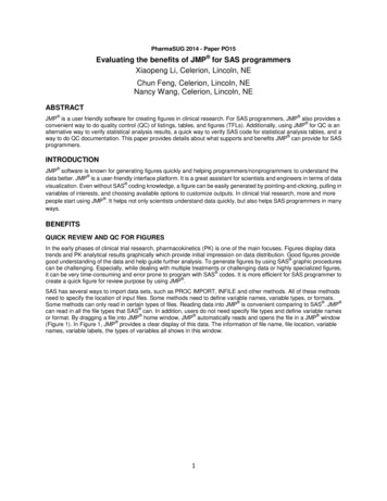
Garcia‑Torres et al. Parasites & Vectors(2022) 15:253Statistical analysisContinuous variables were summarized by mean, range,and SD. For association and risk factor analysis for infection, geographic data were grouped into Mediterraneanand non-Mediterranean countries; breed data weregrouped into domestic short haired (DSH) and nonDSH (Siamese, Siberian, crossbreed, unknown breeds);lifestyle was divided into indoor or outdoor; and clinical signs were grouped into cutaneous, mucocutaneous,ocular, respiratory and systemic. To group clinical signs,we followed the classification used by the clinicians whoauthored each case report; lymphadenomegaly, fever,lethargy, poor body condition, pallor, hepato-splenomegaly, weight loss and abdominal distension were considered systemic signs. Comorbidities were defined asdiseases other than FeL, other medical conditions, prescribed medication, and pathogens that could modify theimmune responseIn the statistical evaluation of diagnostic method reliability, cytology and/or histopathology were considered asgold standards for the direct identification of parasites inaccordance with experts’ recommendations for FeL. Anindirect fluorescent antibody technique (IFAT) had beenused in the majority of cases, so to enable the statisticalanalysis of positive quantitative serological results, all thedata were transformed according to the same titer scalebased on World Organization for Animal Health recommendations [83] and divided into titer ranges of 1/40to 1/80, 1/80 to 1/160, 1/160 to 1/320, 1/320to 1/640, and 1/640. For those cases where quantitative serologic data were not supplied, and only a positiveor negative result from a test such as western blot, theresults were grouped as qualitative.All the statistical analyses were performed using IBMSPSS version 20 for Mac. Univariate analyses were performed, and the results are presented as the number ofaffected cats in relation to the total number of cats forwhich the finding was described. Data were evaluatedfor normal distribution using a Kolmogorov–Smirnovtest. In the univariate analyses, when only two independent continuous variables were compared, an independent-sample t-test was performedaccording to the datadistribution. For the categorical variables, a chi-squaretest of association was used, and for paired samples theWilcoxon test was used. SPSS was used to calculate theexpected frequencies. The rule used was that, at most,only 20% of expected frequencies should be less than 5.The Pearson correlation (r) test of association was usedfor survival and age analyses. A Kaplan–Meier survivalanalysis with log ranks was used to test for significant differences between survival curves for treatment applied.The treatments were grouped as follows: (i) allopurinol, (ii) allopurinol plus meglumine antimoniate, (iii)Page 3 of 13allopurinol plus miltefosine, (iv) meglumine antimoniate,(v) other, and (vi) no treatment. When survival data werenot exactly defined, the highest known values were usedas the survival data for the analyses. P 0.05 was considered the critical level of significance.ResultsCase selectionThe online literature search identified 552 potentially relevant publications. A total of 355 duplicate publicationswere excluded. After the initial screening of the data,based on title and/or abstract evaluation, another 130publications were excluded. A further 25 were excludedduring a second selection process based on the full-textevaluation of the 67 remaining publications. A total of42 articles (63 cats) were finally found to be eligible forinclusion in this review and the data subjected to statistical analysis (Fig. 1; Table 2).Geographic regionAll the cases were of domestic cats living in Europe (59),Brazil (1), Vietnam (1), Reunion Island (1), and Algeria(1). The European cases were from Spain (24), Italy (16),Portugal (9), France (6), Switzerland (3), and Cyprus (1).There was a statistically significant association betweenlocation and the number of cases, with a higher prevalence in Mediterranean (56/63, 88.9%) compared tonon-Mediterranean (7/63, 11.1%) countries (χ2 38.111,df 1, P 0.001) (Table 3).Total number of studiesidentified (n 552)Exclusion of duplicates(n 355)Studies selected for title andabstract reading (n 197)Full-text articles assessedfor eligibility (n 67)Exclusion after screening oftitle and abstract (n 115)Exclusion of species otherthan L. infantum (n 15)Exclusion after full readingof articles (n 25)Papers selected for review andstatistical analysis (n 42)Cases included for review andstatistical analysis (n 63)Fig. 1 Flow chart showing the search and selection process for theinclusion of articles in this study
Garcia‑Torres et al. Parasites & Vectors(2022) 15:253Page 4 of 13Table 2 Cases included in the statistical analysis according to continent, country, type of study and number of cats describedContinentCountryType of study (n)No. of catsReferencesEuropeItalyCase reports (6), case series (1), system‑atic review (1)16[7, 26, 27, 32, 36, 44, 49, 55]FranceCase reports (6)6[19, 21, 22, 29, 39, 40]SpainCase reports (13), case series (1)24[23–25, 31, 33, 34, 37, 38, 46, 51–54, 56]PortugalCase reports (6), case series (1)9[20, 35, 42, 43, 45, 48, 50]SwitzerlandCase report (1), case series (1)3[30, 41]CyprusCase report (1)1[47]AfricaAlgeriaCase report (1)1[17]Reunion IslandCase report (1)1[18]South AmericaBrazilCase report (1)1[28]AsiaVietnamCase report (1)1[16]Table 3 Univariate analysis of the association betweengeographic region, breed, sex, and lifestyle of cats withleishmaniosisχ2 (df)Table 4 Comorbidities in cats with leishmaniosisComorbiditiesNo. of cats (froma total of 53)%CovariateLevelsn (%)GeographicregionNon-Mediterra‑nean7/63 (11.1)Mediterranean56/63 (88.9) 38.111 (1)Siamese4/63 (6.3)Siberian1/63 (1.6)Crossbreed1/63 (1.6)Unknown3/63 (4.8)Yes1120.8DSH54/63 (85.7) 170.571 (4) 0.001*No4279.2Male29/62 (46.7)Female33/62 (53.2) 0.258 (1)Indoor5/22 (22.7)Outdoor17/22 (77.3) 6.545 (1)BreedSexLifestyleP-value 0.001*Present3566.0Absent1834.0FIV veFIV veFeLV veFeLV ve2037.73362.347.54992.5Corticosteroid treatmentOther medical conditions and pathogens0.611 0.011*Bartonella henselae23.8Candidatus Mycoplasma haemominutum23.8Feline coronavirus35.6Mediterranean Mediterranean region, DSH domestic short hairedToxoplasma spp.47.5*P 0.05Hepatozoon us cell carcinoma23.8The age of the cats at clinical presentation was knownin 56/63 cases, and ranged from 2 to 21 years (mean 7.9 4.1 years). Breed was described for 60 out of the 63cases, and was as follows: 54 DSH (85.7%), four Siamese(6.3%), one Siberian (1.6%) and one crossbreed (1.6%)cat. DSH were more likely to be infected than nonDSH breeds (χ2 170.571, df 4, P 0.001). Sex wasreported in 62 out of 63 cases, and there was a slightlyhigher prevalence in females (53.2%) than in males(46.7%), although this difference was not statisticallysignificant (χ2 0.258, df 1, P 0.611). Lifestyle wasknown for 22 cats, of which 17 were outdoor (77.3%)and five indoor cats (22.7%). There was an associationDiabetes mellitus11.9Epidermoid carcinoma11.9FIV Feline immunodeficiency virus, FeLV feline leukemia virusbetween an outdoor lifestyle and infection (χ2 6.545,df 1, P 0.011) (Table 3).Information on comorbidity status was available for53 cats (Table 4); of these 35 (66.0%) had comorbidities and 18 (34.0%) did not. Of the 35 cats with comorbidities, 22 (62.9%) had only one comorbidity, whereas13 (37.1%) had two or more. Positive feline immunodeficiency virus (FIV) antibody status was the mostprevalent comorbidity, but the association between
Garcia‑Torres et al. Parasites & Vectors(2022) 15:253this and leishmaniosis was not statistically significant(χ2 0.277, df 1, P 0.599).Clinical presentationPage 5 of 13Table 5 Clinical signs and lesions described in cats (n 63) withleishmaniosisClinical signs and lesionsFrequencyNo. of cats%The clinical signs and lesions that were reported are givenin Table 5. The most frequent clinical signs were cutaneous (44/63; 69.8%), followed by systemic (35/63; 55.5%),ocular (22/63; 34.9%), mucocutaneous (18/63; 28.6%)and respiratory (8/63; 12.7%). Many of the cats showeda combination of clinical signs (37/63; 58.7%). There wasa statistically significant association between dermatological signs and FIV (χ2 7.185, df 1, P 0.007), andbetween ocular signs and the neutered status of females(χ2 17.814, df 3, P 0.001). However, no other statistically significant associations were found between groupsof clinical signs and age, sex, breed, or comorbidities(Table 6). Lymph node size was described for 45/63 catsfrom physical examination; the percentage of cats withlymph nodes of normal size (27/45; 60.0%) was greaterthan that of cats with lymphadenomegaly (18/45; 40.0%).When clinicopathological abnormalities were reported,hyperproteinemia was the most frequent (19/41; 46.3%),followed by anemia (16/48; 33.3%), neutrophilia (9/48,18%), thrombocytopenia (8/48, 16.6%), proteinuria (7/46;15.2%) and azotemia (7/47; 14.9%). Hypergammaglobulinemia was the most frequent alteration detected byserum protein electrophoresis (27/38; 71.0%) followed byhypoalbuminemia (9/47; 19.1%). Other reported clinicopathological alterations were neutropenia (4/48, 8.3%),eosinophilia (3/48, 6.25%), pancytopenia (1/48, 2%) andan increase in alanine transaminase level (2/48, 4.1%).Keratoconjunctivitis11.6Ulcerative keratitis11.6Mucocutaneous1828.6Diagnostic methodsStomatitis/gingivostomatitis1117.4Cytology, for the detection of Leishmania amastigotes, was the most common first line or preferred diagnostic option of practitioners for the diagnosis of FeL(28/63, 44.4%), followed by histopathology (20/63, 31.7%),serology (17/63, 26.9%) and PCR (3/63, 4.7%). There wasno statistically significant association (χ2 8.980, df 2,P 0.062) between results from the PCR and those fromcytological and/or histopathological examination (widelyused as confirmatory tests for FeL and considered goldstandards here). However, a statistically significant association between cytology and/or histopathology andseropositivity (χ2 26.913, df 14, P 0.020) was found.Of the complete diagnostic procedures (both firstline and additional diagnostic tests), antibody detectiontechniques were performed most (48/63; 76.2%), andcomprised IFAT (28/48), qualitative serology (11/48),enzyme-linked immunosorbent assay (5/48), and directagglutination (4/48). Antibody tests were positive for45/48 cats (93.7%) and negative for 3/48 (6.3%); therewas a statistically significant association between L.Cutaneous4469.8Ulcerative dermatitis2031.7Nodular dermatitis1422.2Alopecia914.3Desquamative dermatitis711.1Crusty dermatitis57.9Pruritus23.1Bloody cyst23.1Papular dermatitis11.6Systemic3555.5Lymph node enlargement1828.5Anorexia/hyperorexia1422.2Weight ar blepharitis69.5Ulcerative blepharitis34.7Glossitis34.7Nasal ulcers34.7Nasal pustules23.1Nasal depigmentation11.6Oral ulcers11.6Respiratory812.7Nasal erse sneezing11.6Dyspnea11.6Bronchitis11.6infantum antibody positive status and diagnosis of FeLby cytology and/or histopathology (χ2 36.750, df 2,P 0.001). In cats seropositive according to quantitative tests (35/45, 77.8%), the titers ranged from 1/40to 1/80 in 5/35 (14.3%), from 1/80 to 1/160 in
Garcia‑Torres et al. Parasites & Vectors(2022) 15:253Page 6 of 13Table 6 Analysis of association (χ2) between sex, age, breed, comorbidities (any), FIV, FeLV, and steroid treatment with groups ofclinical rySystemicSexχ2 6.065, df 3P 0.108χ2 0.386, df 3,P 0.943χ2 17.814, df 3,P 0.001*χ2 3.285, df 3,P 0.350χ2 1.444, df 3,P 0.695χ2 4.534, df 4,P 0.339χ2 4.200, df 4,P 0.380χ2 3.230, df 4,P 0.520χ2 8.126, df 4,P 0.087χ2 2.663, df 4,P 0.616χ2 7.185, df 1,P 0.007*χ2 1.150, df 1,P 0.283χ2 0.070, df 1,P 0.791χ2 0.090, df 1,P 0.764χ2 1.539, df 1,P 0.215AgeBreedComorbiditiesFIVFeLVSteroid treatmentt(63) 1.206, P 0.233χ2 1.914, df 1,P 0.167χ2 0.638, df 1,P 0.424χ2 0.117, df 1,P 0.732t(63) 1.525, P 0.133χ2 0.005, df 1,P 0.945χ2 0.155, df 1,P 0.694χ2 2.622, df 1,P 0.105t(63) 1.906, P 0.062χ2 0.015, df 1,P 0.901χ2 0.277, df 1,P 0.599χ2 0.011, df 1,P 0.916t(63) 0.370, P 0.713χ2 0.105, df 1,P 0.746χ2 0.658, df 1,P 0.417χ2 0.300, df 1,P 0.584t(63) 0.912, P 0.366χ2 0.010, df 1,P 0.922χ2 1.002, df 1,P 0.317χ2 0.895, df 1,P 0.344For abbreviations, see Table 4*P 0.05Table 7 Frequency of tested tissues and positive results frompolymerase chain reaction (PCR)Table 8 Frequency of tested tissues and parasite detectionusing cytologyTested tissueTested tissuePCR (n 38)PCR ve (n 36, 94.7%)Blood18/38, 47.3%16/18, 88.8%Skin samples6/38, 15.7%6/6, 100%Unknown origin6/38, 15.7%6/6, 100%Lymph node4/38, 10.5%4/4, 100%Bone marrow4/38, 10.5%4/4, 100%Ocular samples2/38, 5.2%2/2, 100%Spleen2/38, 5.2%2/2, 100%Respiratory samples1/38, 2.6%1/1, 100%3/35 (8.6%), from 1/160 to 1/320 in 5/35 (14.3%),from 1/320 to 1/640 in 5/35 (14.3%), and were 1/640 in 17/35 cats (48.5%). No statistically significantassociation was found between serological titers andcategory of clinical signs or clinicopathological abnormalities (P 0.05). Qualitative positive serology wasreported for 10/45 cats (22.2%).Polymerase chain reaction (using blood, lymph node,bone marrow, spleen tissue; ocular, lung or skin samples) was performed for 38 of the 63 cats (60.3%), andwas positive for 36 of them (94.7%) (Table 7). Cytologywas performed for 44/63 cats (69.8%) and histopathology for 35/63 cats (55.5%) using different types of tissue. Cytology and histopathology were positive forLeishmania amastigotes in 39/44 cats (88.6%) and 31/35(88.6%), respectively (Tables 8, 9). With respect toCytology (n 44)Cytology ve (n 39,88.6%)Skin lesions20/44, 45.4%18/20, 90%Lymph node18/44, 40.9%16/18, 88.8%Ocular lesions7/44, 15.9%7/7, 100%Bone marrow6/44, 13.6%5/6, 83.3%Spleen2/44, 4.5%2/2, 100%Liver1/44, 2.2%1/1, 100%Blood1/44, 2.2%1/1, 100%Respiratory lesions1/44, 2.2%1/1, 100%Table 9 Frequency of tested tissues and parasite detectionusing histopathologyTested tissueHistopathology(n 35)Histopathology ve (n 31,88.6%)Skin lesions22/35, 62.8%20/22, 90.9%Ocular lesions5/35, 14.2%4/5, 80%Respiratorylesions4/35, 11.4%3/4, 75%Oral lesions2/35, 5.7%2/2, 100%Bone marrow2/35, 5.7%1/2, 50%Spleen2/35, 5.7%2/2, 100%Kidney1/35, 2.8%1/1, 100%serological titers, no statistically significant associationwas found between PCR, cytology or histopathology
Garcia‑Torres et al. Parasites & Vectors(2022) 15:253with any category of clinical sign or clinicopathologicalabnormality (P 0.05).TreatmentMedical treatment was administered in 45/63 cats(71.4%); 16/63 (25.4%) did not receive any treatment, andtreatment was not stipulated for 2/63 (3.2%). Allopurinol was used in 37/45 cats (74.4%), followed by meglumine antimoniate in 13/45 (28.9%) and miltefosine inonly one cat of the 45 (2.2%). Although the dosages varied, the most frequent were 10 mg/kg twice a day (BID)per os (PO) for at least 6 months for allopurinol (20/37cats; 54.0%), 50 mg/kg once a day (SID) subcutaneously(SC) for 30 days for meglumine antimoniate (5/13 cats;38.5%), and 2 mg/kg SID PO for 28 days for miltefosine in the only cat in which it was used. Allopurinol wasused as monotherapy in 28/37 cats (75.7%) and in combination with meglumine antimoniate or miltefosinein 8/37 (21.6%) and 1/37 (2.7%) cats, respectively. Meglumine antimoniate was used as monotherapy in 5/13cats (38.5%) and in combination with allopurinol in 8/13(61.5%). Adverse effects associated with treatment werereported in 10/45 cats (22.2%), and were mainly associated with allopurinol (7/10), and affected the kidney(4/10), skin (2/10) and liver (1/10).Page 7 of 13OutcomeSurvival time ranged from 0 to 2700 days, with a meanof 432 days ( 575). Mean survival time was significantlylonger for treated cats than non-treated cats (520 daysand 210 days, respectively) (χ2 15.311, df 1, P 0.002)(Fig. 2). However, no significant association was foundbetween survival time and any other variable (Table 10).DiscussionThe results of this study indicate that leishmaniosis should be included in the differential diagnosis ofsick cats living in, or with a history of travel to, areaswhere CanL is endemic [1–3, 84]. Prevalences of FeL inendemic areas as shown by positive PCR range from 0to 100% (mean 21.3%), whilst those indicated by positive serology range from 0 to 70.5% (mean 13.7%) [1, 10,85]. Prevalences determined by both of these types oftests are lower for cats than dogs (63% and 27% for PCRand serology, respectively) [85]. There are also fewerclinical feline cases than canine cases in the literature[8–11, 59]. Thus, all this suggests that the prevalenceof FeL could be about half that of CanL for the samegeographical areas [60, 63, 74, 79–81]. Furthermore,as previously reported for CanL [86, 87], increasedmovement of pet cats between countries, especiallyinside Europe, could lead to clinical cases of FeL beingFig. 2 Kaplan–Meier survival curves of cats treated with allopurinol, allopurinol plus meglumine antimoniate, allopurinol plus miltefosine,meglumine antimoniate, other, or no treatment. Median survival time of the treated cats was as follows: allopurinol (28/63; 619 days); allopurinolplus meglumine antimoniate (8/63; 614 days); allopurinol plus miltefosine (1/63; 45 days); meglumine antimoniate (5/63; 1372 days); other suchas fluconazole, metronidazole, spiramycin or lomidine (3/63; 660 days); no treatment (10/63; 411 days)
Garcia‑Torres et al. Parasites & Vectors(2022) 15:253Table 10 Analysis of association (χ2) between treatment, age,breed, sex, lifestyle, comorbidities (any), clinical signs (any),clinicopathological abnormalities (any), PCR positive test,serological titer and survivalVariableSurvivalTreatment (yes/no)χ2 15.311, df 1, P 0.002*AgeBreedSexLifestyleComorbidities (any)Cutaneous clinical signsMucocutaneous clinical signsOcular clinical signsRespiratory clinical signsSystemic clinical PCR veSerological titerr(63) -0.028, P 0.856χ2 17.165, df 4, P 0.144χ2 3.112, df 3, P 0.960χ2 0.889, df 1, P 0.889χ2 0.381, df 1, P 0.944χ2 1.693, df 1, P 0.638χ2 3.100, df 1, P 0.377χ2 3.742, df 1, P 0.291χ2 1.098, df 1, P 0.778χ2 6.014, df 1, P 0.111χ2 0.669, df 1, P 0.881χ2 1.048, df 1, P 0.790χ2 1.495, df 1, P 0.683χ2 2.252, df 1, P 0.522χ2 1.927, df 1, P 0.588χ2 5.555, df 1, P 0.135χ2 2.233, df 1, P 0.526χ2 9.932, df 2, P 0.128χ2 18.920, df 7, P 0.590*P 0.05diagnosed in areas that are not endemic for L. infantum[30, 41]. However, although there have been epidemiological studies on FeL in areas that are also endemic forCanL, such as Brazil [1, 84], only one case from Brazilwas included in the current study. Possible reasons forthis include the non-detection of other cases from Brazil due to the criteria used in this study, and/or perhapsbecause feline medicine is less developed there, and/orbecause most knowledge on leishmaniosis is focusedon dogs. Thus, even in areas that are non-endemic forL. infantum, leishmaniosis should be considered as adifferential diagnosis in cats with clinical signs or pathological alterations consistent with this disease.Few epidemiological studies have reported significantassociations between L. infantum infection in cats andtheir access to outdoors, that they are male, or their agewhen they are adults [60, 64]. Most of the cats affectedby leishmaniosis in the present study were DSH withoutdoor access, and had a mean age of 7.9 years. In contrast to other publications [60, 65, 66], we found thatmore female cats were diagnosed with leishmaniosis thanmale cats, although this difference was not statisticallysignificant.Page 8 of 13It is well recognized that susceptibility to progressive Leishmania infection and the development of clinical signs in dogs is mostly linked to an imbalance in theadaptive immune response, and probably associatedwith a predominant Th2 response and an impaired Th1immune response [88]. However, in contrast to CanL,to the best of our knowledge, no prospective controlledstudies have been published on immune mechanismsinvolved in the pathogenesis of FeL. Some investigations have suggested that cats may have a better immuneresponse against L. infantum because the Th2 responseplays a protective role [81, 89], or because there are otherfactors in seropositive cats that can control the development of patent leishmanios
SPSS version 20 for Mac. Univariate analyses were per-formed, and the results are presented as the number of aected cats in relation to the total number of cats for which the nding was described. Data were evaluated for normal distribution using a Kolmogorov-Smirnov test. In the univariate analyses, when only two independ-