Transcription
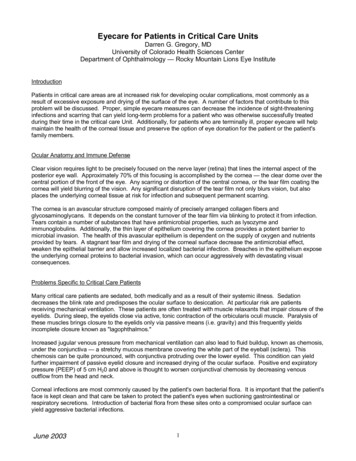
Eyecare for Patients in Critical Care UnitsDarren G. Gregory, MDUniversity of Colorado Health Sciences CenterDepartment of Ophthalmology — Rocky Mountain Lions Eye InstituteIntroductionPatients in critical care areas are at increased risk for developing ocular complications, most commonly as aresult of excessive exposure and drying of the surface of the eye. A number of factors that contribute to thisproblem will be discussed. Proper, simple eyecare measures can decrease the incidence of sight-threateninginfections and scarring that can yield long-term problems for a patient who was otherwise successfully treatedduring their time in the critical care Unit. Additionally, for patients who are terminally ill, proper eyecare will helpmaintain the health of the corneal tissue and preserve the option of eye donation for the patient or the patient'sfamily members.Ocular Anatomy and Immune DefenseClear vision requires light to be precisely focused on the nerve layer (retina) that lines the internal aspect of theposterior eye wall. Approximately 70% of this focusing is accomplished by the cornea — the clear dome over thecentral portion of the front of the eye. Any scarring or distortion of the central cornea, or the tear film coating thecornea will yield blurring of the vision. Any significant disruption of the tear film not only blurs vision, but alsoplaces the underlying corneal tissue at risk for infection and subsequent permanent scarring.The cornea is an avascular structure composed mainly of precisely arranged collagen fibers andglycosaminoglycans. It depends on the constant turnover of the tear film via blinking to protect it from infection.Tears contain a number of substances that have antimicrobial properties, such as lysozyme andimmunoglobulins. Additionally, the thin layer of epithelium covering the cornea provides a potent barrier tomicrobial invasion. The health of this avascular epithelium is dependent on the supply of oxygen and nutrientsprovided by tears. A stagnant tear film and drying of the corneal surface decrease the antimicrobial effect,weaken the epithelial barrier and allow increased localized bacterial infection. Breaches in the epithelium exposethe underlying corneal proteins to bacterial invasion, which can occur aggressively with devastating visualconsequences.Problems Specific to Critical Care PatientsMany critical care patients are sedated, both medically and as a result of their systemic illness. Sedationdecreases the blink rate and predisposes the ocular surface to desiccation. At particular risk are patientsreceiving mechanical ventilation. These patients are often treated with muscle relaxants that impair closure of theeyelids. During sleep, the eyelids close via active, tonic contraction of the orbicularis oculi muscle. Paralysis ofthese muscles brings closure to the eyelids only via passive means (i.e. gravity) and this frequently yieldsincomplete closure known as "lagophthalmos."Increased jugular venous pressure from mechanical ventilation can also lead to fluid buildup, known as chemosis,under the conjunctiva — a stretchy mucous membrane covering the white part of the eyeball (sclera). Thischemosis can be quite pronounced, with conjunctiva protruding over the lower eyelid. This condition can yieldfurther impairment of passive eyelid closure and increased drying of the ocular surface. Positive end expiratorypressure (PEEP) of 5 cm H20 and above is thought to worsen conjunctival chemosis by decreasing venousoutflow from the head and neck.Corneal infections are most commonly caused by the patient's own bacterial flora. It is important that the patient'sface is kept clean and that care be taken to protect the patient's eyes when suctioning gastrointestinal orrespiratory secretions. Introduction of bacterial flora from these sites onto a compromised ocular surface canyield aggressive bacterial infections.June 20031
Nursing AssessmentAssessment of a critical care patient's eyes should routinely be done by each nursing shift. Checking the eyes ofpatients who are not sedated and ventilated will allow nurses to gain an appreciation of the normal appearance ofthe eye. Most ocular problems in critical care patients arise from lagophthalmos, which generally increases inseverity with increased sedation. It is important that eyelid position be carefully assessed. Using a penlight orflashlight will help reveal poor lid closure, which might be masked by the eyelashes. Excessive exposure canlead to breakdown of the corneal epithelium, known as keratopathy. This can be recognized as an irregularreflection of light from a penlight off the corneal surface. Lubricating ointments can also disrupt this reflection, buta gentle rinse of the ocular surface with sterile saline will rinse the ointment away. This irregular reflection of lightindicates an inadequate tear film and a breakdown in the integrity of the corneal epithelium. This places thecornea at risk for infection.Exposure keratopathy can lead to keratitis — a term for any corneal inflammation, infectious or otherwise.Bacterial keratitis in the critical care setting has an increased association with aggressive Gram(-) bacteria suchas Pseudomonas. Once these bacteria gain entry into the subepithelial corneal collagen, rapid destruction ofcorneal tissue can occur. An infectious corneal ulcer will appear as a whitish area on the corneal surface. It isoften accompanied by a purulent discharge. Severe dryness can also yield whitish opacities of the cornea.Therefore, an ophthalmology consultation is recommended for any new, white lesions noted on the cornea.Following infection, the orderly arrangement of corneal collagen fibers is disrupted and the resultant scar tissueyields permanent vision loss, which may require a cornea transplant for visual rehabilitation. The risk of this direconsequence may be greatly diminished if the early signs of lagophthalmos and exposure keratopathy arerecognized and treated.Nursing InterventionsPrevention of ocular complications in critical care areas begins with an awareness of the potentially devastatingconsequences of corneal infections. Both the evaluation and treatment of ocular surface exposure and drynessare relatively simple and minimally labor-intensive, but nevertheless effective.Each assessment of the patients by the nursing staff should include and evaluation of the adequacy of blinkingand eyelid closure. If the eyes appear closed, careful inspection with a penlight or flashlight should be performedto assure the lids are, in fact, completely closed. As mentioned before, the eyelids may hide the fact that the lidsare not completely closed, allowing the ocular surface to dry out.In a sedated patient with seemingly closed eyes, the upper eyelids should be manually elevated to allowinspection of the cornea. A piece of gauze helps with manual traction of the eyelids if they are oily. Does the tearfilm appear uniform or is the light reflection irregular? Sterile saline may be used to rinse away any mucous orlubricant ointment, thus allowing better assessment of the corneal surface. Any patient with signs of exposurekeratopathy, lagophthalmos, or a decreased blink rate (normal blinking occurs every 5-10 seconds) should betreated every 4-6 hours with an ocular lubricating ointment such as Lacrilube. Ointments moisturize the ocularsurface more effectively than drops. The ointment should be placed along the internal surface of the lower eyelid,which should then be manually closed to spread the ointment over the ocular surface. Once again, if any whitishcorneal lesions or purulent discharge develops prompt ophthalmologic consultation is indicated to rule out cornealinfection. A small amount of non-purulent mucous buildup is common in cases of exposure keratopathy.In cases of significant exposure, whether from decreased blinking or poor lid closure, lubricant ointment should beapplied every 4 hours. Prolapsed, chemotic conjunctiva can further worsen the exposure problems. In addition tolubricant ointment, polyethylene cling wrap (i.e. Saran Wrap) may be placed over the skin in a strip from temple totemple, wide enough to cover the lower forehead, bridge of the nose, and upper cheeks. This will create a"moisture chamber" over the eyes. A small amount of petroleum jelly on the skin of the brow, temples and cheekswill create a tighter seal by the cling wrap, but still allow easy removal for inspection and application of ointment.The cling wrap should be changed each shift to lessen the risk of infection. Additionally, visitors to the patientshould be briefed on the need for the cling wrap to avoid any undue alarm.June 20032
Selected Bibliography1. Cortese, D., Capp, L., McKinley, S. “Moisture Chamber Versus Lubrication for the Prevention of CornealEpithelial Breakdown.” American Journal of Critical Care 1995; 4: 425-8.2. Cunningham, C., Gould, D. “Eyecare for the Sedated Patient Undergoing Mechanical Ventilation: theUse of Evidence-Based Care.” International Journal of Nursing Studies 1998; 35: 32-40.3. Dua, H. “Bacterial Keratitis in the Critically Ill and Comatose Patient.” Lancet 1998; 351: 387-8.4. Farrek, M., Wray, F. “Eyecare for Ventilated Patients.” Intensive and Critical Care Nursing 1993; 9:137-41.5. Hernandez, E., Mannis, M. “Superficial Keratopathy in Intensive Care Unit Patients.” American Journalof Ophthalmology 1997; 124: 212-6.6. Lenart, S., Garrity, J. “Eye Care for Patients Receiving Neuromuscular Blocking agents or Propofolduring Mecahnical Ventilation.” American Journal of Critical Care 2000; 9: 188-91.7. McClellan, K. “Mucosal Defense of the Outer Eye.” Survey of Ophthalmology 1997; 42: 233-46.8. Mercieca, F., Suresh, P., Mortan, A., et al. “Ocular Surface Disease in Intensive Care Unit Patients.” Eye1999; 13: 231-6.9. Parkin, B., Turner, A., Moore, E., et al. “Bacterial Keratitis in the Critically Ill.” British Journal ofOphthalmology 1997; 81: 1060-3.10. Suresh, P., Mercieca, F., Morton, A., et al. “Eye Care for the Critically Ill.” Intensive Care Medicine 2000;26: 162-6.June 20033
Provided by the Rocky Mountain Lions Eye Bank in consultation with the University of Colorado Dept. of OphthalmologyEye Care Decision Tree for Unconscious / Sedated Patients on a VentilatorUsing a penlight, assess the degree of ocular surfaceexposure. A close examination is necessary to see beneaththe lashes, which can hide incomplete eyelid closure. Thisassessment should be made each nursingshift or every 8 hours for each patient.No treatment is indicated.Briefly open and close the lidsmanually to assess the ocularsurface.YESAre the lidscompletelyclosed?NOObservation of whitish corneallesions or purulent dischargewarrants an ophthalmologyconsult.Is any of thecornea exposedand the blinkrate 1 blink per10 seconds?YESNO1. Apply lubricating ointmentinside the lower lid every 4hours.A small part of the cornea canbe seen under the lashes2. Apply a polyethylenemoisture chamber (i.e. GladWrap). Apply in a strip templeto-temple, covering the lowerforehead, bridge of the nose andupper cheeks. Petroleum jellyon the skin of the brow, cheeksand temples will create a seal.Change the chamber every 8hours.3. An ophthalmology consult isstrongly recommended.1. Apply lubricating ointmentinside the lower lid every 4hours.2. Apply a polyethylenemoisture chamber (i.e. GladWrap). Apply in a strip templeto-temple, covering the lowerforehead, bridge of the nose andupper cheeks. Petroleum jellyon the skin of the brow, cheeksand temples will create a seal.Change the chamber every 8hours.Observation of whitish corneallesions or purulent dischargewarrants an ophthalmologyconsult.Is thereconjunctivalchemosis orprolapse?There is an accumulation of fluidbeneath the conjunctiva. Theconjunctiva may protrude, keepingthe lids from closing at all. Thecornea is exposed.YESNOOnly the white part of the eye isvisible under the lashes.Apply lubricating ointment insidethe lower lid every 4 hours.No part of the cornea is visible, but the visiblewhite part of the eye is swollen with fluids builtup under the conjunctiva. Often protrudesbeneath the lid.Observation of whitish corneallesions or purulent dischargewarrants an ophthalmologyconsult.
Application of a Polyethylene Moisture ChamberMake sure the patient s face is clean and that lubricating ointments have been instilled prior to applicationof a moisture chamber.1. Apply a small amount of petroleum jelly to your fingertip.2. Apply the petroleum jelly from temple to temple, acrossthe forehead, bridge of the nose and upper cheekbones.(indicated by the red line in the photo)3. Apply a 4-6 inch wide strip of polyethylene (GladWrap, Saran Wrap, Clingwrap) over the area. Press filminto the petroleum jelly at the forehead, then smootharound the petroleum jelly line. The chamber should notbe tight over the eyes.4. The chamber should be changed every 8 hours. It may beeasily lifted or removed for eye examination and for family visitsin cases where the chamber causes family members increasedanxiety. Every effort should be made to explain the role of themoisture chamber in preserving the patient's sight.Provided by the Rocky Mountain Lions Eye BankTel: 800-444-7479
Ocular Health of Patients in Critical Care UnitsPatients in critical care settings are at increased risk for developing ocular complications, mostcommonly as a result of excessive exposure and drying of the surface of the eye. A number offactors contribute to this problem. Sedation and muscle relaxants decrease the blink rate and predispose the ocular surface todesiccation. At particular risk are patients receiving mechanical ventilation. During sleep theeyelids close via active, tonic contraction of the orbicularis oculi muscle. Paralysis of thesemuscles leads to eyelid closure only by passive means (i.e. gravity) and this frequently meansincomplete closure of the eyelids known as “lagophthalmos.” Increased jugular venous pressure from mechanical ventilation can also lead to fluid buildup,known as chemosis, under the conjunctiva. The conjunctiva is a stretchy mucous membranecovering the white part of the eye (sclera). This chemosis can be quite pronounced, withconjunctiva protruding over the lower eyelid. This condition can lead to further impairmentof passive eyelid closure and increased drying of the ocular surface. Positive end expiratorypressure (PEEP) of 5 cm H20 and above is thought to worsen conjunctival chemosis bydecreasing venous outflow from the head and neck. Proper, simple eye care measures can decrease the incidence of sight-threatening infections andscarring that can yield long-term problems for a patient who was otherwise successfully treatedduring their time in the critical care unit. Additionally, for patients who are terminally ill, proper eye care will help maintain the health ofthe corneal tissue and preserve the option of eye donation for the patient or the patient’s familymembers.Nursing Interventions Keep the patient’s face clean. Corneal infections are most commonly caused by thepatient’s own bacterial flora. Protect the patient’s eyes with a towel while suctioning gastrointestinal orrespiratory secretions—even in closed systems. Introduction of bacteria from these sitesonto a compromised cornea can lead to very aggressive infections. Follow the eye care protocol. See the reverse side for instructions on administeringlubricating ointments.
How to Apply Eye Lubricating OintmentsWash hands thoroughly with soap and water!Gently pull the lower lid downward to expose theconjunctival sac and form a pocket. You may use a cottontipped swab to roll the upper lid open to access the lower lidif necessary.Squeeze a ribbon of ointment into the pocket just inside thelower lid. Avoid touching the tip of the medication bottle ortube to the eye to avoid the transfer of micro-organisms tothe medication and injury to the eye. Do not push on theeyeball or touch the eye. Use the cheekbone and/or foreheadto steady your hands.Very gently, pinch the lower lid and move gently upward todisperse some of the ointment onto the cornea.Close the eyes completely. A cotton swab can be helpful toroll the upper lid downward. Do not pull on the edge of thelid. Do not tape closed. Ensure that no part of the cornea isvisible beneath the eye lid. Repeat every 8 hours.
Post-mortem Eye Care for the Potential Eye DonorTear production stops at the time of death, so it doesn't take long for the cornea to dry out.Another problem is that lubricating ointments used prior to death tend to congeal. This canpull the delicate epithelial covering of the cornea away.For all deaths that are potential eye donors, the following eye care regimen should beperformed. A common question is "How long do we have to do this eye care after death?"It's a difficult question to answer. The Rocky Mountain Lions Eye Bank is sensitive to theneeds of the donor's family, so their needs must come first. A saline rinse prior to givingthe family time with their loved one is a good idea to keep some moisture in the eye. Afterthe family is finished, another rinse followed by the remaining steps should be performed.Rinse both eyes with sterile saline or balanced saltsolution. This will help remove any residue fromlubricating ointments used prior to death. This step canbe done prior to family viewing and then repeated beforeproceeding after the family has finished its bedside rituals.Close both eyelids using your fingers. Do not useforceps or other instruments, as they can easily damagethe lids.Cover both closed eyes with a compress (2x2 or 4x4gauze) soaked in saline. The compress should be dripping wet if possible. Do not press the gauze hard onto theeyes, just set it firmly on top of the lids ensuring it coversthe lid line.Elevate the head using a pillow or head block. This helpsfluids to drain from the head, minimizing the chance ofbleeding or bruising during the eye tissue recoveryprocedure.Provided by the Rocky Mountain Lions Eye BankPh: 800-444-7479www.corneas.org
Sample Eye Care Policy/ProcedureProvided by the Rocky Mountain Lions Eye BankEye Care for Ventilated PatientsI.Purpose:This protocol has been established to address the increased risk of ocular complicationsfor patients who are unconscious or sedated and on mechanical ventilation.II.DefinitionsA. Chemosis: a condition in which the membranes that line the eyelids and surface ofthe eye (conjunctiva) are swollen. The outer surface covering appears to have fluid in it.Often, the conjunctiva becomes so swollen that the eyes cannot close properly.B. Cornea: the clear dome-like tissue over the central portion of the front of the eye.The cornea accomplishes approximately 70% of the focusing power of the eye.C. Conjunctiva: the thin, transparent tissue that covers the outer surface of the eye. Itbegins at the outer edge of the cornea, covering the visible part of the sclera, and liningthe inside of the eyelids. It is nourished by tiny blood vessels that are nearly invisible tothe naked eye. The conjunctiva also secretes oils and mucous that moisten andlubricate the eye.D. Lagophthalmos: the inability to close, or poor closure, of the upper eyelid.Lagophthalmos is a form of facial paralysis affecting the orbicularis muscle in the eyelid.Complications associated with lagophthalmos include:Severe dry eye and discomfortCorneal ulceration (damage to the cornea-the clear tissue covering the front of youreye)Decrease or loss of visionUnsatisfactory appearance
E. Sclera: commonly known as "the white of the eye." It is the tough, opaque tissuethat serves as the eye's protective outer coat. Six tiny muscles connect to it around theeye and control the eye's movements. The optic nerve is attached to the sclera at thevery back of the eye.III.PolicyA. All patients who are mechanically ventilated and who are unconscious or aresedated with neuromuscular blockers fall under this protocol.IV.ProcedureA. Once per nursing shift, each eye should be checked for exposure keratopathy orkeratitis. Using a penlight or strong flashlight, lift the lid of each eye and examine thecornea. Note: the presence of lubricating ointments may cause irregular reflection oflight from the cornea. Rinse ointments away with sterile saline prior to examination ofthe cornea.1. Notify the attending physician if any whitish corneal lesions or purulentdischarge is observed.2. Notify the attending physician if the reflection of the penlight beam is irregularor dull off the cornea surface, or if the cornea appears dull or opaque. Irregularreflection of the penlight beam off the surface of the cornea may be an indicationof exposure keratopathy. (Checking the eyes of patients who are not sedatedand ventilated will allow nurses to gain appreciation of the normal appearance ofthe eye.)B. After examination of the corneal surface, the lids should be allowed to close. If thelids do not close, manually close them. Assess the degree of lagophthalmos asdescribed in section C below.C. Every 8 hours, each eye should be assessed for the degree of lagophthalmos usinga penlight or strong flashlight. Close examination is necessary as eyelashes may maskpoor lid closure.1. If eyelids are fully apposed, no treatment is indicated.2. If any portion of the cornea is visible and the blink rate is less than 1 blinkevery 10 seconds, instill ocular lubricating ointment (i.e.: Lacrilube ) every 4hours. In addition, a polyethylene moisture chamber should be applied andchanged every 8 hours. See Appendix A and B for instructions for theproper instillation of ocular ointments and application of a polyethylenemoisture chamber. Inform the attending physician.3. If no portion of the cornea is visible, but there is a presence of conjunctivalchemosis, instill ocular lubricating ointment (i.e.: Lacrilube ) every 4 hours. Inaddition, a polyethylene moisture chamber should be applied and changed every
8 hours. See Appendix A and B for instructions for the proper instillation ofocular ointments and application of a polyethylene moisture chamber. Inform theattending physician.4. If only a portion of the sclera is visible (no portion of the cornea is visible andthere is no chemosis), instill ocular lubricating ointment (i.e.: Lacrilube ) every 4hours.APPENDIX AIllustrated Instructions for Application of Lubricating OintmentsAPPENDIX BIllustrated Instructions for the Application of a Polyethylene Moisture Chamber
Suresh, P., Mercieca, F., Morton, A., et al. "Eye Care for the Critically Ill." Intensive Care Medicine 2000; 26: 162-6. Provided by the Rocky Mountain Lions Eye Bank in consultation with the University of Colorado Dept. of Ophthalmology Eye Care Decision Tree for Unconscious / Sedated Patients on a Ventilator


