Transcription
Affinati et al. Clinical Diabetes and 121-y(2021) 7:8CASE REPORTOpen AccessSevere hyperglycemia and insulinresistance in patients with SARS-CoV-2infection: a report of two casesAlison H. Affinati1, Amisha Wallia2 and Roma Y. Gianchandani1*AbstractBackground: Severe insulin resistance is an uncommon finding in patients with type 2 diabetes but is oftenassociated with difficult to managing blood glucose. While severe insulin resistance is most frequently seen in thesetting of medication side effects or rare genetic conditions, this report of two cases highlights the presence ofsevere insulin resistance in the setting of severe COVID-19 and explores how this may contribute to the poorprognosis of patients with diabetes who become infected with SARS-CoV-2.Case presentation: Here we present the cases of two African-American women with pre-existing type 2 diabeteswho developed severe COVID-19 requiring mechanical ventilation and concurrent severe insulin resistance withtotal daily insulin dose requirements of greater than 5 unit/kg. Both patients received aggressive insulin infusionand subcutaneous insulin therapy to obtain adequate glucose management. As their COVID-19 clinical courseimproved, their severe insulin resistance improved as well.Conclusions: The association between critical illness and hyperglycemia is well documented in the literature,however severe insulin resistance is not commonly identified and may represent a unique clinical feature of theinteraction between SARS-CoV-2 infection and type 2 diabetes.Keywords: Diabetes, COVID-19, Severe Insulin ResistanceIntroductionSARS-CoV-2 infection is associated with severe hyperglycemia, which has been associated with poor patientoutcomes [1–4]. While hyperglycemia is a feature ofmany severe infections, severe insulin resistance is lessfrequently documented. More commonly, severe insulinresistance is seen in the setting of medications or raregenetic disorders [5]. Here we present two cases of patientswith severe Coronavirus Disease (COVID-19) who presented with insulin requirements greater than 5 units/kg.We use these cases as a starting point to explore the* Correspondence: romag@med.umich.edu1Department of Internal Medicine, Division of Metabolism, Endocrinologyand Diabetes, University of Michigan, Domino’s Farms (Lobby G, Suite 1500),24 Frank Lloyd Wright Drive, MI 48106 Ann Arbor, USAFull list of author information is available at the end of the articlepossible interactions between glucose homeostasis,SARS-CoV-2 infection and insulin treatment.Case report 1A 41-year old African-American woman with a historyof type 2 diabetes for at least 2 years, obesity (body massindex (BMI), 44 kg/m2), hypertension, hyperlipidemiaand sleep apnea presented to the emergency departmentat an outside hospital with acute worsening of shortnessof breath which had begun one week prior to presentation. On presentation she was hypoxic with associatedconfusion and was emergently intubated and mechanically ventilated. Her diabetes history was notable for aworsening of glucose control over the past two years,with her hemoglobin A1c increasing from 8.4 % to 2018to 12.8 % in August of 2019 in the setting of medication The Author(s). 2021 Open Access This article is licensed under a Creative Commons Attribution 4.0 International License,which permits use, sharing, adaptation, distribution and reproduction in any medium or format, as long as you giveappropriate credit to the original author(s) and the source, provide a link to the Creative Commons licence, and indicate ifchanges were made. The images or other third party material in this article are included in the article's Creative Commonslicence, unless indicated otherwise in a credit line to the material. If material is not included in the article's Creative Commonslicence and your intended use is not permitted by statutory regulation or exceeds the permitted use, you will need to obtainpermission directly from the copyright holder. To view a copy of this licence, visit http://creativecommons.org/licenses/by/4.0/.The Creative Commons Public Domain Dedication waiver ) applies to thedata made available in this article, unless otherwise stated in a credit line to the data.
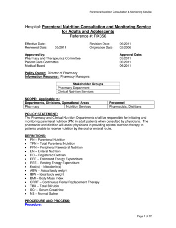
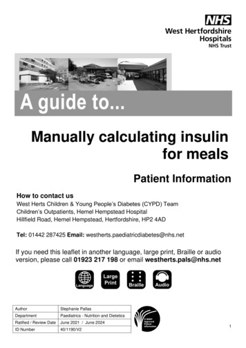
Affinati et al. Clinical Diabetes and Endocrinology(2021) 7:8non-adherence and ongoing depression. At the time ofadmission, her hemoglobin A1c was 11.6 %. Chart reviewindicated that she was prescribed Lantus 60 units daily,metformin 1000 mg twice daily, glimepiride 4 mg twicedaily with meals and 1.5 mg dulaglutide weekly as anoutpatient. Physical exam was notable for acanthosisnigricans.On admission, laboratory testing was notable for asodium of 130 mEq/L, potassium 5.0 mEq/L, chloride 93mmol/L, bicarbonate 16 mmol/L (anion gap 21), bloodglucose 760 mg/dL, creatinine 1.65 mg/dL (eGFR 44),lactic acid of 8.33 mmol/L and serum β-hydroxybutyrateof 2.69 mmol/L. Arterial blood gas showed a pH of 7.36,pCO2 of 31 mmHg and pO2 of 55 mmHg, consistentwith a mixed anion gap metabolic acidosis and respiratory alkalosis. SARS-CoV-2 PCR testing was positive.See Table 1 for additional laboratory values, includinginflammatory markers, which were initially elevated onadmission.Due to the elevated serum ketones and anion-gapmetabolic acidosis, she was treated for diabetic ketoacidosis (DKA) with a continuous insulin infusion at 7 units/hr(0.05 units/kg/hr) along with a normal saline infusion. Herblood glucose levels remained elevated in the 400–500 mg/dL range while her anion gap decreased to 15 butdid not resolve. Over the next 36 h her insulin infusionrate peaked at 34.5 units/hr (0.26 units/kg/hr) and she wasstarted on 50 units of insulin glargine and 50 units ofregular insulin every 6 h in addition to the insulin infusionto facilitate weaning off of the insulin drip. During the first36 h following transfer to our hospital, she had an averageinsulin requirement of 5 units/kg/day. Her insulin infusionwas weaned off within 48 h and her subcutaneous insulinrequirements continued to be elevated at 3.24 units/kg/day. Following extubation on day 6, her insulin requirements dropped to 211 units per day (1.64 units/kg) andshe continued to require over 200 units of subcutaneousinsulin daily for the next 13 days (Fig. 1). Her insulin requirements improved as she was weaned off of heatedhigh flow nasal cannula and transitioned from tube feedsto a diabetic diet. Prior to discharge to a subacute rehabilitation center, she was transitioned to her home regimenof 60 units Lantus, metformin, glimepiride and dulaglutide. While in the hospital she had well-controlled bloodsugars on this regimen. Two months after discharge, whilestill at the subacute rehabilitation center, her hemoglobinA1c was 6.0 %. Her kidney function had returned to herbaseline (eGFR 90).Page 2 of 5Table 1 Laboratory values measured on admissionLaboratory Test3WBC (cells x10 /uL)Patient 1Patient 219.918.34Neutrophils (cells x103/uL)17.314.12Lymphocytes (cells x103/uL)0.82.57Monocytes (cells x103/uL)1.91.2813.711.6Sodium (mEq/L)130141Potassium (mEq/L)54.6Chloride (mEq/L)93100Bicarbonate (mEq/L)1627BUN (mg/dL)1974Hemoglobin (g/dL)Serum ChemistriesCreatinine (mg/dL)1.652.01Glucose (mg/dL)760272Anion Gap2114Calcium (mg/dL)8.79.2Albumin (g/dL)3.93.8Alkaline Phosphatase (IU/L)10598AST (units/L)8144ALT (units/L)4469Total Bilirubin (mg/dL)1.1 0.2Arterial pH7.367.4Arterial CO2 (mmHg)3156Arterial O2 (mmHg)5568Arterial Blood GasInflammatory MarkersESR (mm/hr)85120CRP (mg/L)128.5121.7CK (units/L)-82Fibrinogen (mg/dL)669295 (transfer)LDH (units/L)Admission810885Transfer-871Procalcitonin (ng/mL)Admission-0.72Transfer0.170.21D-Dimer (ng/mL)Admission1547969Transfer12.19 35Admission98744Transfer-595.3352166Ferritin (ng/mL)Triglycerides (mg/dL)Case 2A 47-year old African-American woman with a historyof pre-diabetes on metformin, obesity (BMI 39 kg/m2),hypertension and hyperlipidemia presented with cough,dyspnea, hypoxia and fever and was emergently intubated.AdmissionTransferHemoglobin A1c34729511.60%7.30%
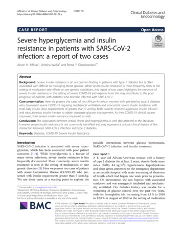
Affinati et al. Clinical Diabetes and Endocrinology(2021) 7:8Page 3 of 5Fig. 1 Glucose levels and total daily insulin dose during the first 10 days following admission. Glucose levels (black circles, upper panel) and totaldaily dose of insulin (black bars, lower panel) by days following admission. Respiratory interventions indicated by arrows. Blue bars representduration of 1 mg/kg methylprednisolone treatment for ARDS. Green bars represent duration of tube feedsShe was found to be SARS-CoV-2 positive. She wasinitially treated with hydroxychloroquine, azithromycin,40 mg methylprednisolone twice daily and high dosevitamin C with blood glucoses recorded in the 300 s. Herhemoglobin A1c was 7.3 % on admission.Regarding her diabetes history, she was diagnosed withpre-diabetes in 2016 with a hemoglobin A1c of 5.9 %and started on metformin therapy. Her hemoglobin A1cranged between 5.5 and 6.2 % until her admission forCOVID-19.Following admission, her course was complicated by significant difficulty with oxygenation, pneumomediastinumdue to barotrauma and acute kidney injury. She was ultimately transferred to our institution for extra-corporealmembrane oxygenation (ECMO) from an outside hospital.On arrival at our hospital, she received 1 dose of sarilumab(human monoclonal antibody against interleukin-6 (IL-6))and was started on IV methylprednisolone. She rapidly developed worsening hyperglycemia with glucose levels above400 mg/dL in the setting of methylprednisolone treatmentand was started on an insulin infusion. Subcutaneous insulin was initiated simultaneously to assist in weaning off theinsulin infusion as rapidly as possible. This protocol has recently been published in Gianchandani et al. [6]. Over thenext 24 h she required insulin drip rates up to 45 units/hrin addition to 20 units insulin glargine twice daily and 16units regular insulin every 6 h (see Fig. 1). The subcutaneous insulin was titrated up over 4 days until she was receiving 30 units of insulin glargine twice a day and 56 units ofregular insulin every 6 h with drip rates between 2 and 10units/hr. Her 24-hour insulin requirements peaked at 6.45units/kg/day. After discontinuation of methylprednisoloneher insulin requirements decreased significantly, her insulin infusion was titrated off and subcutaneous insulin wasdecreased to Lantus 25 units twice daily and regular insulin48 units every 6 h. Despite this improvement, she didcontinue to require over 200 units of insulin daily viasubcutaneous injection even after discontinuation of themethylprednisolone. Six days after methylprednisolonediscontinuation, her insulin requirements dropped precipitously, which coincided with ECMO decannulation. Insulinwas discontinued completely 12 days later. She continuedto require mechanical ventilation for over 2 months andwas discharged to a long-term care facility for furtherventilator weaning. Six months after discharge from thehospital, her creatinine had not yet returned to baseline,but had improved to 1.12 (eGFR 58). Her hemoglobin A1cat that time was 5.9 % without any diabetes medications.She had successfully been liberated from the ventilator andreturned home.DiscussionOver the last year, a picture has emerged that illustratesthe increased risk of severe infection and death in patients with diabetes who develop COVID-19. Almost30 % of patients with diabetes who present to the hospital with COVID-19 require intubation and over 10 %will die within a week [1]. Patients with diabetes aremore likely to require hospitalization and ICU admissionand have a higher risk of death than those withoutdiabetes [2–4]. Here we present the cases of two patientswith a history of diabetes that developed COVID-19,difficult to control hyperglycemia and severe insulinresistance, prompting us to consider the mechanisms
Affinati et al. Clinical Diabetes and Endocrinology(2021) 7:8underlying the poor prognosis for patients with diabetesand how glucose control and insulin treatment mightinteract with this infection.Prior to the COVID-19 pandemic, there was already awealth of data demonstrating that hyperglycemia and insulin resistance during critical illness is associated withworse outcomes [7–10]. One of the remarkable featuresof the patients presented here is the degree of insulin resistance, with both patients requiring more than 5 units/kg/day shortly after admission. This meets the criteriafor extreme insulin resistance, defined as more than 3units/kg/day. Extreme insulin resistance is most oftenseen in the setting of rare genetic disorders such as lipodystrophy and severe insulin resistance syndrome, use ofmedications such as glucocorticoids or endocrinopathiessuch as Cushings [5].As dexamethasone is one of the few treatmentsthat decreases mortality in severe COVID-19 infection [11, 12], this is likely a significant contributorto the severe hyperglycemia in many patients withCOVID-19. However, it is not the only cause, as thefirst patient described in this report developed severehyperglycemia and insulin resistance despite neverreceiving steroids. Similarly, the second patient continued to have severe insulin resistance for 6 daysfollowing discontinuation of steroids.Acute infections also lead to increased insulin resistancethrough enhanced secretion of the counter-regulatoryhormones cortisol, glucagon and growth hormone [13]. Inaddition to the hormonal response to stress, cytokineexpression may independently contribute to increasinginsulin resistance during infection [14]. Thus, the robustinflammatory response in patients with SARS-CoV-2could be a significant contributor to their severe insulinresistance [6], as both of our patients had elevated inflammatory markers that coincided with increasing insulindemands (Table 1).The interaction between insulin resistance, severehyperglycemia and inflammation is likely to be particularly important in the setting of SARS-CoV-2 infection,as the robust inflammatory response has been linked topoor outcomes [15]. In patients with diabetes whopresent with COVID-19, the inflammatory marker Creactive protein (CRP) has been identified as one of thestrongest risk factors for mortality [2], while patientswith prolonged hyperglycemia have higher IL-6 and Ddimer levels and a higher risk of progressing to severedisease [16]. While inflammation leads to hyperglycemiaand insulin resistance, hyperglycemia and insulin resistance also exacerbate inflammation, impair immune cellfunction and promote epithelial cell dysfunction, even inotherwise healthy volunteers [17–21].Treatment of hyperglycemia in patients with COVID-19has been particularly difficult as many centers havePage 4 of 5attempted to limit insulin infusions to preserve PPE andminimize health care provider exposure, as treatment withinsulin infusion require hourly finger stick blood glucosechecks. Given the extraordinary doses of insulin requiredin the patients presented here, it is valuable to considerthe potential effects of insulin beyond its hypoglycemic effects, and in particular, its role in inflammation.Insulin’s role in inflammation is likely to be contextdependent. When evaluated in the setting of obesityrelated insulin resistance, high doses of insulin may bepro-inflammatory. In both obese mice and humans,hyperinsulinemia results in elevated adipose tissue cytokine levels [22–25], while this does not occur in healthy,non-obese and non-insulin resistant patients [26]. Incontrast, many studies have demonstrated that insulintreatment in the setting of sepsis is anti-inflammatory.In multiple animal models of infection, insulin treatmentdecreases systemic cytokines independent of glucoselevels [27–29]. This finding holds true in human studiesas well. In healthy patients with both type 1 and type 2diabetes, insulin infusion (with dextrose to maintain euglycemia) leads to decreased reactive oxygen species, systemiccytokine gene expression and serum CRP levels [30–32].ConclusionsBased on our review of the literature and exemplified bythe cases presented here, it is likely that hyperglycemiain patients with COVID-19 infection worsens the dramatic inflammation and cytokine storm that accompanies severe infection. Insulin treatment reduces bloodglucose levels and may have an anti-inflammatory actionin the setting of COVID-19 sepsis.AbbreviationsCOVID-19: Coronavirus Disease 2019; DKA: Diabetic ketoacidosis;ECMO: Extra-corporeal membrane oxygenation; CRP: C-reactive protein; IL6: Interleukin-6AcknowledgementsNot applicable.Authors’ contributionsAHA drafted the manuscript. AHA and RYG were involved in the care of atleast one of the patients described; all authors reviewed and edited themanuscript; and have approved the final version of the article.FundingAHA is supported by grant F32 DK122660 from the National Institutes ofDiabetes and Digestive and Kidney Diseases. AW has received funding fromNovo Nordisk and research support from United Health Group and Eli Lilly.Availability of data and materialsData sharing is not applicable to this case report as no datasets weregenerated or analyzed during the current study.DeclarationsEthics approval and consent to participateNot applicable.
Affinati et al. Clinical Diabetes and Endocrinology(2021) 7:8Consent for publicationNot applicable.Competing interestsThe authors declared that they have no competing interests.Author details1Department of Internal Medicine, Division of Metabolism, Endocrinologyand Diabetes, University of Michigan, Domino’s Farms (Lobby G, Suite 1500),24 Frank Lloyd Wright Drive, MI 48106 Ann Arbor, USA. 2Division ofEndocrinology, Metabolism and Molecular Medicine, Northwestern UniversityFeinberg School of Medicine, IL, Chicago, USA.Received: 18 January 2021 Accepted: 27 April 2021References1. For the CORONADO investigators. Cariou B, Hadjadj S, Wargny M, PichelinM, Al-Salameh A, et al. Phenotypic characteristics and prognosis ofinpatients with COVID-19 and diabetes: the CORONADO study. Diabetologia[Internet]. 2020 May 29 [cited 2020 Jun 1]; Available from: 00125-020-05180-x.2. Chen Y, Yang D, Cheng B, Chen J, Peng A, Yang C, et al. ClinicalCharacteristics and Outcomes of Patients With Diabetes and COVID-19 inAssociation With Glucose-Lowering Medication. Diabetes Care. 2020;43(7):1399-407.3. Kumar A, Arora A, Sharma P, Anikhindi SA, Bansal N, Singla V, et al. Isdiabetes mellitus associated with mortality and severity of COVID-19? Ameta-analysis. Diabetes Metab Syndr Clin Res Rev. 2020;14(4):535–45.4. Zhu L, She Z-G, Cheng X, Qin J-J, Zhang X-J, Cai J, et al. Association ofBlood Glucose Control and Outcomes in Patients with COVID-19 and Preexisting Type 2 Diabetes. Cell Metab. 2020;31(6):S1550413120302382.5. Ovalle F. Clinical approach to the patient with diabetes mellitus and veryhigh insulin requirements. Diabetes Res Clin Pract. 2010;90(3):231–42.6. Gianchandani R, Esfandiari NH, Ang L, Iyengar J, Knotts S, Choksi P, et al.Managing Hyperglycemia in the COVID-19 Inflammatory Storm. Diabetes.2020;69(10):dbi200022.7. Bonizzoli M, Zagli G, Lazzeri C, Degl’Innocenti S, Gensini G, Peris A. Earlyinsulin resistance in severe trauma without head injury as outcomepredictor? A prospective, monocentric pilot study. Scand J Trauma ResuscEmerg Med. 2012;20:69.8. Capes SE, Hunt D, Malmberg K, Gerstein HC. Stress hyperglycaemia andincreased risk of death after myocardial infarction in patients with andwithout diabetes: a systematic overview. Lancet. 2000;4(9206):773–8.9. Lazzeri C, Bonizzoli M, Cianchi G, Ciapetti M, Socci F, Peris A. The prognosticrole of peak glycemia and glucose variability in trauma: a single-centerinvestigation. Acta Diabetol. 2020;57(8):931-5.10. Brealey D, Singer M. Hyperglycemia in critical illness: a review. J Diabetes SciTechnol. 2009;1(6):1250–60.11. Tomazini BM, Maia IS, Cavalcanti AB, Berwanger O, Rosa RG, Veiga VC, et al.Effect of Dexamethasone on Days Alive and Ventilator-Free in Patients WithModerate or Severe Acute Respiratory Distress Syndrome and COVID-19:The CoDEX Randomized Clinical Trial. JAMA. 2020;324(13):1307.12. The RECOVERY Collaborative Group. Dexamethasone in HospitalizedPatients with Covid-19 — Preliminary Report. N Engl J Med. 2020;384:NEJMoa2021436.13. Rizza RA, Cryer PE, Gerich JE. Role of glucagon, catecholamines, and growthhormone in human glucose counterregulation. Effects of somatostatin andcombined alpha- and beta-adrenergic blockade on plasma glucoserecovery and glucose flux rates after insulin-induced hypoglycemia. J ClinInvest. 1979;64(1):62–71.14. Aljada A, Ghanim H, Assian E, Dandona P. Tumor necrosis factor-alphainhibits insulin-induced increase in endothelial nitric oxide synthase andreduces insulin receptor content and phosphorylation in human aorticendothelial cells. Metab Clin Exp. 2002;51(4):487–91.15. Blanco-Melo D, Nilsson-Payant BE, Liu W-C, Uhl S, Hoagland D, Møller R,et al. Imbalanced Host Response to SARS-CoV-2 Drives Development ofCOVID-19. Cell. 2020;181(5):S009286742030489X.16. Sardu C, D’Onofrio N, Balestrieri ML, Barbieri M, Rizzo MR, Messina V, et al.Outcomes in Patients With Hyperglycemia Affected by Covid-19: Can WeDo More on Glycemic Control? Dia Care. 2020;43(7):dc200723.Page 5 of 517. Ieronymaki E, Theodorakis EM, Lyroni K, Vergadi E, Lagoudaki E, Al-QahtaniA, et al. Insulin Resistance in Macrophages Alters Their Metabolism andPromotes an M2-Like Phenotype. J Immunol. 2019;15(6):1786–97.18. Marik PE, Raghavan M. Stress-hyperglycemia, insulin andimmunomodulation in sepsis. Intensive Care Med. 2004;30(5):748–56.19. Nielson CP, Hindson DA. Inhibition of polymorphonuclear leukocyterespiratory burst by elevated glucose concentrations in vitro. Diabetes. 1989;38(8):1031–5.20. Perner A, Nielsen SE, Rask-Madsen J. High glucose impairs superoxideproduction from isolated blood neutrophils. Intensive Care Med. 2003;29(4):642–5.21. Ghanim H, Aljada A, Daoud N, Deopurkar R, Chaudhuri A, Dandona P. Roleof inflammatory mediators in the suppression of insulin receptorphosphorylation in circulating mononuclear cells of obese subjects.Diabetologia. 2007;50(2):278–85.22. Pedersen DJ, Guilherme A, Danai LV, Heyda L, Matevossian A, Cohen J, et al.A major role of insulin in promoting obesity-associated adipose tissueinflammation. Mol Metab. 2015;4(7):507–18.23. Zhang AMY, Wellberg EA, Kopp JL, Johnson JD. Hyperinsulinemia inObesity, Inflammation, and Cancer. Diabetes Metab J. 2021. Online ahead ofprint.24. Westerbacka J, Cornér A, Kannisto K, Kolak M, Makkonen J,Korsheninnikova E, et al. Acute in vivo effects of insulin on geneexpression in adipose tissue in insulin-resistant and insulin-sensitivesubjects. Diabetologia. 2006;49(1):132–40.25. Krogh-Madsen R, Plomgaard P, Keller P, Keller C, Pedersen BK. Insulinstimulates interleukin-6 and tumor necrosis factor-alpha gene expression inhuman subcutaneous adipose tissue. Am J Physiol Endocrinol Metab. 2004;286(2):E234–8.26. Tannous A, Bradford AP, Kuhn K, Fought A, Schauer I, Santoro N. Arandomised trial examining inflammatory signaling in acutely inducedhyperinsulinemia and hyperlipidemia in normal weight women-thereprometabolic syndrome. PLoS One. 2021;16(3):e0247638.27. Hagar JA, Edin ML, Lih FB, Thurlow LR, Koller BH, Cairns BA, et al.Lipopolysaccharide Potentiates Insulin-Driven Hypoglycemic Shock. JImmunol. 2017;15(10):3634–4328. Holger JS, Dries DJ, Barringer KW, Peake BJ, Flottemesch TJ, Marini JJ.Cardiovascular and metabolic effects of high-dose insulin in a porcineseptic shock model. Acad Emerg Med. 2010;17(4):429–35.29. Brix-Christensen V, Andersen SK, Andersen R, Mengel A, Dyhr T, AndersenNT, et al. Acute hyperinsulinemia restrains endotoxin-induced systemicinflammatory response: an experimental study in a porcine model.Anesthesiology. 2004;100(4):861–70.30. Dandona P, Ghanim H, Green K, Sia CL, Abuaysheh S, Kuhadiya N, et al.Insulin infusion suppresses while glucose infusion induces Toll-like receptorsand high-mobility group-B1 protein expression in mononuclear cells of type1 diabetes patients. Am J Physiol Endocrinol Metab. 2013;304(8):E810-818.31. Ghanim H, Korzeniewski K, Sia CL, Abuaysheh S, Lohano T, Chaudhuri A,et al. Suppressive effect of insulin infusion on chemokines and chemokinereceptors. Diabetes Care. 2010;33(5):1103–8.32. Ghanim H, Mohanty P, Deopurkar R, Sia CL, Korzeniewski K, Abuaysheh S,et al. Acute modulation of toll-like receptors by insulin. Diabetes Care. 2008;31(9):1827–31.Publisher’s NoteSpringer Nature remains neutral with regard to jurisdictional claims inpublished maps and institutional affiliations.
decreased to Lantus 25 units twice daily and regular insulin 48 units every 6 h. Despite this improvement, she did continue to require over 200 units of insulin daily via subcutaneous injection even after discontinuation of the methylprednisolone. Six days after methylprednisolone discontinuation, her insulin requirements dropped precipi-