Transcription
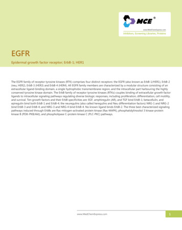
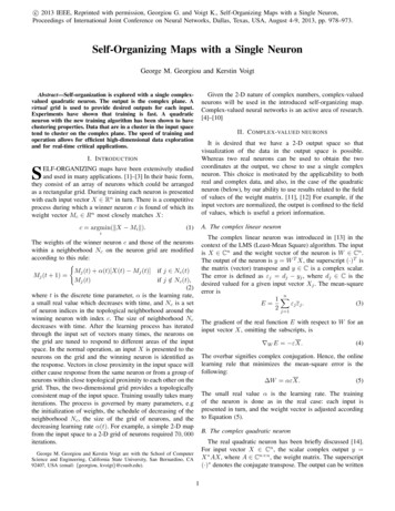
Neuron, Vol. 20, 371–380, March, 1998, Copyright 1998 by Cell PressVoltage-Gated Ion Channelsand Electrical ExcitabilityClay M. Armstrong* and Bertil Hille†‡* Department of PhysiologyUniversity of PennsylvaniaPhiladelphia, Pennsylvania 19104† Department of Physiology and BiophysicsUniversity of WashingtonSeattle, Washington 98195Our ability to do gymnastics, to perceive a colorful world,and to process language relies on rapid communicationamong neurons. Such signaling, the fastest in our bodies, involves electrical messages produced as ion channels in cell membranes open and close. Various ionchannels mediate sensory transduction, electrical “computations,” propagation over long distances, and synaptic transmission. Here, we focus on voltage-gated ionchannels, the family of channels that includes the familiar Na1, K1, and Ca2 1 channels of nerve and muscleexcitability. In the computer metaphor of the brain, thevoltage-gated ion channels are like the transistors oflogical circuits, detecting, amplifying, and reshapingelectrical messages. Our basic understanding of theseproteins maintains the framework and rigor established50 years ago by Hodgkin and Huxley (1952), enriched bymuch new molecular information and by insights gainedfrom patch-clamp methods.Although we have had full amino acid sequences ofvoltage-gated channels for over a decade, we still lackeven modest resolution three-dimensional information.All three-dimensional diagrams in the literature derivefrom functional studies without the benefit of crystallography or NMR. Figure 1A represents widely acceptedfunctional information, much of it deriving from earlybiophysical and pharmacological work that will be described in this article. An overriding conclusion is thation channels are aqueous pores. Proceeding throughthe pore from the outside, an ion would find a wide outervestibule, a narrow selectivity filter, a voluminous innervestibule, and finally, at the cytoplasmic end, the gatingmachinery that closes the pore. Highly charged voltagesensors control activation of the channel but are lessimportant for inactivation. All of the voltage-gated channels have a 4-fold symmetry, with the pore formed at thecentral line of contact of four channel-forming domains.Unlike ligand-gated channels of fast chemical synapses,much more of the mass of the voltage-gated channelslies on the intracellular side of the membrane than onthe extracellular side.In this review, we first consider how this field wasinitiated six decades ago and then see how functionsof voltage-gated ion channels have been uncovered andmapped onto the linear amino acid sequence of theprotein. The rigor of the original analysis set the tonefor a new discipline that now produces more than 5000papers a year.‡ To whom correspondence should be addressed.ReviewEarly Biophysics and Voltage Clamp RevealedVoltage-Gated Membrane PermeabilitiesThe period from 1939 to 1952 was a heroic time in thestudy of membrane biophysics. During this period,Hodgkin and Huxley explained the propagated actionpotential. Their definitive description of ionic permeability changes in the axon membrane in 1952 was closelypreceded by five important discoveries. Four of themoccurred shortly before the Second World War. Hodgkinshowed that local circuit currents from an excited regionof nerve are needed to bring the next region into activity.This meant that depolarization is the natural stimulusfor action potential propagation. J. Z. Young (1936) rediscovered the giant axon of the squid. It provided thefirst convenient way to place electrodes and even electrode arrays inside an excitable cell. Cole and Curtis(1939) showed that the membrane of the squid giantaxon increases its conductance 40-fold during an actionpotential, in apparent agreement with Bernstein’s earliertheory of membrane breakdown. Hodgkin and Huxley(1939) discovered with intracellular electrodes that thepeak of the action potential overshoots 0 mV by a significant margin. The overshoot was a serious problem forthe Bernstein theory, and Hodgkin and Katz (1949) eventually provided the crucial resolution: the overshoot isdetermined by the Na1 equilibrium potential and mustbe due to Na1 entry during the action potential.With this background, Hodgkin and Huxley sought tounderstand how excitation regulates the entry of Na1ions. They developed the voltage clamp to measureion movements as electric currents. The clamp recordsrevealed an inward current followed by an outward current during step depolarizations. In a major conceptualleap, Hodgkin and Huxley (1952) deduced that thesemembrane currents could be assigned to Na1 and K1permeability mechanisms whose conductances arefunctions of time and membrane potential. The assumption of separable permeability components and the realization that membrane potential is the controlling variable were the paradigm shifts that opened a new fieldof inquiry. Their five seminal papers systematically extracted kinetic constants for an empirical description ofthe conductance changes, showed that this detailedkinetic description—the Hodgkin-Huxley model—suffices to explain all of the classical properties of actionpotential excitation and propagation, and even offereda plausible physical basis for the control by membranepotential. The stage was brilliantly set.Consider a few of the findings of these papers. Today,we like to emphasize that ion channels have two majorproperties: permeation and gating. This separation wasclear in the original papers. With respect to permeation,the principal emphasis was that each component ofcurrent obeyed Ohm’s law with a reversal potential at theNernst potential for Na1 and K1 ions. When extracellularNa1 concentration was changed, they argued that theflux in either direction across the membrane was proportional to the concentrations on either side. The clearimplication was that ion movement was strictly diffusiondown an electrochemical gradient without additionalforces.
Neuron372Figure 1. Structure of Voltage-Gated Ion ChannelsFunctional components (A) and peptide folding (B) are shown diagrammatically with P regions in red and the S4 segment in pink. Abbreviations:S. F., selectivity filter; V. S., voltage sensor; O. V., outer vestibule; and I. V., inner vestibule. The folding diagram is for an Na1 channel withfour similar domains (I–IV). A K 1 channel is a homo-tetramer of subunits similar to a single Na1 channel domain.With respect to gating, much more was discovered.Notably, the permeabilities to Na1 and to K1 had quitedifferent time courses. The Na1 conductance rose andinactivated quickly during a depolarization, and in theaxon the K1 conductance rose more slowly without inactivation. Changing ion concentrations changed thedirection of current flow but not the time course: “Thechanges in membrane permeability appear to dependon membrane potential, and not on membrane current.”In addition, Hodgkin and Huxley succeeded in describing the permeability changes as a set of chemical reactions whose rate constants are a function of voltage.The rate of change of each permeability depended quitesteeply on the membrane potential, and a 10-fold increase in opening occurred with membrane potentialdepolarizations as small as 7–12 mV.What did Hodgkin and Huxley say about mechanism?About permeation, they said very little except that fluxwas downhill. They did not mention the concept of anaqueous pore, nor did they use the word channel. Aboutgating, a word they also did not use, they argued, “Details of the mechanism will probably not be settled forsome time, but it seems difficult to escape the conclusion that the changes in ionic permeability depend onthe movement of some component of the membranewhich behaves as though it had a large charge or dipolemoment.” These charged controlling “particles” wereenvisioned to move under the influence of the electricfield in the membrane and to “allow ions to pass whenthey occupy particular sites in the membrane.” The distribution of the particles was compared to a Boltzmanndistribution, exactly as is done today, and the steepnessof the voltage dependence required that as many assix electronic charges move fully across the membraneduring activation. To account for the sigmoid time courseof activation of the conductances, the model supposedthe movement of three gating particles for Na1 and fourfor K1. These hypothetical particles combined the functions that we now separately assign to the voltage sensors, the gates, and the conducting pore. Their multiplicity in the model was a harbinger of the modern findingof four structurally separate voltage sensors in eachchannel. The novelty, depth, and durability of these insights are a monument to the powerful physical intuitionof these two great scientists.Channels Become MoleculesCloning of the pore-forming a subunits of voltage-gatedNa1, K1, and Ca21 channels was reported in 1984–1988.It showed that, fundamentally, these channels are allmembers of the same gene superfamily with the sameoverall structure. This good news confirmed that thereshould be much mechanistic similarity among the channels and that we were free to generalize—as we hadalready been doing in the biophysical work of the previous decades.The Na1 and Ca21 channel clones, first reported by theNuma laboratory, predict large peptides (.2000 aminoacids) containing four homologous repeats (domainsI–IV), each of which has a motif with six putative transmembrane segments, S1–S6 (Figure 1B). The cloned K1channel was first reported by the laboratories of Janand Jan (Tempel et al., 1987), Tanouye (Kamb et al.,1987), and Pongs (Pongs et al., 1988). It is about onefourth as large as an Na1 channel and contains only onecopy of the S1–S6 motif. As might be expected, K1channels were later shown to be tetramers of the poreforming subunit (MacKinnon, 1991), making them structurally quite similar to Na1 and Ca21 channels. In all ofthree clones, segments S1, S2, S3, S5, and S6 are quitehydrophobic, and each is long enough to span the membrane as a helix. In the S4 segments, however, everythird residue is a positively charged arginine or lysine,for a total of seven positive charges in KV1.1 and five toeight positive charges in the various S4 domains of Na1and Ca21 channels. The S4 segment was almost immediately recognized as a candidate for the voltage sensor—one of Hodgkin and Huxley’s controlling particles. Aftera brief consignment to the cytoplasm, it joined the membrane-crossing segments where it could experience theelectric field of the membrane as required. The greatimportance of another amphipathic loop between segments S5 and S6 (called P or H5 and shown in red inFigure 1A) was recognized only later. It was first assigned its present status as part of the outer vestibuleand the conducting pore on the basis of thoughtful molecular modeling (Conti and Guy, 1990).The folding topology in Figure 1B has much to supportit. To mention only some of the evidence, the N terminusis known from the experiments of Aldrich and colleagues
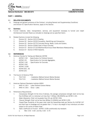
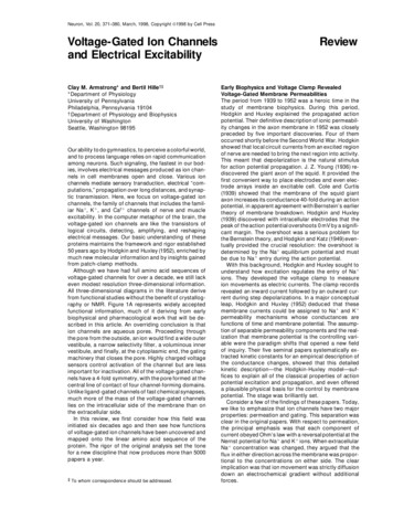
Review: Armstrong and Hille373Figure 2. Ionic Currents through a SingleChannel Sum to Make Classic MacroscopicCurrents(A) is a single sodium channel from Sigworthand Neher (1980), and (B) is a single potassium channel from Zagotta et al. (1988).to be cytoplasmic, and in Shaker B (ShB) K1 channelsthis terminus contains the molecular machinery for fast“N-type” inactivation gating (Hoshi et al., 1990). Thesidedness of many residues of these channels has beenidentified by mutating an individual residue to cysteineand then testing whether one of Karlin’s thiol-reactivecompounds, e.g., MTSET (Akabas et al., 1992), must beapplied to the extracellular or the cytoplasmic face tolabel the cysteine. One particularly illuminating set ofexperiments showed that S4 extends across the membrane: a cysteine introduced into the N-terminal end ofS4 can be labeled only from the outside, and a cysteineintroduced into the other end of S4 can be labeled onlyfrom the inside, both in Na1 (Yang and Horn, 1995; Yanget al., 1996) and K1 channels (Larsson et al., 1996). Thegeneral hairpin topology of the P region was workedout by mutating residues that altered sensitivity to poreblockers. The key was that a threonine residue in themiddle of the P region is crucial for block by intracellulartetraethylammonium ion (TEA1), whereas residues bothbefore and after this threonine determine the sensitivityto block by extracellular TEA1 and charybdotoxin (Yellenet al., 1991; and see Hartmann et al., 1991). Many otherinsightful experiments support the proposed topology(e.g., Shih and Goldin, 1997), and we are aware of nocontradictions.Conduction and SelectivityHodgkin and Huxley conceptually separated ion conduction from what we now call gating, but the nature ofthe conducting path, whether ion carrier or ion pore,lipid or protein, did not become clear for many years.Selective block and channel conductance estimatesfrom experiments with the channel blockers tetrodotoxin (TTX1) and TEA1 led each of us to conclude bythe early seventies that Na1 and K1 channels must beseparate aqueous pores and that transport based on acarrier such as valinomycin was too slow to be considered. The final proof was made possible 10 years laterby the the patch clamp (Hamill et al., 1981), which conferred the ability to measure current through a single ionchannel (Figure 2). We found this beautiful and exactly asanticipated.What is the nature of the conducting pore? Becauseof its simple tetrameric structure, the K1 channel hasbeen a favorite for experimentation. This channel’s maintask is letting K1 ions out and keeping Na1 ions fromgoing in. To explain the .50:1 selectivity, one can invokeonly the geometry of the pore and the energetics ofinteraction of the ions with water and with the residuesof the pore. We both found by ion substitution studiesin the early seventies that the pore narrows somewhereto a bore of only 3.0 Å—just accommodating Rb 1 ions.As the crystal radius of Na1 ions is smaller than that ofK1 ions, Na1 would fit through any hole that K1 can.Therefore, the discrimination has to be blamed on theunfavorable energetics of stripping most of the waterfrom the more strongly hydrated Na1 ion in a small holewithout providing much favorable interaction with thechannel in return.This constriction of the pore is now attributed to thefour 20-residue P regions, which are thought to dip partway into the membrane from the extracellular side andthen loop back out (red loops in Figure 1B). These residues narrow the pore, forming the lining of the outervestibule and the narrow selectivity filter (Figure 3A)but not the inner vestibule. Within each P region is aremarkably conserved “signature sequence”, -TXXTXGYG- (-Thr-X-X-Thr-X-Gly-Tyr-Gly ), found in at least 50cloned K1-selective channels, including the much smallerinward rectifier channels. The signature sequence isthought to be the heart of the selectivity filter. How doesit work? Thus far, the most illuminating experiment hasbeen negative. The sequence of the Shaker K1 channelP region (TMTTGYG) contains three threonine residueswhose -OH groups seemed prime candidates for complexing with and selecting K1 ions. However, all hydroxylgroups except the one on the first threonine were foundto be unnecessary for a selective channel, and evidenceregarding the first was inconclusive (Heginbotham etal., 1994). As an alternate explanation, the authors speculated that each subunit contained a sharp loop (perhaps at GYG), resulting in an exposed backbone carbonyl whose oxygen served to complex the K1 ion.Another possibility is that K1 channels have more thanone selective site, and no single mutation will completelydestroy selectivity.Many ingenious experiments have explored the poreregion of K1 channels, beginning in Miller’s lab with theuse of charybdotoxin to probe the geometry of the poremouth (MacKinnon and Miller, 1989). Other experiments
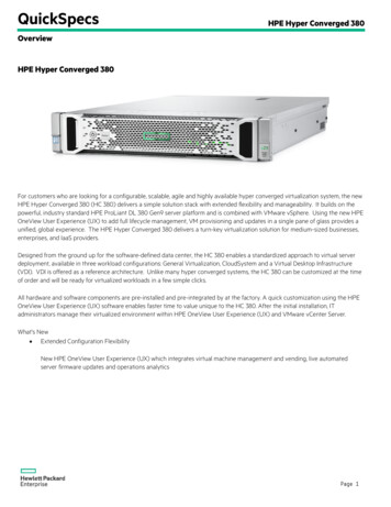
Neuron374Figure 3. Pore Regions at Varying Degrees of Abstraction(A) Cross section of the P region, S5 (containing L396), and S6 (containing V474) of a K1 channel, as visualized by Durell and Guy (1996).White dots mark the P loops.(B) The Na1 channel selectivity filter, from Hille (1971). At left, the 3 3 5 Å pore is surrounded by carbonyl and carboxyl oxygens, which, atright, bind a partially dehydrated Na1 ion sufficiently well to allow permeation.(C) A molecular model of the Na1 channel P region, from Lipkind and Fozzard (1994). Two charges from the outer (Glu 387 and 945) and inner(Asp 384 and Glu 942) charged rings are shown, and a TTX molecule occupies the outer vestibule. The selectivity filter is at the inner ring.White dots mark the backbone of the P loop.have involved replacement of selected residues by cysteine, followed by oxidation to produce disulfide bondsor reaction with Karlin’s thiol reagents or Ag1 (Yellen etal., 1994; Lü and Miller, 1995; Kürz et al., 1995; Krovetzet al., 1997). Thus far, these studies are in a general waycompatible with the picture shown in Figures 1 and 3A.The outer part of the P region is a funnel that reachesits narrowest part near the signature sequence. ExternalTEA1 blocks the wider part of the funnel, and its bindingis strongly enhanced by a mutation in the outer poreregion (T449Y; Figure 3A). The part of the pore internal tothe selectivity filter is formed by the S5 and S6 segments(Choi et al., 1993; Kirsch et al., 1993; Lopez et al., 1994;Holmgren et al., 1997). Ions in the outer vestibule alreadyfeel strong selectivity (Neyton and Miller, 1988). It seemslikely that the outer vestibule selects on the basis ofhydrated radius, whereas the narrow selectivity filterselects on the basis of the energy of stripping the iondown to its crystal radius (Hille, 1973). Unlike a K1 ion,which can shed some surrounding water molecules,TEA1 cannot shed its covalently linked ethyl groups.Thus TEA1, which is about the size of a hydrated K1ion, can occupy the outer and inner vestibules but cannot enter the selectivity filter.Similar studies have been done with Na1 channels.To generate the upstroke of the action potential, thesechannels have three selectivity tasks: letting Na1 go in,keeping K1 from going out, and preventing Ca21 ionsfrom getting stuck in the pore and interfering with Na1permeation. Early studies defined an outer vestibule asthe receptor for potent block by extracellular tetrodotoxin and an inner vestibule as the receptor for blockby local anesthetics. These studies also provided cleararguments for an acid group—a negative charge—withinthe pore that could interact with permeant cations andwith tetrodotoxin. The initial evidence was that the conductance of Na1 channels drops sharply when the bathing pH is lowered, as if an essential acid group (Hille,1968) located quite near the extracellular end of thepore (Woodhull, 1973) becomes neutralized, preventingcations from passing. The narrow part of the pore admitscations up to the size of the aminoguanidinium ion (requiring at least a 3 3 5 Å rectangular hole; Figure 3B),yet K1 ions permeate only 1/12 as well as Na1 (Hille,
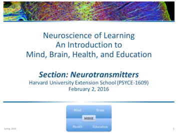
Review: Armstrong and Hille3751971, 1972). Why don’t K1 ions (2.7 Å diameter) glidethrough such an aperture as easily? Hille’s working hypothesis was that as ions passing through the apertureare partially dehydrated, they are stabilized by compensatory direct interaction with the negative charge in theselectivity filter. In this view, small ions like Li1 and Na1are well stabilized by the negative charge because theycan get close to it, and larger ions like K1 and Rb1cannot. Subsequent cloning showed that acid groupsare indeed present in the P regions of all Na1 channels(Figure 3C), and mutagenesis proved that they contribute significantly to conductance, selectivity, and tetrodotoxin binding (Terlau et al., 1991; Heinemann et al.,1992). Nevertheless, thus far we don’t know how to makequantitative tests of the selectivity hypothesis.The P regions of Na1 and Ca21 channels have similarsequences, and illuminating experiments have beenperformed to determine the basis for their Na1 or Ca21preference. Both channels have two rings of chargeencircling the pore, each ring containing four residues,one from each homologous domain (Figure 3C). Theouter ring is entirely negative and is thought to be relatively distant from the pore axis. The inner ring, two tothree residues deeper into the pore, is composed ofDEKA (Asp Glu Lys Ala) in the Na1 channel and EEEE(Glu Glu Glu Glu) in the Ca21 channel. In general, it seemsappropriate that the channel conducting doubly chargedCa21 should have more negative charge. With this inmind, Heinemann et al. (1992) succeeded in convertinga Na1 channel into a Ca2 1-preferring channel by pointmutations that increased the negativity in the inner ring.From their experiments, the importance of the inner ringwas immediately apparent. The mutated channels lostNa1/K1 discrimination (see also Favre et al., 1996) andbecame quite Ca21 permeable. In addition, Ca21 interference with monovalent permeability became severe, because Ca21 binds tightly to the added negative charge.The importance of the EEEE ring in Ca21 channels wasexamined in detail by Tsien and colleagues (Ellinor etal., 1995), yielding a detailed mechanism for Ca21/Na1discrimination by calcium channels. Divalent/monovalent selection thus seems well understood, whereasmonovalent/monovalent selectivity is at best understood in principle.Activation GatingVoltage-gated channels are exquisitely sensitive tosmall changes in membrane potential. Hodgkin andHuxley realized that Na1 and K1 channel opening oractivation must result from movement of charges withinthe membrane. Their work predicted “gating current,”a small charge movement generated (in more recentterms) by the voltage-driven conformational changesthat open the channels. Theoretically speaking, therewas no alternative, and in due course the expected current was detected, first in connection with excitationcontraction coupling in muscle (Schneider and Chandler,1973) and subsequently in nerve membranes (Figure 4A;Armstrong and Bezanilla, 1973; Keynes and Rojas,1974). By 1980, it seemed probable that the channelswere composed of protein, leading one of us to proposeFigure 4. Gating Current Gives Evidence for Slow Steps during Activation-Deactivation(A) The upper trace of each pair is Ig, and INa is below. At 210 mV,Ig has a slow component that parallels the activation of channels(as monitored by INa). From Armstrong and Gilly (1979).(B) For a K 1 channel, gating currents associated with deactivation(downward tails at pulse end) are much slower for large depolarizations that open many channels. From Stefani et al. (1994).that the gating particles of Hodgkin and Huxley were infact charged membrane helices—specifically, a negatively charged helix that moved inward relative to a positively charged helix, yielding a lot of charge movement,and hence voltage sensitivity, with relatively little physical motion (Armstrong, 1981). Each of four or five proposed subunits would contain such a pair of helices.When the Na1 channel was cloned a few years later,the positively charged helix—the S4 segment—was immediately visible. The expected negatively charged helixdoes not exist, and it is still not entirely clear how thenecessary counter charges for stabilizing the amphipathic S4 helix in the membrane are provided.The likelihood that S4 was the long-sought voltagesensor was immediately apparent, but experimental proofhas come more slowly. A first strategy was to createmutant channels with neutral residues replacing someof the positive charges in S4 (Stühmer et al., 1989).These experiments provided suggestive support, butthey were complicated by the failure of channels withmany neutralized residues to express and by drasticand often unanticipated changes in gating propertieswhen this sensitive helix was altered in any way. Recently, cysteine mutagenesis and cysteine labeling withthiol-reactive compounds has provided good evidencethat the S4 segments in both Na1 and K1 channels moveas expected following voltage changes (Yang and Horn,
Neuron3761995; Larsson et al., 1996; Yang et al., 1996). Cysteineresidues introduced at certain points in S4 show statedependent labeling; i.e., a given cysteine may be labeledonly from the ouside when the channel is open and onlyfrom the inside when it is closed. The labeling patterns ofboth Na1 and K1 channels are consistent with outwardmovement of S4 in response to a positive change ofmembrane potential.The details of how S4 moves may not be clear forsome time. It is likely that all four S4 units must movein order to open the channel and that there are severalnonconducting, partially activated states in which somebut not all S4 units have moved. In behavior, this corresponds closely to the Hodgkin-Huxley model of the K1channel with its four gating particles. But does each S4have only two possible positions, off and on, or does ithave more? This cannot yet be answered with precision.Biophysical evidence suggests that several fast stepsin the activation process are followed by a slow onethat is quite different. Gating current recorded from asquid axon at 240 mV decays to an undetectable levelbefore the ionic current begins to rise (Figure 4A; Armstrong and Gilly, 1979); at 210 mV, the gating currenthas two distinct components, a fast one and a slowercomponent (large enough to see at this voltage) thatparallels the rise of Na1 inward current. From examiningK1 channels, Aldrich and colleagues concluded that theS4 segments of the individual domains moved separately at first, followed by a “concerted” step involvingall four domains (Zagotta et al., 1994). Suggestively, thislast step can be separated from the early ones by amutation at the inner end of S4 (Schoppa et al., 1992).Gating current measurements from K1 channels alsostrongly support the concept of a late, slow voltagesensing step during activation (Stefani et al., 1994). InFigure 4B, for example, the gating current recorded asa mutant K1 channel closes (inward tail at pulse end) ismuch slower after a depolarization that opens the gatesof many channels (110 mV) than after a smaller depolarization that opens very few (230 mV). Apparently, thegate-opening step is reversed much more slowly thanthe preceding transitions among closed states. The precise meaning of these tantalizing hints is a puzzle thatremains to be worked out.Movement of the S4 segments provides the impetusfor conformational change, but where is the physicalgate that moves to open the channel? That the gate isinternal was shown long ago by experiments with TEA1(Armstrong, 1971). TEA1 and its relatives (QA1) occupythe inner vestibule of the channel, but they can get tothis blocking site only if the gate is open. Consistentwith this, Yellen and colleagues found (1) that mutationof residues in S6, which lines the inner vestibule, stronglyaffects binding of internally applied TEA1 derivatives(Holmgren et al., 1997); and (2) that cysteines substitutedinto S6 can be labeled only when the gate is open (Liuet al., 1997). Thus, the gate must be inside where it canprotect the inner vestibule, as in Figure 1A. Interestingly,the gate of a QA1-occupied channel can close rathereasily, trapping QA1 in the vestibule. Evidently, the vestibule must be large enough to accommodate the blockereven with the gate closed (Holmgren et al., 1997).What forms the gate is not yet clear. A reasonablecandidate would be the S4–S5 linker, the internally located 13-residue segment joining S4 to S5. Cysteinesintroduced into this segment are easier to label whenthe channels are partially or fully activated (Holmgrenet al., 1996), consistent with the idea that they form flapsof the gate but perhaps falling short of proving the case.Whatever its identity, does each monomer form onefourth of the gate? If this is the case, and the flaps arewithdrawn one by one, it is hard to see why the channeldoes not conduct after a single one is withdrawn. Thereare several alternatives. One is that there are four flapsand they are withdrawn simultaneously in a single, concerted step. Another possibility is that a gate flap fromone subunit is sufficient to close the channel and isoverlapped by flaps from the other subunits, as in Figure1A (a “stacked hands” model). A final possibility is thatopening the channel involves both flap movement andassociated changes in the P region. At least one gatingchange, called C-type inactivation, is associated with aclearly demonstrated constriction of the outer mouth ofthe pore (Liu et al., 1996). Thus far, however, there are noclear changes in the P region associated with activationgating.Inactivation GatingSodium channels open in response to depolarization,admitting Na1 and driving the membrane voltage positive during the upstroke of the action potential. Theythen inactivate spontaneously (stop conducting), making it easy for the K1 channels to restore the membranepotential to rest. Thus, Na1 channels and also someK1 channels have a second gating factor, inactivationgating, that is mechanistically simpler and quite differentfrom activation: after the activation gate opens, a cytoplasmically located portion of the channel peptide diffuses into the mouth of the inner vestibule of the poreand blocks conduction (Figure 5). The movement of thispeptide is relatively slow, allowing the average Na1channel, for example, enough conduction time to complete the rising phase of the action potential. The apparent voltage dependence of inactivation is primarily derived from the voltage dependence of the activationthat precedes it rather than from highly charged voltagesensors devoted to inactivation itself.Can it be that simple? Not quite, but let us beginsimply. The picture developed in several steps. Ancientexperiments showed that squid K1 channels could begiven a rather close semblance of inactivati
(1939) showed that the membrane of the squid giant tic transmission. Here, we focus on voltage-gated ion axon increasesitsconductance 40-foldduringan action channels, the family of channels that includes the famil- potential, inapparentagreement withBernstein's earlier iar Na1,K1, and Ca21 channels of nerve and muscle theory of membrane .