Transcription
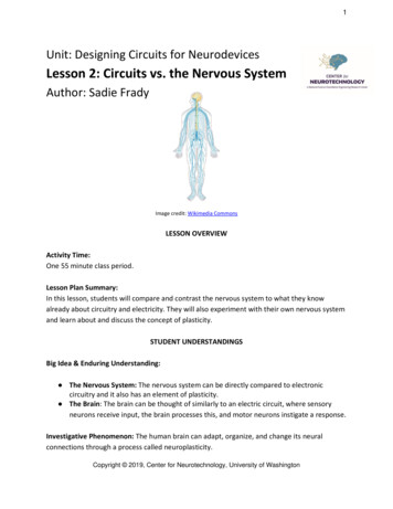
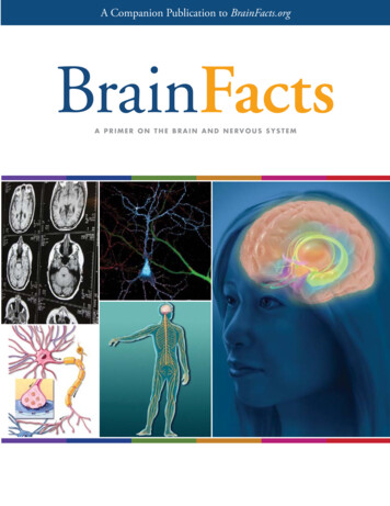
The Nervous SystemFunctions of the Nervous System1. Gathers information from both inside and outside the body - Sensory Function2. Transmits information to the processing areas of the brain and spine3. Processes the information in the brain and spine – Integration Function4. Sends information to the muscles, glands, and organs so they can respond appropriately – MotorFunctionIt controls and coordinates all essential functions of the body including all other body systemsallowing the body to maintain homeostasis or its delicate balance.The Nervous System is divided into Two Main Divisions: Central Nervous System (CNS) andthe Peripheral Nervous System (PNS)Divisions of the Nervous System1
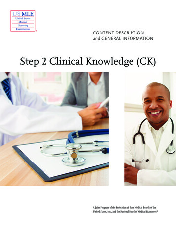
Basic Cells of the Nervous SystemNeuron Basic functional cell of nervous system Transmits impulses (up to 250 mph)Parts of a Neuron Dendrite – receive stimulus and carries it impulsestoward the cell bodyCell Body with nucleus – nucleus & most ofcytoplasmAxon – fiber which carries impulses away from cell bodySchwann Cells- cells which produce myelin or fat layer in the Peripheral Nervous SystemMyelin sheath – dense lipid layer which insulates the axon – makes the axon look grayNode of Ranvier – gaps or nodes in the myelin sheathImpulses travel from dendrite to cell body to axonThree types of Neuronso Sensory neurons – bring messages to CNSo Motor neurons - carry messages from CNSo Interneurons – between sensory & motor neurons in theCNSImpulses A stimulus is a change in the environment with sufficientstrength to initiate a response.Excitability is the ability of a neuron to respond to the stimulus and convert it into a nerve impulseAll of Nothing Rule – The stimulus is either strong enough to start and impulse or nothing happensImpulses are always the same strength along a given neuron and they are self-propagation – once itstarts it continues to the end of the neuron in only one direction- from dendrite to cell body to axonThe nerve impulse causes a movement of ions across the cell membrane of the nerve cell.Synapseo Synapse - small gap or space between the axon of one neuron and the dendrite of another - theneurons do not actually tough at the synapseo It is junction between neurons which uses neurotransmitters to start the impulse in the secondneuron or an effector (muscle or gland)o The synapse insures one-waytransmission of impulsesNeurotransmittersNeurotransmitters – Chemicals inthe junction which allow impulses tobe started in the second neuron2
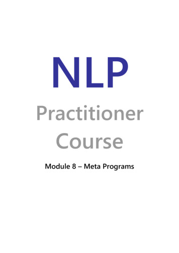
Reflex ArcComponents of a Reflex ArcA. Receptor - reacts to a stimulusB. Afferent pathway (sensory neuron) - conducts impulses to the CNSC. Interneuron - consists of one or more synapses in the CNS (most are in the spine)D. Efferent pathway (motor neuron) conducts impulses from CNS to effector.E. Effector - muscle fibers (as in the Hamstring muscle) or glands responds by contracting or secreting aproduct.Spinal reflexes - initiated and completed at the spinal cord level. Occur without the involvement of higher braincenters.Central Nervous System BrainooooSpineoBrain stem – medulla, pons, midbrainDiencephalon – thalamus & hypothalamusCerebellemCerebrumSpinal CordMeningesMeninges are the three coverings aroundthe brain & spine and help cushion, protect,andnourish the brain and spinal cord. dura mater is the most outer layer, verytough arachnoid mater is the middle layer andadheres to the dura mater and hasweblike attachments to the innermostlayer, the pia mater pia mater is very thin, transparent, buttough, and covers the entire brain,following it into all its crevices (sulci) and spinal cord cerebrospinal fluid, which buffers, nourishes, and detoxifies the brain and spinal cord, flows throughthe subarachnoid space, between the arachnoid mater and the pia mater3
Regions of the BrainCerebellum – coordination of movement andaspectsof motor learningCerebrum – conscious activity includingperception, emotion, thought, and planningThalamus – Brain’s switchboard – filters and thenrelays information to various brain regionsMedulla – vital reflexes as heart beat and respirationBrainstem – medulla, pons, and midbrain(involuntary responses) and relays information fromspine to upper brainHypothalamus– involved in regulating activitiesinternal organs, monitoring information from theautonomic nervous system, controlling the pituitary gland and its hormones, and regulating sleep andappetiteCerebrum Is the largest portion of the brain encompassesabout two-thirds of the brain mass It consists of two hemispheres divided by afissure – corpus callosumIt includes the cerebral cortex, the medullarybody, and basal gangliacerebral cortex is the layer of the brain oftenreferred to as gray matter because it has cellbodies and synapses but no myelino The cortex (thin layer of tissue) is graybecause nerves in this area lack theinsulation or white fatty myelin sheath thatmakes most other parts of the brain appearto be white.o The cortex covers the outer portion (1.5mmto 5mm) of the cerebrum and cerebellumo The cortex consists of folded bulges calledgyri that create deep furrows or fissures called sulcio The folds in the brain add to its surface area which increases the amount of gray matter and thequantity of information that can be processedMedullary body – is the white matter of the cerebrum and consists of myelinated axonsoCommisural fibers – conduct impulses between the hemispheres and form corpuscallosumoProjection fibers – conduct impulse in and out of the cerebral hemispheresoAssociation fibers – conduct impulses within the hemispheresBasal ganglia – masses of gray matter in each hemisphere which are involved in the control ofvoluntary muscle movements4
Lobes of the Cerebrum Frontal – motor area involved inmovement and in planning &coordinating behaviorParietal – sensory processing, attention,and languageTemporal – auditory perception, speech,and complex visual perceptionsOccipital – visual center – plays a role inprocessing visual informationSpecial regions Broca’s area – located in the frontal lobe – important in the production of speechWernicke’s area – comprehension of language and the production of meaningful speechLimbic System – a group of brain structures (aamygdala, hippocampus, septum, basal ganglia, andothers) that help regulate the expression of emotions and emotional memoryBrain WavesBrain waves are rhythmic fluctuation of electric potentialbetween parts of the brain as seen on anelectroencephalogram (EEG). To measure brain waves electrodes are placed ontothe scalp using the EEG.There are four types of brainwaves:ooooBetaAlphaThetaDelta5
Peripheral Nervous SystemCranial nerves 12 pair Attached to undersurface of brainSpinal nerves 31 pair Attached to spinal cordSomatic Nervous System (voluntary) Relays information from skin, sense organs &muscles to CNSBrings responses back to skeletal muscles forresponsesskeletalvoluntaryAutonomic Nervous System (involuntary) Regulates bodies involuntary responsesRelays information to internal organsTwo divisionso Sympathetic nervous system – in times ofstress§ Emergency response§ Fight or flighto Parasympathetic nervous system – when body is at rest or with normal functions§ Normal everyday conditions6
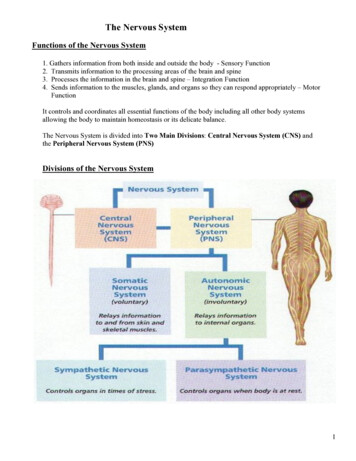
Major Sense OrgansSensation and perception Vision – Eye Hearing – Ear Taste – Taste receptors (new) Smell – Olfactory system Skin – Hot, cold, pressure, painSense OrgansEye – the organ used to sense lightThree layers –1. Outer layer consists of sclera and cornea2. Middle layer consists of choroid, ciliarybody and iris3. Inner layer consists of retinaFunctions of the major parts of the eye:Sclera or Scleroid Layer – (white of eye) a tough protective layer of connective tissue that helps maintainthe shape of the eye and provides an attachment for the muscles that move the eyeCornea - the clear, dome-shaped part of the sclera covering the front of the eye through which light entersthe eyeAnterior Chamber – a small chamber between the cornea and the pupilAqueous Humor - the clear fluid that fills that anterior chamber of the eye and helps to maintain the shapeof the cornea providing most of the nutrients for the lens and the cornea and involved in wastemanagement in the front of the eyeChoroid Layer - middle layer of the eye containing may blood vesselsCiliary Body - the ciliary body is a circular band of muscle that is connected and sits immediately behindthe iris- produces aqueous humor, changes shape of lens for focusing, andIris - the pigmented front portion of the choroid layer and contains the blood vessels - it determines the eyecolor and it controls the amount of light that enters the eye by changing the size of the pupil (an albinoonly has the blood vessels – not pigment so it appears red or pink because of the blood vessels)Lens - a crystalline structure located just behind the iris - it focuses light onto the retinaPupil - the opening in the center of the iris- it changes size as the amount of light changes (the more light,the smaller the hole)Vitreous - a thick, transparent liquid that fills the center of the eye - it is mostly water and gives the eye itsform and shape (also called the vitreous humor)Retina - sensory tissue that lines the back of the eye. It contains millions of photoreceptors (rods for black& white and cones for color ) that convert light rays into electrical impulses that are relayed to thebrain via the optic nerveOptic nerve - the nerve that transmits electrical impulses from the retina to the brainCommon eye defects include – myopia or nearsightedness where the eyeball is too long or the cornea is toosteep; hyperopia or far sightedness where the eyeball is short or lens cannot become round enough:cataracts where the lens becomes fogged; presbyopia where the muscles controlling the bulging of thelens become weak as we age; nyctalopia or night blindness where vision is impaired in dim light and inthe dark due to pigment rhodospin in the rods not functioning properly7
Images the cornea and the lens help to produce the image on the retinaimages formed by the lens are upside down and backwards when they reach the retinatwo types of receptors on the retinaRods – 125 million on a single retina – extremely sensitive to all wavelengths of visible light butdo not distinguish different color – in dim light only rods are activated where one can see objectsbut not as sharp images and are not able to distinguish their color – most dense in peripheralview – nighttime vision Rods have a pigment called rhodospinAs amount of light increases, the cones – 7 million on a single retina – mainly in central view arestimulated and the color becomes clear – daytime visionThere are three types of cones which distinguish the three colors – blue, red, greenFovea – point of central focus – great density of cones - center of the eye's sharpest vision andthe location of most color perception - the layers of the retina spread aside to let light fall directlyon theconesLight stimulates rods and cones and sends impulse via optic nerve to brain areas for visionThe Optic Nerve exits the eye just off center near the Fovea - the Optic Nerve exits is referred toas the Blind Spot due to the lack of the receptors in this areaThe two Optic Nerves come together at the Optic Chiasm located just under the hypothalamus a crucial part of vision and perception must happen - cross-over of information from the right eyecrosses over to the left side and visa versa happens here at the Optic ChiasmInformation from each eye mustbe processed in both halves of thebrainInformation leaves the chiasm viathe optic tract.Reorganized optic tract leaves theOptic Chiasm and passes onto thelateral geniculate nucleusAt the lateral geniculate nucleithe information is separated,organized, and relayed todifferent areas of the visualcortexThe different zones of the visualcortex process the differentaspects of vision and information,taken from both visual fields, isprocessed and an image isperceived.8
EAROuter Ear & ear canal – brings sound into eardrumEardrum – vibrates to amplify sound & separates inner and middle earMiddle ear has 3 small bones or Ossicles anvil, stirrup, stapes – amplify sound (small bones) whichvibrate soundEustachian tube – connects middle ear to throat and equalizes pressure on eardrumCochlea – in inner ear – has receptors for sound & sends signals to brain via Auditory NerveProcess of hearing: Sound waves enter your outer ear and travel through your ear canal to the middle ear. The ear canal channels the waves to your eardrum, a thin, sensitive membrane stretched tightly overthe entrance to your middle ear. The waves cause your eardrum to vibrate. It passes these vibrations on to the hammer, one of three tiny bones in your ear. The hammervibrating causes the anvil, the small bone touching the hammer, to vibrate. The anvil passes thesevibrations to the stirrup, another small bone which touches the anvil. From the stirrup, the vibrationspass into the inner ear. The stirrup touches a liquid filled sack and the vibrations travel into the cochlea, which is shapedlike a shell. Inside the cochlea, a vestibular system formed by three semicircular canals that are approximately atright angles to each other and which are responsible for the sense of balance and spatial orientation.It has chambers filled with a viscous fluid and small particles (otoliths) containing calciumcarbonate. The movement of these particles over small hair cells in the inner ear sends signals to thebrain that are interpreted as motion and acceleration. The brain processes the information from theear and lets us distinguish between different types of sounds.9
Taste and Smell – Chemical ReceptorsTaste buds The mouth contains around 10,000 taste buds, most ofwhich are located on and around the tiny bumps on yourtongue. Every taste bud detects five primary tastes:o Souro Sweeto Bittero Saltyo Umami - salts of certain acids (for examplemonosodium glutamate or MSG) Each of your taste buds contains 50-100 specialisedreceptor cells. Sticking out of every single one of these receptor cells isa tiny taste hair that checks out the food chemicals inyour saliva. When these taste hairs are stimulated, they send nerveimpulses to your brain. Each taste hair responds best to one of the five basictastes.Smell Receptors or Olfactory receptors Humans able to detect thousands of different smells Olfactory receptors occupy a stamp-sized area in the roof of the nasal cavity, the hollow space inside thenose Tiny hairs, made of nerve fibers, dangle from all your olfactory receptors. They are covered with alayer of mucus. If a smell, formed by chemicals in the air, dissolves in this mucus, the hairs absorb it and excite yourolfactory receptors. A few molecules are enough to activate these extremely sensitive receptors. Olfactory Hairs easily fatigued so you do not notice smells Linked to memories - when your olfactory receptors are stimulated, they transmit impulses to your brainand the pathway is directly connected to the limbic system - the part of your brain that deals withemotions so you usually either like or dislike a smell Smells leave long-lasting impressions and are strongly linked to your memories Much of what we associate as taste also involves smell – that is why hot foods “taste” differentthan “cold” foods10
Skin receptors:Your skin and deeper tissues contain millions of sensory receptors.Most of your touch receptors sit close to your skin's surface.Light touch Meissner's corpuscles areenclosed in a capsule ofconnective tissue They react to light touch and arelocated in the skin of your palms,soles, lips, eyelids, externalgenitals and nipples these areas of your body areparticularly sensitive.Heavy pressure Paccinian corpuscules sensepressure and vibration changesdeep in your skin. Every square centimeter of yourskin contains around 14 pressurereceptorsPain skin receptors register pain pain receptors are the mostnumerous each square centimeter of yourskin contains around 200 painreceptorsTemperature skin receptors register warmth and cold each square centimeter of your skin contains 6 receptors for cold and 1 receptor for warmth Cold receptors start to perceive cold sensations when the surface of the skin drops below 95 º F. Theyare most stimulated when the surface of the skin is at 77 º F and are no longer stimulated when thesurface of the skin drops below 41 º F. This is why your feet or hands start to go numb when they aresubmerged in icy water for a long period of time. Hot receptors start to perceive hot sensations when the surface of the skin rises above 86 º F and aremost stimulated at 113 º F. Beyond 113 º F, pain receptors take over to avoid damage being done to theskin and underlying tissues. thermoreceptors are found all over the body, but cold receptors are found in greater density than heatreceptors – most of the time of our environment is colder than our body temperature The highest concentration of thermoreceptors can be found in the face and ears so your nose and earsalways get colder faster than the rest of your body on a chilly winter day11
Disorders of the Nervous System – symptoms, prevention, treatment Epilepsy - common and diverse set of chronic neurological disorders characterized by seizures.Seizures - the physical findings or changes in behavior that occur after an episode of abnormalelectrical activity in the brain and are caused by abnormal electrical discharges in the brainAlzheimer’s Disease - a degenerative disease of the brain that causes dementia, which is a gradualloss of memory, judgment, and ability to function. - the most common form of dementia- affects anestimated 1 in 10 people over age 65Multiple Sclerosis - an autoimmune disease that affectsthe brain and spinal cord (central nervous system) body's immune system eats away at the protectivemyelin sheath that covers the axons of the neurons andinterferes with the communication - MS can affectvision, sensation, coordination, movement, and bladderand bowel control.Parkinson’s Disease - disorder of the brain that leads toshaking (tremors) and difficulty with walking,movement, and coordination. People with Parkinson'sdisease have low brain dopamine concentrations.Shingles (herpes zoster) - painful, blistering skin rash due to the varicella-zoster virus, the virus thatcauses chickenpox – the virus remains inactive (becomes dormant) in certain nerves in the body.Shingles occurs after the virus becomes active againCerebral Palsy - group of disorders that can involve brain and nervous system functions such asmovement, learning, hearing, seeing, and thinking resulting from damage to certain parts of thedeveloping brainGlaucoma - a group of eye conditions that lead to damage to the optic nerve due to increasedpressure in the eye - the eye’s drainage system becomes clogged so the intraocular fluid cannot drainand as the fluid builds up, it causes pressure to build within the eye. High pressure damages thesensitive optic nerve.Pink eye (Conjunctivitis) – infection of the conjunctiva of the eyeEffects of Drugs on the Nervous System Alcohol - central nervous system depressant – cell membranes are highly permeable to alcohol soonce in the bloodstream it can diffuse into almost all body tissues. It is absorbed in the stomach so itgets into the blood stream quickly and slows down function of the nervous systemCaffeine - acts as a central nervous system stimulant - caffeine suppresses melatonin for up to 10hours and also promotes adrenalin. Melatonin is strongly associated with quality sleep, whileadrenalin is the neurotransmitter associated with alertness.Nicotine - small doses of nicotine have a stimulating action on the central nervous system – it ishighly addictive nicotine's effects on the brain cause an increased release of neurotransmittersassociated with pleasure. The brain quickly adjusts to repeated nicotine consumption by decreasingthe amount of neurotransmitters released. The effect of this increased tolerance is that the smokermust continue to use nicotine in order to avoid the feelings of discomfort associated with withdrawalfrom the drug. Irritability and anxiety often ensue during nicotine withdrawal.Marijuana - THC, the main active ingredient in marijuana, binds to membranes of nerve cells in thecentral nervous system that have protein receptors. After binding to nerve cells, THC initiates achemical reaction that produces the various effects of marijuana use. One of the effects issuppression of memory and learning centers (called the hippocampus) in the brain.12
3 Reflex Arc Components of a Reflex Arc A. Receptor - reacts to a stimulus B. Afferent pathway (sensory neuron) - conducts impulses to the CNS C. Interneuron - consists of one or more synapses in the CNS (most are in the spine) D. Efferent pathway (motor neuron) conducts impulses from CNS to effector. E. E