Transcription
ArticleDying “from” or “with” COVID-19 during the Pandemic:Medico-Legal Issues According to a Population PerspectiveFabio De-Giorgio 1,2,*, Vincenzo M. Grassi 1,2, Eva Bergamin 1,2, Alessandro Cina 2,3, Franca Del Nonno 4,Daniele Colombo 4, Roberta Nardacci 5, Laura Falasca 5, Celeste Conte 1,2, Ernesto d’Aloja 6, Gianfranco Damiani 2,7and Giuseppe Vetrugno 1,2Department of Health Care Surveillance and Bioethics, Section of Legal Medicine, Università Cattolica delSacro Cuore, 00168 Rome, Italy; vincenzograssi@live.com (V.M.G.); eb95@live.it (E.B.);celestemgconte@gmail.com (C.C.); giuseppe.vetrugno@policlinicogemelli.it (G.V.)2 Fondazione Policlinico Universitario A. Gemelli, IRCCS, 00168 Rome, Italy;alessandro.cina@policlinicogemelli.it (A.C.); Gianfranco.Damiani@unicatt.it (G.D.)3 Department of Diagnostic Imaging, Oncological Radiotherapy and Hematology, Diagnostic Imaging Area,Università Cattolica del Sacro Cuore, 00168 Rome, Italy4 Pathology Unit, National Institute for Infectious Diseases “Lazzaro Spallanzani”, IRCCS, 00149 Rome, Italy;franca.delnonno@inmi.it (F.D.N.); daniele.colombo@inmi.it (D.C.)5 Laboratory of Electron Microscopy, National Institute for Infectious Diseases “Lazzaro Spallanzani”, IRCCS,00149 Rome, Italy; roberta.nardacci@inmi.it (R.N.); laura.falasca@inmi.it (L.F.)6 Department of Medical Sciences and Public Health, University of Cagliari, 09124 Cagliari, Italy;ernestodaloja@gmail.com7 Department of Life Sciences and Public Health, Università Cattolica del Sacro Cuore, 00168 Rome, Italy* Correspondence: fabio.degiorgio@unicatt.it; Tel.: 39-06-3550-7031; Fax: 39-06-3550-70331Citation: De-Giorgio, F.; Grassi,V.M.; Bergamin, E.; Cina, A.; DelNonno, F.; Colombo, D.; Nardacci,R.; Falasca, L.; Conte, C.; d’Aloja, E.;et al. Dying “from” or “with”COVID-19 during the Pandemic:Medico-Legal Issues According to aPopulation Perspective. Int. J.Environ. Res. Public Health 2021, 18,8851. https://doi.org/10.3390/ijerph18168851Academic Editors: Michael D.Freeman, Maurice Zeegers, HenryOtgaar and Paul B. TchounwouReceived: 5 July 2021Accepted: 19 August 2021Published: 22 August 2021Publisher’sNote:MDPIstaysneutral with regard to jurisdictionalclaimsinpublishedAbstract: There is still a lack of knowledge concerning the pathophysiology of death amongCOVID-19-deceased patients, and the question of whether a patient has died with or due to COVID19 is still very much debated. In Italy, all deaths of patients who tested positive for SARS-CoV-2 aredefined as COVID-19-related, without considering pre-existing diseases that may either contributeto or even cause death. Our study included nine subjects from two different nursing homes (Cases1–4, Group A; Cases 5–9, Group B). The latter included patients who presumably died from COpoisoning due to a heating system malfunction. All subjects tested positive for COVID-19 both anteand post-mortem and were examined using post-mortem computed tomography prior to autopsy.COVID-19 was determined to be a contributing cause in the deaths of four out of nine subjects(death due to COVID-19; i.e., pneumonia and sudden cardiac death). In the other five cases, forwhich CO poisoning was identified as the cause of death, the infection presumably had no role inexitus (death with COVID-19). In our attempt to classify our patients as dying with or due to COVID19, we found the use of complete assessments (both histological analyses and computedtomography examination) fundamental.mapsandKeywords: COVID-19; post-mortem computed tomography; CO intoxication; pneumonia, postmortem computed tomography; causalityinstitutional affiliations.Copyright: 2021 by the authors.Licensee MDPI, Basel, Switzerland.This article is an open access articledistributed under the terms andconditions of the Creative mons.org/licenses/by/4.0/).1. IntroductionIn December 2019, an outbreak of lower respiratory tract infection cases was detectedin Wuhan City in the Hubei Province of China. In the absence of a known etiologicalagent, the first cases were classified as “pneumonia of unknown etiology.” Followingcareful investigations by the local CDC (Center for Disease Control and Prevention), theetiology of the disease was attributed to severe acute respiratory syndrome coronavirus 2(SARS-CoV-2). The novel coronavirus disease was named “coronavirus disease 19”(COVID-19) and declared a pandemic by the World Health Organization (WHO) in MarchInt. J. Environ. Res. Public Health 2021, 18, 8851. /journal/ijerph
Int. J. Environ. Res. Public Health 2021, 18, 88512 of 132020. To date, over 127.3 million cases and over 2.7 million deaths have been confirmedworldwide [1]. COVID-19 infection represents a complex challenge and continuous threatto public health. Although many studies regarding this topic can be found in the literature,there is still a general lack of knowledge concerning the pathophysiology of death amongdeceased patients. Undoubtedly, COVID-19-related signs and symptoms are extremelyvariable, and the clinical course of the disease can vary from person to person;nonetheless, studies concerning the causes and underlying mechanisms of death areinsufficient, and this automatically translates into an unrealistic assessment of infectionrelated mortality rates. As a consequence, problems may arise both from anepidemiological point of view and from a clinical point of view, with doctors having toface unpleasant situations; indeed, a lack of information regarding the potential causes ofdeath can hinder the implementation of correct prevention and treatment measures inspecific patients. In this regard, autopsies can provide clinically relevant insights andsupport, and should be mandatory in order to identify not only the cause of death, butalso the underlying infection-related mechanisms and patterns of organ damage (i.e., withthe use of biopsies). Needless to say, this can only be achieved by following specificguidelines and recommendations on the best standards of practice and safety measures[2]. Studies have demonstrated that autopsy personnel have a minimal risk of acquiringinfection, provided specific protocols are followed and precautions are taken. Thus, in thecase of COVID-19-positive bodies, autopsies should be considered safe and, in fact,encouraged, because they have been shown to confirm the presence of COVID-19 evenafter extensive periods of time post-mortem [3,4].As mentioned, the clinical course of COVID-19 is highly heterogeneous; the majorityof patients are either asymptomatic or experience only mild symptoms (i.e., fever, drycough, shortness of breath) [2]. In contrast, in some cases, the disease causes severerespiratory symptoms, often requiring oxygenation and admission to an intensive careunit (ICU), with symptoms and signs of multi-organ manifestations. This extensiveinvolvement can be partially explained by the high tropism demonstrated by the virustowards angiotensin-converting enzyme II (ACE2). ACE2 receptors are ubiquitouslyexpressed in the body (lungs, vessels, liver, kidneys, spleen, skin, muscle, and nervousstructures), and their binding to SARS-CoV-2 represents an essential step for the viralinvasion of human cells [5]. In a study by Zou et al., the authors identified the organs andspecific cell types (with ACE2 expression) at risk for SARS-CoV-2 infection: lung (type IIalveolar cells), heart (myocardial cells), kidney (proximal tubule cells), ileum andesophagus (epithelial cells), bladder (urothelial cells), venous and arterial endothelialcells, and arterial smooth muscle cells [5,6]. Such multi-organ involvement can be studiedand evaluated by analyzing the course of the disease from a clinical and radiological pointof view, and by integrating these findings with the valid support of complete post-mortemexaminations and histological analysis, which provide valuable information through theanalysis of transformative phenomena and organ damage.In the literature, there are several reports concerning the increased risk ofcardiovascular (CV) complications connected to COVID-19. The two conditions appear tobe closely related; acute respiratory infections (including COVID-19) are known to act astriggering factors for CV diseases (CVD) [7,8] and, vice versa, pre-existing CVD and othercomorbidities usually increase the incidence of infectious diseases [9]. Indeed, SARS-CoV2 infection has been associated with cases of acute myocardial infarctions (MIs),myocarditis, arrhythmias, and venous thromboembolism [10].Herein, we report nine cases of SARS-CoV-2 infection-positive patients in twodifferent groups in order to discuss these cases in terms of “deaths with COVID-19 or deathsdue to COVID-19”.2. Materials and MethodsBetween March 2020 and January 2021, two groups of corpses arrived at our institutefrom two different nursing homes where COVID-19 outbreaks were suspected. Our study
Int. J. Environ. Res. Public Health 2021, 18, 88513 of 13was carried out on a total of 9 cases, including patients from 2 different nursing homes.Based on the provenance of the bodies, we divided the subjects into two groups: Casesfrom 1 to 4 were assigned to Group A (these corpses which arrived at our institute fromnursing home A) and Cases from 5 to 9 were assigned to Group B (these corpses whicharrived at our institute from nursing home B) (Table 1). The conducted investigations,including total-body CT examination and complete autopsy (macroscopic andmicroscopic examination, toxicological analyses), were authorized by the JudicialAuthority.The study was approved by the Institutional Research Ethics Committee (ID 3862).Table 1. Clinical characteristics of the study population.GroupsCase #Age1Group AGender72F279F349091FF589F687FGroup B789998971FComorbidity Diabetes; Chronic cerebral vasculopathy. Right parietal ischemic stroke; Epilepsy; Secondary parkinsonism; Senile dementia.Not availableNot available Diabetes; Hypo-mobility. Dilated cardiomyopathy (ICD); Hypertension; Atrial fibrillation; COPD; Obesity; Chronic renal failure. COPD; Hypertension; Chronic renal failure; Diabetes; Polyarthritis. Hypertension;F Polyarthritis;F Cerebrovascular leukoencephalopathy.Not availableThe mean age in Group A was 83 years (range: 72–91 years). The mean age in GroupB was 87 years (range: 71–99 years). All patients in both groups were female. In Group B,all 5 patients presumably died at night from carbon monoxide poisoning due to a heatingsystem malfunction; the cadavers were found in the same nursing home. All 9 subjectshad undergone routine COVID-19 testing (in order to exclude a cluster in Group A, andas a screening organized by the National Health Service in Group B) and, in all cases,positivity to the virus was confirmed post-mortem by means of RT-PCR on swabsperformed at the level of the right and left bronchi and right and left pulmonaryparenchyma. All cases were examined, prior to autopsy, with the use of post-mortemtotal-body CT. Afterwards, all corpses underwent a complete autopsy, including thecollection of specimens for histological evaluation based on standard H&E staining.2.1. Toxicological AnalysisToxicological analyses were carried out for all Group B cases. In 2/5 cases, peripheralblood and urine samples were analyzed. In the remaining 3 cases, samples were collected
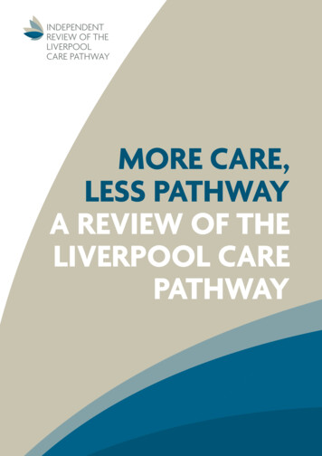
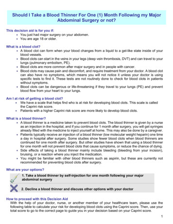
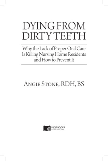
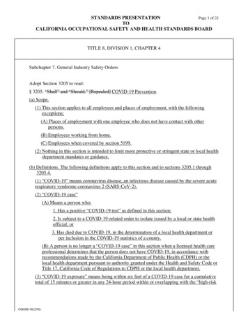
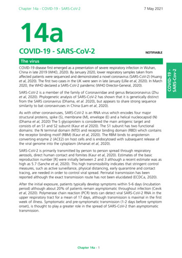
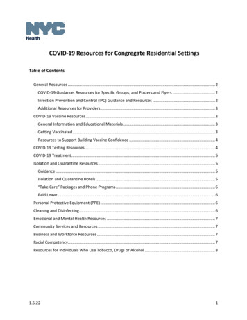
Int. J. Environ. Res. Public Health 2021, 18, 88514 of 13from the peripheral blood and vitreous humor, because urine was either contaminated ornot available.The presence of ethanol was investigated by adding sodium chloride and an internalstandard n-propanol to 0.2 mL of peripheral blood. The sample was then analyzed usingheadspace gas chromatography. A reference sample at a concentration of 1 g/L was alsoexamined (parameters: Finnigan trace GC gas chromatograph with FID detector,Supelcowax10 column 20 m, id 0.25 mm phase 0.25 um. Temperature: 60 C (1’), 35 C/minat 220 C, 5 min). Moreover, in order to evaluate the presence of toxic substances or drugs,urine and blood samples were analyzed using liquid chromatography and highresolution mass spectrometry.The percentage of carboxyhemoglobin was calculated via spectrophotometry on awhole blood sample using the “Huffner–Heilmeyer” coefficient method. In total, 20 μL ofblood was hemolyzed with 5 mL of 1% ammonia solution and directly subjected tospectrophotometric analysis (“Evolution 201” Thermo Double Beam Spectrophotometer).2.2. Post-Mortem CTEach corpse was examined with the use of PMCT prior to autopsy. The scans wereconducted on a Somatom Scope 16-slice CT scanner, Siemens Healthineers Italia, andcharacterized by the following parameters: 130 kV, 150 mA, 2.4 mm slice thickness. Thecorpses were placed on a horizontal CT table in a supine position with their arms at theirsides. They were fully clothed and wrapped in body bags. Whole-body CT scans wereobtained. No contrast agent was used in this procedure.3. ResultsThe results have been summarized in Table 2. All corpses from Group A displayedfindings of interstitial pneumonia, ischemic heart disease (chronic ischemic heart disease,N 3; recent ischemic necrosis and myocardial fibrosis, N 1), and some degree of cardiachypertrophy. Case 1, in which signs of recent ischemic necrosis were observed, alsoshowed evidence of micro-thrombosis and myocarditis upon histopathologicalexamination. In this case, the cause of death was identified as SCD, sustained by therupture of an atherosclerotic plaque in a subject with signs of multiorgan involvement(i.e., lung). Cases 2 to 4, on the other hand, died of COVID-19 pneumonia and displayedsuggestive pulmonary findings, including pulmonary edema, fibrous thickening of thesepta, and basal lung hepatization (Figure 1). Concerning Group B cases, even though allpatients tested positive for COVID-19 both ante- and post-mortem, the cause of death wasCO intoxication. All corpses showed similar autopsy and histological findings, includingcharacteristic cherry-red hypostasis and internal visceral cherry-red coloration,pulmonary edema and congestion with intra-alveolar hemorrhages, and various levels ofatherosclerosis (Figure 2).Typical CT imaging of COVID-19, including bilateral multi-lobar ground glassopacities (GGO), were seen in all cases (Figure 3). A peripheral localization of GGO wasfound only in Group A, Case 3. In Group A, no gravity-dependent distribution of GGOwas identified in Cases 1, 3 or 4; in Group B, it was only found in Case 6. Alveolarconsolidations were found in two cases for both groups. Lymphadenopathies wereidentified in only one case. Pleural effusions were commonly found in both groups (threeof four in Group A and five of five in Group B). CT findings are reported in Table 3.
Int. J. Environ. Res. Public Health 2021, 18, 88515 of 13Table 2. Post-mortem findings and causes of death.GroupsCase #Autopsy 1 2 Group A 3 4 5 6Group B7 8 9 HistologyCause of Death Interstitial pneumonia;Eccentric cardiac hypertrophy; LAD Micro-thrombosis;chronic atherosclerosis with thrombosis;Sudden cardiac death in Myocarditis;Apical emphysema, compact basalCOVID-19 Recent ischemic necrosis in myocardial fibrosis;parenchyma, edema. Dilation of the hepatic centrilobular veins.Concentriccardiachypertrophy;moderate atherosclerosis; Interstitial pneumonia;COVID-19 pneumoniaPulmonary edema, basal hepatization, Chronic ischemic heart disease.fibrous thickening of the septa.Concentriccardiachypertrophy; Interstitial pneumonia and honeycombmoderate atherosclerosisappearance;COVID-19 pneumoniaPulmonary edema, basal hepatization, Chronic ischemic heart disease;fibrous thickening of the septa; Dilation of the hepatic centrilobular veins.Septalhypertrophy;slightatherosclerosis; Interstitial pneumonia;COVID-19 pneumoniaPulmonary oedema, fibrous thickening Chronic ischemic heart disease.of the septa.Cherry-red hypostasis and internalvisceral cherry-red coloration; Chronic ischemic heart disease;CO intoxication (COHbConcentric cardiac hypertrophy; slight Giant cell bronchiolitis, nary edema and congestion.Cherry-red hypostasis and internalvisceral cherry-red coloration; Chronic ischemic heart disease;CO intoxication (COHbSlight atherosclerosis;50%) COPD; endo-alveolar hemorrhages.Pulmonary edema and congestion.Cherry-red hypostasis and internalvisceral cherry-red coloration; Chronic ischemic heart disease;Septalhypertrophy;moderateCO intoxication (COHb Interstitial lung fibrosis; endo-alveolaratherosclerosis; aortic ectasia;55%)hemorrhages.Pulmonary oedema, congestion, andcompact basal parenchyma.Cherry-red hypostasis and internalvisceral cherry-red coloration; Chronic ischemic heart disease;CO intoxication (COHbConcentriccardiachypertrophy; Pulmonaryedema;intra-alveolar40%)moderate atherosclerosis; aortic ectasia;hemorrhages; bronchial necrosis.Pulmonary edema and congestion.Cherry-red hypostasis and internal Chronic ischemic heart disease;visceral cherry-red coloration; Pulmonary fibrosis and edema, intra-alveolar CO intoxication (COHbModerate atherosclerosis; rophages.Pulmonary edema and congestionTable 3. Post-mortem CT findings.CT FindingGround glass opacities-Peripheral-Bilateral-Multilobar-no gravitational distribution“Crazy paving” patternAlveolar consolidationsLymphadenopathiesPleural effusionCase 1xCase 2xxxxxxxxxCase 3xxxxxxxxxCase 4xCase 5xCase 6xCase 7xCase 8xCase 9xxxxxxxxxxxxxxxxxxxxxxThere was a suspicion of CO intoxication; therefore, toxicological analyses wereperformed in all the Group B cases, with the aim of determining both the cause andmanner of death. The search for ethanol via headspace gas chromatography yieldednegative results in all five cases. Concerning toxic substances/drugs, we found promazine
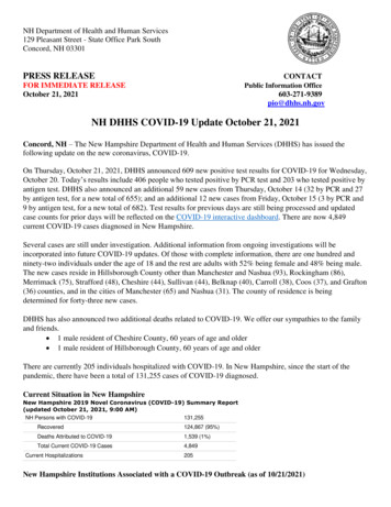
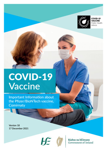
Int. J. Environ. Res. Public Health 2021, 18, 88516 of 13in the therapeutic range (N 1), benzodiazepines and metabolites in the therapeutic range(i.e., flurazepam and alprazolam) (N 1), and quetiapine and metabolite at subtherapeuticlevels (N 1). In terms of carboxyhemoglobin (CO-Hb) percentages, these were found tobe 40% (N 2), 45–50% (N 1), and 50–55% (N 2). These values appear to be compatiblewith severe carbon monoxide poisoning. According to the literature, values higher than40–50% are almost invariably associated with a clinical picture of severe poisoning,including lethargy, coma, motor incoordination, memory, and praxia disorders [11], aswell as the possible instability of cardiac, respiratory, and neurological functions, up todeath [12].Figure 1. Group A: lung macroscopic and microscopic findings: (A,B) cut surface showing theconsolidation of lobes and red congested areas, with thickening of the interstitial septa andpulmonary edema; (C) interstitial fibrosis H&E, 200 ; (D) numerous inflammatory cells consistingmostly of lymphocytes infiltrating into alveolar septa and clustering around capillary vessels,H&E, 200 .Figure 2. Group B: pulmonary macroscopic and microscopic findings: (A,B) cherry-red coloration,pulmonary edema, and congestion; compact basal parenchyma; (C) desquamative necrosis of thebronchial epithelium, H&E, 400 ; (D) bronchi surrounded by inflammatory cells, H&E, 400 .
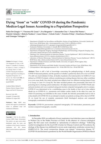
Int. J. Environ. Res. Public Health 2021, 18, 88517 of 13Figure 3. Post-mortem lung CT (parenchymal window) showing “ground glass” opacities in asubject of group A (A) and in one of group B (B). In A, the distribution of opacities was bilateral,peripheral, and without gravitational gradient. This pattern is considered typical in COVID-19pulmonary infection. In B, the “ground glass” opacities showed a more central and gravitationaldistribution, as seen in post-mortem CT lung changes.4. DiscussionThe mortality rate of SARS-CoV-2 infection varies from country to country, probablyas a result of various factors such as different public health services and adopted policies[13]. In Italy, the Italian National Institute of Health (ISS, Istituto Superiore di Sanità) hasestablished a surveillance system with the aim of gathering data concerning all COVID19 cases throughout the country [14,15]. COVID-19-associated deaths were defined asdeaths in patients who had tested positive for the virus (fatality rate: number of deaths inpersons who tested positive for SARS-CoV-2 divided by the number of SARS-CoV-2cases) [14].Initially, the case–fatality rate in Italy was higher than that observed in othercountries [15], and this could be explained by different factors. First of all, it is importantto consider the fact that Italy has the highest percentage of elderly population in Europe,and the second-highest percentage worldwide after Japan; in this regard, it has beenproven that a strong correlation exists between the severity and risk of death of SARSCoV-2 infection and age [16]. In addition, COVID-19 mortality was found to be dependenton the presence of serious comorbidities, which are rather prevalent in the Italianpopulation [17]. Finally, the high case–fatality rate may also be explained by the methodsused to identify COVID-19–related deaths by the Italian NHS. Indeed, in Italy, all deathsof patients who tested positive for SARS-CoV-2 are defined as COVID-19-related, withouttaking into account pre-existing diseases that may either contribute or even cause death[15]. Hence, this system may have led to an overestimation of the overall case–fatality rate.In the context of legal medicine, the question of whether a patient has died with ordue to COVID-19 is very much debated, especially considering the implications that thisparticular matter has in terms of professional responsibility. Coroners are frequentlyasked to assess whether COVID-19 played a direct role in a patient’s death, as well aswhether the medical staff’s treatment and management were appropriate. Indeed,medical malpractice cases are not uncommon. Early on in the pandemic, we witnessed anincrease in the risk of infection, as well as the overall number of positive cases; one of thereasons for this was that many COVID-19-positive patients were relocated to nursing
Int. J. Environ. Res. Public Health 2021, 18, 88518 of 13homes and assisted living facilities due to a shortage of hospital beds [18–20].Furthermore, data regarding the disease, its treatment, and preventive measures werelimited. As a result, dealing with professional liability issues has represented a complextask for coroners; if we then consider the question of “dying with or due to COVID-19”,the matter certainly becomes even more problematic. In this setting, despite the high riskof infection for personnel, autopsies play a critical role, because they determine theseverity of direct viral damage and assess whether organ failure plays a part in a patient’sdeath. Multiple data sources, such as ante- and post-mortem microbiological testing, CTscans, complete forensic autopsy, and histological analyses, may be relevant in this regard[20].Differences in terms of surveillance methods also play a fundamental role when itcomes to answering the query. It may be helpful to consider a “death due to COVID-19” asone resulting from a compatible illness, in either a suspected or confirmed SARS-CoV-2positive case, in the absence of a clear alternative cause, such as trauma [21]. Thus, it issafe to indicate as the underlying cause of death when the latter takes part in a causalchain that leads to death [22]. In a study by Cobos-Siles et al., the authors evaluated 128individuals with the aim of differentiating between patients whose death was directlyassociated with the development of COVID-19 complications and SARS-CoV-2-positivepatients who died of causes unrelated to COVID-19 complications [23]. Among thecomplications associated with COVID-19, the authors included acute respiratory distresssyndrome (ARDS) or hyperinflammation response, acute respiratory failure, severe lunginjury on chest X-ray/CT, thromboembolic events, and septic shock. On the other hand,“deaths with COVID-19” were defined as those deaths in which the disease acted as aprecipitating factor, leading to the decompensation of underlying pathologies. Accordingto the authors, only 20% of patients died of unrelated complications [24]. Similarly, Slateret al. examined 162 COVID-19-positive cases and found the disease to be the direct causeof death in 150 (93%) patients [25]. Grippo et al. analyzed a total of 5311 death certificatesof persons who had tested positive for SARS-CoV-2 infection [26]. According to theauthors, COVID-19 was the underlying cause of death in 88% of cases.Regarding our study population, Case 1 died of sudden cardiac death (SCD)sustained by the rupture of an atherosclerotic plaque (middle third of the anteriordescending coronary). The subject tested positive for COVID-19 and displayed signs ofmultiorgan involvement (lung, heart, liver). The underlying mechanisms that couldexplain Case 1’s clinical picture are as follows: (a) T cell- and monocyte-mediatedhyperinflammation and cytokine storm that leads to myocarditis; (b) respiratory failureand hypoxia, especially in the setting of ARDS and severe infections, resulting in thedamage of cardiac muscle cells; (c) down-regulation of angiotensin-converting enzyme 2(ACE2) expression and subsequent protective signaling pathways in cardiac myocytes;(d) blood hypercoagulability and the development of coronary microvascularthromboses; (e) diffuse endothelial lesions and inflammation of the endothelium invarious organs (including the heart) as either a direct consequence of SARS-CoV-2 viralinvolvement and/or resulting from the host’s inflammatory response; and (f)inflammation and/or stress causing coronary plaque rupture or supply–demandmismatch, leading to myocardial ischemia/infarction [27,28].In a study by Sheth et al., the authors hypothesized that the cytokine storm may playa role in the pathogenesis of acute coronary syndromes. In particular, the authorsidentified two possible pathophysiological mechanisms: on the one hand, systemicinflammation caused by the viral infection can determine a rapid formation of a coronaryplaque or an acute modification of an existing plaque, causing the rupture of its surfacewith exposure of the underlying components that determine the formation of a thrombus;on the other hand, systemic viral infection can cause direct myocardial damage, boththrough coronary vasoconstriction and the stimulation of platelet activation, as well as anincreased prothrombotic condition, either mediated by the cytokine storm or caused by ahypoxia-related increase in metabolic demand [29]. Based on such considerations, it was
Int. J. Environ. Res. Public Health 2021, 18, 88519 of 13possible to assume that SARS-CoV-2 infection and the consequent onset of COVID-19 mayhave played a con-causal role in the occurrence of the rupture of the atherosclerotic plaqueand, ultimately, in the death of Case 1.In Case 2, there was evidence of a more severe pulmonary involvement. Thus, thelung was the organ that was mainly affected by the infection, which inevitably played acausal role in exitus. Likewise, the deaths in Cases 3 and 4 were attributed to COVID-19related interstitial pneumonia in subjects suffering from chronic ischemic heart disease.As mentioned, most COVID-19-positive patients display mild symptoms or arecompletely asymptomatic. Nonetheless, approximately 20% of affected patientsdeteriorate rapidly and develop severe respiratory diseases, such as pneumonia, withsignificant mortality rates [30]. Studies have determined that a correlation exists betweenthe development of major complications/outcomes and both increasing age and a highnumber of associated comorbidities [30,31]. Advanced age, as for many other diseases andinfections, is a known risk factor for COVID-19 and its complications; it has been observedthat the majority of patients affected by severe COVID-19 pneumonia are elderly and that,of all the predictors of mortality in COVID-19 pneumonia, advanced age is the mostimportant [32,33]. This correlation can be explained by the physiological deterioration ofone’s immune system that occurs with age, as well as by the age-dependent loss of T cellsand T cell subtypes [34,35]. Lymphopenia, increased CRP, and erythrocyte sedimentationwere observed in the laboratory results of COVID-19 patients [36]. The word“lymphopenia” refers to a reduction in the peripheral lymphocyte count (primarily CD4 T and CD8 T cells); this condition increases the risk of secondary bacterial infections[37].Conversely, Cases 5 to 9 refer to deaths from CO poisoning. Indeed, all corpsesdisplayed pulmonary findings indicative of both the cause and mechanism of death.Carboxyhemoglobin in the blood is responsible for the development of characteristicautopsy findings, such as “cherry-red” hypostases (that typically appear atcarboxyhemoglobin concentrations 30%), marked pulmonary edema, parenchymalimbibition, and severe polyvisceral congestion. CO causes harm to the lungs via directhypoxic damage to tissues and pathological alterations that may be secondary to CO’seffect on the heart [38]. Thus, cases of CO intoxication often display intense pulmonaryhyperemia and congestion, acute emphysema, interstitial thickening, focal infiltrations ofpolymorphonuclear leukocytes, focal and diffuse endo-alveolar hemorrhages, andepithelial desquamation [39]. These findings were observed in all our cases (5 to 9) andwas further supported by histopathological examination. The results of our analysis arein accordance with those observed by King et al. [40]. Traces of pigment were appreciablein the majority of subjects. Three out of five showed desquamation of the bronchialepithelium, with the presence of necrotic material; bronchial necrosis (usually mucosalnecrosis) may indicate exposure to toxic substances and/or fumes [41]. ConsideringCOVID-19, lung damage plays a central role; findings typically in
1-4, Group A; Cases 5-9, Group B). The latter included patients who presumably died from CO poisoning due to a heating system malfunction. All subjects tested positive for COVID-19 both ante- and post-mortem and were examined using post-mortem computed tomography prior to autopsy.