Transcription
Petrova et al. Journal of Foot and Ankle Research (2018) ARCHOpen AccessReliability of a novel thermal imagingsystem for temperature assessment ofhealthy feetN. L. Petrova1,2* , A. Whittam3, A. MacDonald4, S. Ainarkar5, A. N. Donaldson1, J. Bevans5, J. Allen4, P. Plassmann6,B. Kluwe7, F. Ring7, L. Rogers3, R. Simpson3, G. Machin3 and M. E. Edmonds1,2AbstractBackground: Thermal imaging is a useful modality for identifying preulcerative lesions (“hot spots”) in diabetic footpatients. Despite its recognised potential, at present, there is no readily available instrument for routine podiatricassessment of patients at risk. To address this need, a novel thermal imaging system was recently developed. Thispaper reports the reliability of this device for temperature assessment of healthy feet.Methods: Plantar skin foot temperatures were measured with the novel thermal imaging device (Diabetic FootUlcer Prevention System (DFUPS), constructed by Photometrix Imaging Ltd) and also with a hand-held infrared spotthermometer (Thermofocus 01500A3, Tecnimed, Italy) after 20 min of barefoot resting with legs supported andextended in 105 subjects (52 males and 53 females; age range 18 to 69 years) as part of a multicentre clinical trial.The temperature differences between the right and left foot at five regions of interest (ROIs), including 1st and 4thtoes, 1st, 3rd and 5th metatarsal heads were calculated. The intra-instrument agreement (three repeated measures)and the inter-instrument agreement (hand-held thermometer and thermal imaging device) were quantified usingintra-class correlation coefficients (ICCs) and the 95% confidence intervals (CI).Results: Both devices showed almost perfect agreement in replication by instrument. The intra-instrument ICCs forthe thermal imaging device at all five ROIs ranged from 0.95 to 0.97 and the intra-instrument ICCs for the hand-heldthermometer ranged from 0.94 to 0.97. There was substantial to perfect inter-instrument agreement between thehand-held thermometer and the thermal imaging device and the ICCs at all five ROIs ranged between 0.94 and 0.97.Conclusions: This study reports the performance of a novel thermal imaging device in the assessment of foottemperatures in healthy volunteers in comparison with a hand-held infrared thermometer. The newly developedthermal imaging device showed very good agreement in repeated temperature assessments at defined ROIs as wellas substantial to perfect agreement in temperature assessment with the hand-held infrared thermometer. In additionto the reported non-inferior performance in temperature assessment, the thermal imaging device holds the potentialto provide an instantaneous thermal image of all sites of the feet (plantar, dorsal, lateral and medial views).Trial registration: Diabetic Foot Ulcer Prevention System NCT02317835, registered December 10, 2014Keywords: Thermal imaging, Diabetic foot ulcer, Temperature, Reliability, Prevention* Correspondence: nina.petrova@nhs.netNL Petrova and A Whittam are joint first authorsG Machin and ME Edmonds are joint senior authors1Diabetic Foot Clinic, King’s College Hospital NHS Foundation Trust, DenmarkHill, London SE5 9RS, UK2Division of Diabetes and Nutritional Sciences, King’s College London,London, UKFull list of author information is available at the end of the article The Author(s). 2018 Open Access This article is distributed under the terms of the Creative Commons Attribution 4.0International License (http://creativecommons.org/licenses/by/4.0/), which permits unrestricted use, distribution, andreproduction in any medium, provided you give appropriate credit to the original author(s) and the source, provide a link tothe Creative Commons license, and indicate if changes were made. The Creative Commons Public Domain Dedication o/1.0/) applies to the data made available in this article, unless otherwise stated.
Petrova et al. Journal of Foot and Ankle Research (2018) 11:22BackgroundDiabetic foot ulcer is a major complication of diabetes [1].In people with diabetic neuropathy, tissue damage canprogress to ulcer, infection and necrosis and ultimatelyresults in amputation [2]. Indeed, in diabetes, almost 85%of all non-traumatic amputations are preceded by a footulcer. The financial cost of foot ulcers and amputations isimmense [3, 4]. Diabetic foot ulcer imposes substantialburden on public and private payers, ranging from 9–13billion in addition to the costs associated with diabetesitself [3]. A recent health economics analysis has reportedthat the total expenditure on healthcare related to footulcer and amputation in people with diabetes for 2014–2015 in England was estimated at 1billion [4]. At least 1in every 140 of the National Health Service (NHS) expenditure in England is spent on footcare for people withdiabetes [4]. This is equivalent to around 0.7–0.8% of theentire NHS budget. Recent data show that at least60,671–75,838 people with diabetes in England have footulcers at any given time (2–2.5% of the diagnosed diabetespopulation), and that the mean weekly cost of caring foreach patient is 208 [4]. Thus timely identification of patients at risk is fundamental to reduce adverse outcomesand reduce costs [5, 6]. It has been estimated that reducing the prevalence of people with diabetic foot ulcers byone third could save the NHS 210 m– 262 m a year [4].Over the last fifteen years there has been an increasedinterest in thermal imaging as a possible modality forearly detection of incipient tissue damage in diabeticfoot patients [7–9]. Clinical trials have indicated thatregular measurement of foot skin temperatures withnon-contact infrared thermometers in high-risk patientscan reduce the incidence of foot ulcers [10]. However, inthese studies, foot temperatures were assessed only atpredefined regions of interest (ROIs) using single spotinfrared thermometers [10, 11] and the low specificity ofthis method is now well recognised [12]. Thus, despitethe evidence that neuropathic foot ulcer is preceded by arise in skin temperature [11] the latter is not routinelymeasured in clinical practice.There is a requirement for a reliable portable device ascertified to medical device regulations to document thermalimages of high risk diabetic foot patients during routinepodiatry assessment. The ideal thermal imager should beuser friendly, widely available, reproducible and accurate[13]. In addition, thermal imaging should not only belimited to the plantar site of the feet as more than half ofthe diabetic foot ulcers (52%) are with non-plantar location[14]. Detailed assessment with such a device can provideinformation of up to several thousand ROIs as opposed toup to 12 ROIs most commonly assessed by podiatristsusing non-contact infrared thermometers. A thermal imaging device would help identify areas of raised temperature(or ‘hotspots’) which others have reported to be indicativePage 2 of 6of pre-ulcerous inflammation [9, 10]. These could bemissed during routine foot examination of the neuropathicdiabetic foot, when signs and symptoms of inflammationare often lacking.To address this need, a novel medical thermal imagingdevice was recently developed [15]. Laboratory testingshowed that the overall temperature uncertainty of thethermal imaging device was 0.2 C (k 2, 95% confidencelimit) for the range 15 C to 45 C which is comparable tothe uncertainty of the CE marked hand-held spotthermometers, (CE is abbreviated from ConformitéEuropéenne, meaning European Conformity), [15]. Theusefulness of this system in temperature assessment of thefeet of healthy volunteers at 33 ROIs (12 plantar, 15dorsal, 3 medial and 3 lateral) has been documented [16].To assess the performance of the novel thermal imagingdevice in the assessment of foot temperatures in healthyvolunteers in comparison with a hand-held infraredthermometer we selected five easily identifiable plantarfoot landmarks (1st and 4th toes, 1st, 3rd and 5th metatarsal heads). The objectives of this study were twofold: firstlyto measure the agreement in replication (three repeatedmeasures) for the thermal imaging device and for thehand-held thermometer (inter-instrument agreement) andsecondly, to measure the agreement between the thermalimaging device and the hand-held-thermometer (intra-instrument agreement) in the assessment of temperatures ofthe feet of healthy volunteers.MethodsParticipantsThe study was carried out at three clinical centres aspreviously described [16]. Male and female volunteerswere recruited if they had intact feet and no previous history of diabetes, foot ulcer or foot surgery either for correction of a foot deformity or following foot trauma.Subjects were excluded if they reported unsteadiness ingait, if they experienced burning pain, aching of the feet orlegs, prickling sensation or numbness of the feet or legs orif they had any discomfort in the calf muscles when walking that was relieved with rest or any health problems affecting their feet and legs. The study was approved byLondon-City Road and Hampstead Research Ethics Committee (REC reference 15/LO/0070) and was carried outin accordance with the Declaration of Helsinki as revisedin 2000. The study was registered on ClinicalTrials.govwebsite (Clinicaltrials.gov identifier NCT02317835). Allsubjects provided written informed consent and screeningand assessment were performed at one study visit.Temperature measurement and data acquisitionTemperature measurements were carried with a novel thermal imaging device (Diabetic Foot Ulcer Preventions System, DFUPS), developed specifically for this investigation
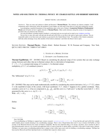
Petrova et al. Journal of Foot and Ankle Research (2018) 11:22by Photometrix Imaging Ltd. in association with theUniversity of South Wales [15, 16] and with a hand-heldinfrared thermometer (Thermofocus 01500A3, Tecnimed,Italy). The thermal imaging device is a battery operatedinstrument with on-board software. The captured footthermal image is downloaded on to a computer for furtheranalysis [15]. Circles with an area equal to 1 cm2 aremanually placed on ROIs of each foot. The Thermofocus isa non-contact spot thermometer, which measures theemitted thermal radiation of a selected ROI of the foot andconverts that measurement into a temperature. The field ofview of the scanned area is nominally 1 cm2.Four thermal imaging systems (one for each clinicalcentre and one as a back-up) and four hand-held infrared thermometers (one for each clinical centre and oneas a back-up) were used in the study. All devices werecharacterised at the National Physical Laboratory (NPL)before usage by the clinical centres, as described previously [15]. In brief, the thermal imaging systems and thehand-held infrared thermometers were evaluated toassess the temperature resolution, the spatial resolutionand performance (repeatability, stability and accuracy).All devices were calibrated under laboratory conditionsin terms of radiance temperature versus the NPL blackbody calibration sources [17] over the range of 15 C to45 C, traceable to the international temperature scale of1990 (ITS-90) with uncertainties of 0.2 C (k 2, 95%confidence limit) quantified in accordance with theinternationally agreed Guide to Uncertainty in Measurement CGM 100 2008 E.pdf ).Participants were assessed after 20 min of barefoot reston a podiatry chair with their legs extended and supported. Three consecutive measurement sequences werecarried out. In each sequence, thermal imaging alternated with hand-held thermometry. Initially a combinedplantar image of the right foot and left foot was capturedwith the thermal imaging device. This was followed byspot thermometry at five predefined ROIs (1st and 4thtoes, and 1st, 3rd and 5th metatarsal heads). The temperatures of each ROI were measured with thehand-held thermometer initially on the right foot andthen on the left foot. The same ROIs of the right footand left foot were manually selected on each thermalimage and the temperatures were recorded (Fig. 1).Statistical methods:Temperature differences between feet (Right Foot-LeftFoot) were calculated for each ROI for the thermal imagingdevice and for the hand-held thermometer, respectively.Each measure was replicated three times. The differencesbetween repeated measurements as well as the differencesbetween instruments (infrared thermal imaging device andhand-held thermometer) were modelled with multilevelPage 3 of 6Fig. 1 A typical example of a combined plantar thermal image of theright and left foot captured with the thermal imaging device in ahealthy volunteer. The white circles show the manually selected ROIslinear regression and random effects analysis of variance.The agreement between the repeated measures at five ROIsfor each instrument (intra-instrument agreement) andbetween the two instruments at the same ROI (inter-instrument agreement) was quantified using intra-class correlation coefficient (ICC) and the 95% confidence intervals(CI) following a multilevel modelling approach (random effects regressions). If a substantial agreement between replications was established, the replications within each ROIwere averaged. Bland and Altman analysis and plots wereused to complement the assessment of any bias betweenthe two instruments [18]. The benchmark limits for agreement followed established classifications [18, 19]. In allcases, for more rigour, in addition to the point estimate, thelower limit of the 95% CI was taken into account.ResultsA total of 105 subjects (52 males and 53 females; agerange 18 to 69 years (mean age 44 11 years (mean SD)), weight range 49 to 136 kg (mean weight 77.5 16.2 kg), height range 1.50 to 1.98 m (mean height 1.70 0.10 m), body mass index (BMI) range 18.2 to 51.8 kg/m2(mean BMI 26.7 5.4 kg/m2) were recruited in the studyat the three clinical centres. Temperature measurementswere carried out by trained operators (one operator percentre) and were taken in controlled room conditions.The mean study room temperature and humidity were 23 0.5 C (mean SD) and 50 8%RH, (mean SD) respectively. In two subjects, the thermal imaging data wasunavailable (the images were not saved after acquisitionand could not be recovered). Repeated measurement datafor both instruments were available for 103 subjects. Themean duration of the temperature assessment (three
Petrova et al. Journal of Foot and Ankle Research (2018) 11:22repeated sequences of alternating thermal imaging andhand-held thermometry) was 3 min 40 s (mean SD).No correction was made for skin emissivity as onlytemperature differences were determined in this study andit was assumed that skin emissivity was the same atequivalent points on the foot.Intra-instrument agreement (agreement in replication)The random effects linear regression analysis indicated thatthere were no significant differences in the temperature assessment between the three replications at all ROIs (1st toep 0.26; 4th toe p 0.97; 1st metatarsal head p 0.93; 3rdmetatarsal head p 0.69 and 5th metatarsal head p 0.98).The intra-instrument agreement for the thermal imagingdevice and for the hand-held thermometer was similar atall five ROIs, as indicated by a non-significantreplication-by-instrument interaction in any of the fivemeasured ROIs: 1st toe p 0.23; 4th toe p 0.97, 1stmetatarsal head p 0.23, 3rd metatarsal head p 0.84 and5th metatarsal head p 0.37.The intra-instrument ICCs for the thermal imagingdevice ranged from 0.95 to 0.97 at the selected ROIs andthe intra-instrument ICCs for the hand-held-thermometerranged from 0.94 to 0.97, (Table 1).Inter-instrument agreement between hand-heldthermometer and thermal imaging deviceRandom effects linear regression, averaging the threereplications at the selected ROIs indicated that the meandifference between instruments (hand-held thermometerminus thermal imaging device) ranged between 0.01 Cand 0.21 C and the inter-instrument ICCs ranged between 0.94 and 0.97, respectively (Table 2).At all five ROIs, Bland and Altman analysis indicatedthat the mean differences between the two instrumentswere very close to zero (Table 3) and the Bland andAltman plots present the limits of agreement for all fiveROIs (Fig. 2).Page 4 of 6Table 2 Measure of agreement between hand-heldthermometer and thermal imaging device at five ROIsROIsP-value ICC (95% C.I.)Mean temperaturedifference ( C) betweeninstrumentsa (95% C.I.)1st toe0.04 ( 0.01, 0.10)0.180.95 (0.93; 0.97)4th toe0.03 ( 0.05, 0.12)0.420.94 (0.92; 0.96)1st metatarsal head 0.01 ( 0.05, 0.04)0.810.97 (0.95; 0.98)3rd metatarsal head 0.11 (0.05, 0.17) 0.001 0.96 (0.94; 0.97)5th metatarsal head 0.21 (0.16, 0.27) 0.001 0.94 (0.91; 0.96)aHand-held thermometer minus thermal imaging devicehealthy volunteers in comparison with a hand-held infrared spot thermometer.Both instruments showed agreement in repeatedtemperature assessment and also agreement betweeninstruments. Logistic regression analysis indicated thatthere were no differences in the repeated temperatureassessment at five ROIs between the two instruments.The inter-instrument ICCs at all ROIs were equal to orabove 0.95 for the novel thermal imaging device andequal to or above 0.94 for the hand-held infrared spotthermometer, indicating almost perfect agreement inreplication by instrument. Moreover, there was substantial to perfect agreement in temperature assessmentbetween the two instruments and the intra-instrumentICCs were equal to or above 0.94 at all five ROIs. Blandand Altman plots showed that only a few points wereoutside the limits of agreement. Based on the benchmark limits for agreement, these analyses demonstratedconsistency of measure.In addition to the reported non-inferior performance intemperature assessment at predefined ROIs, the novelthermal imaging device holds the potential to overcomethe significant limitations of spot thermometry andprovide an instantaneous thermal image of all sites of thefeet (plantar, dorsal, lateral and medial views), [16]. Indeed,the advantages of a full imaging acquisition sequenceincluding plantar, dorsal, medial and lateral views capturedDiscussionThis study reports the performance of a novel thermalimaging device in the assessment of foot temperatures inTable 3 Limits of agreement between hand-held thermometerand thermal imaging device at five ROIsROIsMean temperaturedifference (SD)a CLower Limit(95% C.I.) CUpper Limit(95% C.I.) CTable 1 Intra-instrument agreement in repeated measures atfive ROIs by instrument1st toe0.04 (0.30) 0.54( 0.64 to 0.44)0.62(0.52 to 0.72)4th toe0.03 (0.42) 0.78( 0.93 to 0.64)0.85(0.71 to 0.99)1st metatarsal head 0.01 (0.25) 0.50( 0.58 to 0.41)0.49(0.40 to 0.57)3rd metatarsal head0.11 (0.29) 0.47( 0.57 to 0.37)0.69(0.59 to 0.79)5th metatarsal head0.21 (0.29) 0.35( 0.45 to 0.26)0.78(0.69 to 0.88)ROIsHand-held thermometer Thermal imaging device1st toe0.94 (0.92, 0.96)0.95 (0.93, 0.96)4th toe0.95 (0.93, 0.96)0.95 (0.94, 0.97)1st metatarsal head0.97 (0.96, 0.98)0.97 (0.96, 0.98)3rd metatarsal head 0.96 (0.94, 0.97)0.96 (0.94, 0.97)5th metatarsal head 0.97 (0.95, 0.98)0.97 (0.96, 0.98)Data are presented as ICC (95% C.I.) for each ROI by instrumentaThermal imaging device - Hand-held thermometer
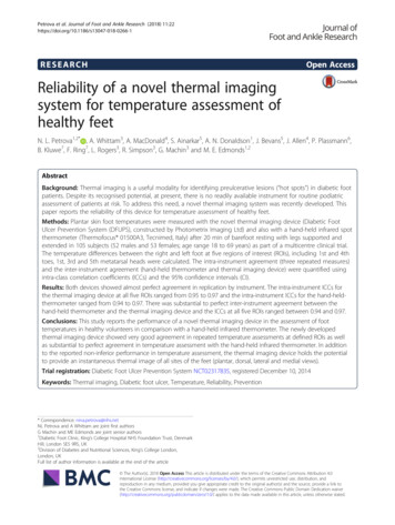
Petrova et al. Journal of Foot and Ankle Research (2018) 11:22abcdPage 5 of 6with DFUPS in the temperature assessment of the feet ofhealthy volunteers have been reported [16]. In addition,thermal imaging with DFUPS does not require any calibration for age, gender, weight, height or BMI and therefore it can be readily implemented in everyday clinicalassessment. The importance of foot skin temperaturemonitoring in the identification of the early signs ofinflammation has been emphasised in the 2015 guidelinesof the International Working Group on the Diabetic Foot[20]. We have recently completed a multicentre clinicaltrial (NCT02579070) in high-risk diabetic foot patients toassess the usefulness of thermal imaging with DFUPS inaddition to standard podiatric treatment to reducediabetic foot ulcer recurrence. In addition to diabetic footulcer prevention, a further study is planned to investigatethe usefulness of DFUPS in the assessment of the acuteCharcot foot.ConclusionsThe newly developed thermal imaging device showed verygood agreement in repeated temperature assessments atdefined ROIs as well as substantial to perfect agreement intemperature assessment with a hand-held infrared thermometer. This device fulfils the requirements of a reproducible and accurate thermal imaging device. It addresses theclinical need of a “portable, reliable and accurate” thermalimaging instrument [8, 13]. We believe that the developedthermal imaging device holds the potential of becoming areal asset in the diabetic foot clinic, to identify potentialpatients at risk of diabetic foot ulcer.AbbreviationsBMI: Body mass index; CE: is abbreviated from Conformité Européenne,meaning European Conformity; CI: Confidence intervals; DFUPS: DiabeticFoot Ulcer Preventions System; ICC: Intra-class correlation coefficient;NHS: National Health Service; NPL: National Physical Laboratory;REC: Research Ethics Committee; ROI: Region of interest; ROIs: Regions ofinterestAcknowledgementsThe authors would like to thank the participants of this study and theproject advisory board for their support and contribution to the study.eFundingThe research was funded by the National Institute for Health Research (NIHR)Invention for Innovation (i4i) programme (An Innovative system for the earlyidentification, monitoring, evaluation and diagnosis of diabetic foot ulcers II-LA0813-20007). The views expressed are those of the authors and not necessarilythose of the NHS, the NIHR or the Department of Health.Availability of data and materialsThe datasets used and/or analysed during the current study are availablefrom NLP on reasonable request.Fig. 2 Bland and Altman plots of agreement between the thermalimaging device and the hand-held thermometer for the 1st toe (a), 4th toe(b), 1st metatarsal head (c), 3rd metatarsal head (d) and 5th metatarsal headAuthors’ contributionsAll authors contributed to the design of the study. NLP, AM and SA recruitedparticipants and collected data. NLP, AW and AND processed and analyseddata. NLP, AW and MEE drafted the manuscript with input from all authors.All authors have read and approved the final manuscript. NLP and AW arejoint first authors. GM and MEE are joint senior authors.
Petrova et al. Journal of Foot and Ankle Research (2018) 11:22Ethics approval and consent to participateApproval for this study was obtained from the London-City Road and HampsteadResearch Ethics Committee (REC reference 15/LO/0070). Each person in the studywas given a participant’s information sheet and all study participants providedwritten informed consent.Consent for publicationConsent for publication containing no personal identifying information wassought and gained from all participants.Competing interestsThe authors declare that they have no competing interests.Publisher’s NoteSpringer Nature remains neutral with regard to jurisdictional claims inpublished maps and institutional affiliations.Author details1Diabetic Foot Clinic, King’s College Hospital NHS Foundation Trust, DenmarkHill, London SE5 9RS, UK. 2Division of Diabetes and Nutritional Sciences,King’s College London, London, UK. 3Temperature and Humidity, NationalPhysical Laboratory, London, UK. 4Microvascular Diagnostics, NorthernMedical Physics and Clinical Engineering, Newcastle upon Tyne Hospitals,Newcastle upon Tyne, UK. 5Community Podiatry Department, Pennine AcuteHospitals NHS Trust, Manchester, UK. 6Photometrix Imaging Ltd, Pontypridd,UK. 7Department of Computing, University of South Wales, Pontypridd, UK.Received: 23 March 2018 Accepted: 16 May 2018References1. Boulton AJM, Vileikyte L, Ragnarson-Tennvall G, Apelqvist J. The globalburden of diabetic foot disease. Lancet. 2005;366(9498):1719–24.2. Edmonds ME. The diabetic foot, 2003. Diabetes Metab Res Rev. 2004;20(Suppl 1):S9–S12.3. Rice JB, Desai U, Cummings AK, Birnbaum HG, Skornicki M, Parsons NB.Burden of diabetic foot ulcers for medicare and private insurers. DiabetesCare. 2014;37(3):651–8.4. Diabetes UK. Improving footcare for people with diabetes and saving money: aneconomic study in England. anuary%25202017%29.pdf Accessed 30 Apr 2018.5. Chammas NK, Hill RL, Edmonds ME. Increased mortality in diabetic footulcer patients: the significance of ulcer type. J Diabetes Res. 2016;2016:2879809.6. Vamos EP, Bottle A, Edmonds ME, Valabhji J, Majeed A, Millett C. Changes inthe incidence of lower extremity amputations in individuals with andwithout diabetes in England between 2004 and 2008. Diabetes Care. 2010;33(12):2592–7.7. Ring F. Thermal imaging today and its relevance to diabetes. J Diabetes SciTechnol. 2010;4(4):857–62.8. Pafili K, Papanas N. Thermography in the follow up of the diabetic foot: bestto weigh the enemy more mighty than he seems. Expert Rev Med Devices.2015;12(2):131–3.9. Frykberg RG, Gordon IL, Reyzelman AM, Cazzell SM, Fitzgerald RH,Rothenberg GM, Bloom JD, Petersen BJ, Linders DR, Nouvong A, FeasibilityNB. Efficacy of a smart mat Technology to predict development of diabeticplantar ulcers. Diabetes Care. 2017;40(7):973–80.10. Armstrong DG, Holtz-Neiderer K, Wendel C, Mohler MJ, Kimbriel HR, LaveryLA. Skin temperature monitoring reduces the risk for diabetic footulceration in high-risk patients. Am J Med. 2007;120(12):1042–6.11. Houghton VJ, Bower VM, Chant DC. Is an increase in skin temperaturepredictive of neuropathic foot ulceration in people with diabetes? Asystematic review and meta-analysis. J Foot Ankle Res. 2013;6:31.12. van Netten JJ, Prijs M, van Baal JG, Liu C, van der Heijden F, Bus SA.Diagnostic values for skin temperature assessment to detect diabetesrelated foot complications. Diabetes Technol Ther. 2014;16(11):714–21.13. Bharara M, Cobb JE, Claremont DJ. Thermography and thermometry in theassessment of diabetic neuropathic foot: a case for furthering the role ofthermal techniques. Int J Low Extrem Wounds. 2006;5:250–60.Page 6 of 614. Prompers L, Huijberts M, Apelqvist J, Jude E, Piaggesi A, Bakker K, EdmondsM, et al. High prevalence of ischaemia, infection and serious comorbidity inpatients with diabetic foot disease in Europe. Baseline results from theEurodiale study. Diabetologia. 2007;50(1):18–25.15. Machin G, Whittam A, Ainarkar S, Allen J, Bevans J, Edmonds M, Kluwe B,Macdonald A, Petrova N, Plassmann P, Ring F, Rogers L, Simpson R. Amedical thermal imaging device for the prevention of diabetic footulceration. Physiol Meas. 2017;38(3):420–30. https://doi.org/10.1088/13616579/aa56b1.16. Macdonald A, Petrova N, Ainarkar S, Allen J, Plassmann P, Whittam A,Bevans J, Ring F, Kluwe B, Simpson R, Rogers L, Machin G, Edmonds M.Thermal symmetry of healthy feet: a precursor to a thermal study ofdiabetic feet prior to skin breakdown. Physiol Meas. 79/38/1/33 .17. Machin G, Chu B. High-quality blackbody sources for infrared thermometryand thermography between-40 and 1000 degrees C. Imaging Sci J. 2000;48(1):15–22.18. Bland JM, Altman DG. Statistical methods for assessing agreement betweentwo methods of clinical measurement. Lancet. 1986;1:307–10.19. Landis JR, Koch GC. The measurement of observer agreement forcategorical data. Biometrics. 1977;33:159–74.20. IWGDF Guidance on the prevention of foot ulcers in at-risk patients withdiabetes. http://www.iwgdf.org/files/2015/website prevention.pdf.
The study was carried out at three clinical centres as previously described [16]. Male and female volunteers were recruited if they had intact feet and no previous his-tory of diabetes, foot ulcer or foot surgery either for cor-rection of a foot deformity or following foot trauma. Subjects were excluded if they reported unsteadiness in