Transcription
Journal of Cancer 2021, Vol. 12IvyspringInternational PublisherResearch Paper6610Journal of Cancer2021; 12(22): 6610-6619. doi: 10.7150/jca.58500FIN56, a novel ferroptosis inducer, triggers lysosomalmembrane permeabilization in a TFEB-dependent mannerin glioblastomaXin Zhang1,2, Yulian Guo1, Hao Li3, Lizhang Han1 1.2.3.Department of Neurosurgery, Qilu Hospital of Shandong University and Institute of Brain and Brain-Inspired Science, Shandong University, Jinan, China.Shandong Key Laboratory of Brain Function Remodeling, Jinan, China.Department of Neurosurgery, Heze third people’s hospital, Heze, China. Corresponding author: Lizhang Han, Department of Neurosurgery, Qilu Hospital of Shandong University and Institute of Brain and Brain-Inspired Science, ShandongUniversity, Jinan, China. E-mail: QL-hanlizhang@sdu.edu.cn. The author(s). This is an open access article distributed under the terms of the Creative Commons Attribution License (https://creativecommons.org/licenses/by/4.0/).See http://ivyspring.com/terms for full terms and conditions.Received: 2021.01.21; Accepted: 2021.08.23; Published: 2021.09.13AbstractObjective: To explore the anti-tumor effect of FIN56, a novel ferroptosis inducer, on glioblastoma and itsunderlying mechanisms.Methods: Two human glioblastoma cell lines, LN229 and U118 were applied in this study. Anti-tumor effectwas measured by CCK-8 assay, EdU assay and cell cycle analysis. Fluorescent probes, immunofluorescence,plasmid transfection, shRNA knocking out, reverse transcription PCR, western blot analysis, and transmissionelectron microscopy were used to study the underlying mechanisms. At last, a subcutaneous nude mice modelwas used to study the anti-tumor effect of FIN56 in vivo. The GraphPad Prism software program was applied forstatistical analysis.Results: FIN56 decreased cell viability, inhibited cell proliferation and caused cell cycle arrest on LN229 andU118 cells. Further study showed that FIN56 induced ferroptosis and induced lysosomal membranepermeabilization in a ferroptosis and transfactor EB dependent manner. Animal study demonstrated that FIN56inhibited glioma growth and caused ferroptosis in vivo.Conclusion: FIN56 is a promising anti-tumor compound.Key words: FIN56; Ferroptosis; Lysosomal membrane permeabilization; GlioblastomaIntroductionGlioblastoma (GBM) is one of the mostmalignant primary tumors in the central nervoussystem [1]. Although a comprehensive treatmentincluding surgical resection, chemo- and radiotherapy exists, the prognosis of patients with GBMstill remains poor [1, 2]. Apoptosis, a programmed celldeath, is one of the most important signalingpathways involved in various anti-cancer treatment[3]. But tumor cells will develop resistance toapoptosis after repeated treatment [4].Except for apoptosis, many programmed celldeath modes have been discovered, including autophagy, necroptosis, pyroptosis and ferroptosis [5].Ferroptosis, a programmed cell death denominatedby Dixon in 2012, is reported to be involved in variousmedical diseases, including neurodegeneration, organfailure, and cancer inhibition [6]. Ferroptosis ischaracterized by intracellular iron accumulation andsubsequent lipid peroxidation [6, 7]. Morphologically,shrunken mitochondria and increased density of themitochondrial membrane can be detected in cells withferroptosis [5]. Cystine-glutamate antiporter (xCT),glutathione peroxidase 4 (GPX4), ferroptosis suppressor protein 1 (FSP1), and GTP cyclohydrolase-1(GCH1) are reported to be involved in the regulationof ferroptosis by limiting the production of lipidperoxides [8].Lysosomal membrane permeabilization (LMP),another programmed cell death mode, is a lysosomedependent cell death process [9]. In LMP, impairedlysosomal membrane induces the release of specificlysosomal enzymes into the cytoplasm, which triggershttp://www.jcancer.org
Journal of Cancer 2021, Vol. 12a cascade of regulated cell death signaling [10].Among all the lysosomal enzymes involved in LMP,cathepsin B and cathepsin D are the most important[11]. Recently, lysosomes are reported to be involvedin the regulation of ferroptosis [12]. Gao et al. showedthat erastin, a ferroptosis inhibitor, induced LMP,which contributed to ferroptosis [13]. Zhou et alreviewed that Stat3-dependent LMP facilitates theinduction of ferroptosis [14]. But until now, fewstudies illustrated the relationship betweenferroptosis and LMP. The underlying mechanisms arestill unknown.FIN56, a novel ferroptosis inducer, triggersferroptosis by increasing the degradation of GPX4[15]. FIN56 also activates squalene synthase, anenzyme involved in the cholesterol synthesis [16]. Butwhether FIN56 has anti-tumor effect is still unknown.In our study, we tried to study the potential role ofFIN56 on GBM. Our results showed that FIN56inhibited GBM cell growth both in vitro and in vivo.Besides, FIN56 increased lipid peroxidation and ROSproduction, which suggested that FIN56 inducedferroptosis in GBM cells. We also found that FIN56triggered LMP. Further studies demonstrated thatFIN56 induced LMP in a transfactor EB(TFEB)-dependent manner. As a result, FIN56 is apotential compound in treating GBM patients.Materials and methodsCell cultureLN229 and U118, two GBM cell lines, wereobtained from the Culture Collection of the ChineseAcademy of Sciences (Shanghai, China) and culturedin Dulbecco’s modified Eagle’s medium (DMEM,ThermoFisher Scientific; Waltham, MA, USA)supplemented with 10% fetal bovine serum (FBS;ThermoFisher Scientific).Cell viability assayGBM cell lines LN229 and U118 cells were platedinto 96-well plates and incubated overnight. Differentdoses of FIN56 (0, 0.1, 0.5, 1.0, 2.0 4.0 and 8.0 µM) wasadded to wells. 24 h later, CCK-8 solution (Dojindo,Kumamoto, Japan) was added to each well. 2 h later,samples were measured at 450 nm on a microplatereader (PerkinElmer; San Jose, CA, USA).EdU assayAn EdU Assay Kit (Ribobio; Guangzhou, China)was used to test cell proliferation after differenttreatment. LN229 and U118 cells were seeded into24-well, flat-bottomed plates and incubatedovernight. Then cells were treated with 1 μM FIN56 orDMSO. 24 h later, cells were incubated with EdUaccording to the manufacturer’s instructions. EdU6611positive cells were calculated under fluorescencemicroscopy (Leica DMi8; Wetzlar, Germany).Cell cycle analysisLN229 and U118 cells were seeded into 6-well,flat-bottomed plates and incubated overnight. Aftertreatment with 1 μM FIN56 or DMSO for 24 h, LN229and U118 cells were collected and fixed with 70%ethanol. Then cells were incubated with propidiumiodide (PI) supplemented with RNase (BectonDickinson, San Diego, CA). Cell cycle analysis wasperformed on a C6 flow cytometer (BD Biosciences;San Jose, CA, USA).Lipid peroxidation and ROS assayLN229 and U118 cells were seeded into 6-well,flat-bottomed plates and incubated overnight. Aftertreatment with 1 μM FIN56 or DMSO for 24 h,intracellular levels of lipid peroxidation/ROS wereassessed using BODIPY581/591 C11/CellRox greendye (ThermoFisher Scientific). BODIPY581/591 C11or CellRox green dye (5 μM; diluted in DMEM with10% fetal bovine serum) was added to LN229 andU118 cells. Then images were obtained under confocalmicroscopy (Leica SP5 Confocal Microscope, Leica).ImmunofluorescenceLN229 and U118 cells were treated with 1 μMFIN56 or DMSO for 24 h. Then cells were fixed with4% paraformaldehyde, permeabilized with 0.3%Triton X-100, and incubated with 4-Hydroxynonenal(4-HNE, a lipid peroxidation marker) primaryantibody (Abcam; Cambridge, UK) and Alexa Fluor594 conjugated goat anti-rabbit secondary antibody(Abcam). Images were taken under a Leica TCS SP5Confocal Laser Scanning Microscope (LeicaMicrosystem).Transmission electron microscopyThe ultrastructure of glioma cells was observedunder transmission electron microscopy after LN229and U118 cells were treated with 1 μM FIN56 orDMSO. Experiments were carried out as describedpreviously [2]. Images were obtained using aJEM-1200EX II electron microscope (JEOL; Tokyo,Japan).Lysosomal membrane stabilityAcridine orange (AO; Sigma-Aldrich, USA)staining and GFP‐fused galectin 3 (Obio, Shanghai,China) transient transfection were used to testlysosomal membrane stability.After being incubated with AO (5 μg/mL) for 15min, LN229 and U118 cells were treated with 1 μMFIN56 or DMSO. Then images were obtained by aLeica DMi8.http://www.jcancer.org
Journal of Cancer 2021, Vol. 12Lipofectamine 2000 reagent (ThermoFisherScientific) was used for transient transfections. 48 hlater, transfected cells were treated with 1 μM FIN56or DMSO for 24 h. Images were taken by a Leica SP5Confocal Microscope.Transfection of shRNALN229 and U118 cells were transfected with 20nM shRNA by using lipofectamine 2000 (ThermoFisher Scientific) according to the manufacturer’sprotocol. Sequences for the shRNAs were thefollowing: negative control, 5’-UUCUCCGAACGUGUCACGUTT-3’; TFEB, 5’-TGTTGGTCATCCAGGCG-3’ (Obio, Shanghai, China). Knocking downefficiency was analyzed by western blot analysis andPCR according to our previous studies [5, 11].Animal studyIn vivo study was carried out by a subcutaneousnude mouse model. Briefly, 105 LN229 cells wereinjected subcutaneously into the right shoulder of 4week-old nude mice (SLAC laboratory animal Center;Shanghai, China). 2 weeks later, nude mice (n 10)were divided into two groups, control group andFIN56 treatment group. Subcutaneous tumors wereharvested 30 days after treatment. Tumors were fixedand paraffin-embedded for embedded tumor tissues were sectioned(4 μm) and mounted onto microscopic slides.Deparaffinized sections were incubated with theprimary antibody at 4 C overnight (4-HNE, Abcam),rinsed with PBS, and incubated with goat anti-rabbitsecondary antibody (Beyotime; Shanghai, China).Images were taken by a Leica DMi8 microscope (LeicaMicrosystems). Staining of cancer cells was scored asfollows: 0, no staining; 1, weak staining in 50% cells;2, weak staining in 50% cells; 3, strong staining in 50% cells; and 4, strong staining in 50% cells.Statistical analysisUnpaired t-tests were performed usingGraphPad Prism software program (Version 6.07; LaJolla, CA, USA). Results are presented as the mean SE. P-values 0.05 were considered statisticallysignificant.ResultsFIN56 inhibits cell growth in GBM cellsFirstly, we tested the anti-tumor effect of FIN56on GBM cells in vitro. CCK-8 was used to test the cellviability after different doses of FIN56 treatment andresults showed that FIN56 decreased the cell viabilityof LN229 and U118 cells in a dose-dependent manner6612(Fig. 1A). The IC50 for LN229 and U118 were 4.2, 2.6µM respectively (Fig. 1A). Normal human astrocytes(NHA) were used as normal control and resultsshowed that glioma cells were more sensitive toFIN56 than NHA (Fig. 1A). Then we used EdU assayto test the cell proliferation after FIN56 treatment. Wefound that FIN56 decreased cell proliferationsignificantly compared with control group (Fig. 1Band 1C). Cell cycle arrest is one of the most importantfactors contributing to the inhibition of cellproliferation. We found that after FIN56 treatment,cell cycle of LN229 and U118 was arrested at GO/G1phases (Fig. 1D and 1E). All these results suggestedthat FIN56 had promising anti-tumor effect on GBMcells.FIN56 induces ferroptosis in GBM cellsPrevious studies recognized FIN56 as aferroptosis inducer. In our study, we tried to figureout whether FIN56 induced ferroptosis in GBM cells.Lipid peroxidation is one of the most importantfeatures of ferroptosis. Lipid peroxidation wasassessed by BODIPY581/591/C11, a free radical sensor.When the levels of lipid peroxidation increase, redfluorescence of BODIPY581/591 /C11 decreases andgreen fluorescence shows up. FIN56 treatmentincreased green fluorescence and red fluorescencedecayed (Fig. 2A). Immunofluorescence revealed thatFIN56 increased the expression of 4 hydroxynonenal(4-HNE), an aldehydic product of lipid peroxidation(Fig. 2B). We also found that FIN56 also increasedROS production compared with control group (Fig.2C). The ultrastructure of GBM cell lines LN229 andU118wasstudiedby transmission electronmicroscope. Fig 2D indicated that FIN56 induced theshrink of the mitochondria and increased the densityof its membrane (red arrows). To test whether thecytotoxic effect is ferroptosis-dependent, LDH releaseassay was carried out and results demonstrated thatFerrostatin-1 (Fer-1), a ferroptosis inhibitor, obviouslyinhibited FIN56-induced cell death (Fig. 2E). All theseresults illustrated that FIN56 induced ferroptosis inGBM cells.FIN56 causes lysosomal membranepermeabilization in a ferroptosis-dependentmannerOur previous study showed that disulfiram, anovel ferroptosis inducer, induced LMP in GBM cells.Here, we tried to figure out whether FIN56 had thesame effect.Acridine orange (AO), a lysosomotropic dye, isenriched in intact lysosomes, which emits redfluorescence. While in impaired lysosomes, theenrichment of AO is interfered, and red fluorescencehttp://www.jcancer.org
Journal of Cancer 2021, Vol. 12intensity decreases. Fig. 3A illustrated that FIN56decreased red fluorescence compared with controlgroup.When the membranes of lysosomes areimpaired, Galectin 3, a β-galactoside binding lectin,accumulates on luminal glycoproteins and isrecognized as a better method to monitor theoccurrence of LMP. LN229 and U118 cells weretransfected with a GFP-fused Galectin 3 (EGFP-Gal3)plasmid and results showed that FIN56 treatmentincreased green fluorescent dots, compared withcontrol group (Fig. 3B and 3C). In the process of6613ferroptosis, levels of lipid peroxidation and ROSincrease. ROS accumulation is one of the mostimportant factors leading to LMP. Our previous studyalso showed that ferroptosis inducer disulfiramcontributed to LMP in a ferroptosis and ROSdependent manner. Here, ferroptosis was inhibited byFer-1 and EGFP-Gal3 dots decreased obviously inFer-1 and FIN56 combined treatment, compared withFIN56 treatment alone (Fig. 3D and 3E). All these datademonstrated that FIN56 triggered LMP in aferroptosis manner in GBM cells.Figure 1. FIN56 inhibits cell growth of GBM cells (A) Growth curves generated with colorimetric data (O.D. 450 nm) from the CCK-8 assay. LN229, U118 and normalhuman astrocytes (NHA) cells were treated with different doses of FIN56 for 24 h. (B) Fluorescence images of EdU of 1 μM FIN56 or DMSO (control) for 24 h in LN229 andU118 cells. (C) Quantification of EdU positive cells in (B). (D) Cell cycle analysis of LN229 and U118 cells treated with 1 μM FIN56 or DMSO (control) for 24 h. (E)Quantification of cell cycle parameters G0-G1, S, and G2-M obtained from flow cytometric analysis in D. **P 0.01; ***P 0.001; scale bars: 50 µm.http://www.jcancer.org
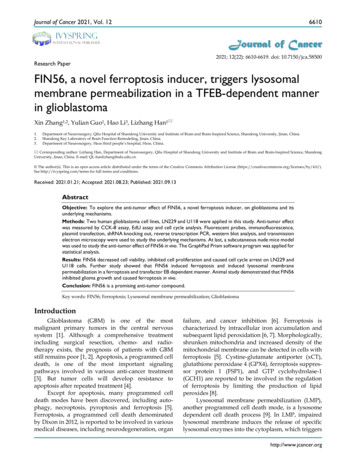
Journal of Cancer 2021, Vol. 126614Figure 2. FIN56 induces ferroptosis in GBM cells. (A) Images of BODIPY581/591 C11 staining of LN229 and U118 cells treated with 1 μM FIN56 or DMSO (control) for24 h. (B) Images of immunofluorescence staining of 4-HNE of LN229 and U118 cells treated with 1 μM FIN56 or DMSO (control). (C) Images of CellRox green staining of LN229and U118 cells treated with 1 μM FIN56 or DMSO (control). (D) Ultrastructure of LN229 cells treated with 1 μM FIN56 or DMSO (control) by transmission electronmicroscopy. Mitochondria were pointed out in DMSO (black arrows) and FIN56 (red arrows) treatment group. **P 0.01; ***P 0.001. Size bars in (A), (B) and (C): 10 µm, in(D): 0.5 µm.FIN56 induces LMP in a TFEB dependentmannerWhen LMP occurs, hydrolases are released fromdamaged lysosomal membrane and cell death isinduced. There are more than 50 acid hydrolases inthe lumen of lysosomes. Among all the hydrolasesinvolved in lysosomal cell death, cathepsin B andcathepsin D are the most important. Our previousstudy found that knocking down transfactor EB(TFEB), the expression of cathepsin B and cathepsin Dwas inhibited [11]. Besides, TFEB played an importantrole in the biogenesis of lysosome. As a result, weassumed that TFEB might contribute to LMP. To testour assumption, we knocked down TFEB with aspecific siRNA and the knocking-down efficiency wasverified by reverse transcription PCR and westernblot analysis (Fig. 4A and 4B). Then we added FIN56in control or TFEB knocking-out LN229 and U118cells. Fig. 4C and 4D showed that after FIN56/siramesine treatment, EGFP-Gal3 dots decreasedsignificantly in TFEB knocking-out LN229 and U118cells compared with control group. We also foundthat after knocking down TFEB, FIN56 didn’t increaseROS production in LN229 and U118 cells (Fig. 4E).These data showed that FIN56 induced LMP in ahttp://www.jcancer.org
Journal of Cancer 2021, Vol. 12TFEB dependent manner. Further study alsoillustrated that when cathepsin B and cathepsin Dwere inhibited by their inhibitor, CA-074 Me,pepstatin A, FIN56 didn’t induce LMP in LN229 andU118 cells (supplementary Fig. 1), which suggestedthat FIN56-induced LMP was cathepsin B- andcathepsin D-dependent.FIN56 inhibited cell growth and inducedferroptosis in vivoFinally, we tested the anti-tumor effect of FIN56in vivo. We implanted LN229 cell subcutaneously in6615nude mice and treated them with or without FIN56.30 days later tumors were harvested and tested byimmunohistochemistry. Results showed that FIN56decreased tumor volume obviously (Fig. 5A and 5B).Ki67 is a marker to test proliferation ability andimmunohistochemistry results demonstrated thatFIN56 treatment decreased Ki67 positive cells (Fig. 5Cand 5D). FIN56 also significantly increased proteinlevels of 4-HNE (Fig. 5E and 5F). All those resultsillustrated that FIN56 had efficient anti-tumor effect invivo.Figure 3. FIN56 causes lysosomal membrane permeabilization in a ferroptosis-dependent manner. (A) Fluorescence images of acridine orange staining of LN229and U118 cells treated with 1 μM FIN56 or DMSO (control). (B) Fluorescence images of EGFP-Gal3 transfection of LN229 and U118 cells treated with 1 μM FIN56 or DMSO(control). (C) Quantification of green puncta in (B). (D) Fluorescence images of EGFP-Gal3 transfection of LN229 and U118 cells treated with DMSO (control), 1 μM FIN56 or,2 µM ferrostatin-1 (Fer-1) or 2 µM ferrostatin-1 (Fer-1) combined with 1 μM FIN56. (E) Quantification of green puncta in (D). **P 0.01; scale bars: 10 µm.http://www.jcancer.org
Journal of Cancer 2021, Vol. 126616Figure 4. FIN56 induces LMP in a TFEB dependent manner. (A) Reverse transcription PCR of LN229 and U118 cells transfected with shControl or shTFEB. (B)Western blot analysis of LN229 and U118 cells transfected with shControl or shTFEB. (C) Fluorescence images of EGFP-Gal3 transfection of LN229 and U118 cells treated withshControl combined with 1 μM FIN56, shTFEB combined with 1 μM FIN56, shControl combined with 5 μM siramesine (SM), shTFEB combined with 5 μM SM. (D) Quantificationof green puncta in (C). (E) Images of CellRox green staining of LN229 and U118 cells treated with shControl combined with 5 μM siramesine (SM), shTFEB combined with 5 μMSM. **P 0.01; ***P 0.001; size bars: 10 µm.DiscussionAlthough more and more attention has beenpaid to the studies of glioma, few breakthroughs havebeen achieved. Apoptosis is considered as the mostimportant signaling pathway of various methods ofanti-tumor treatment [4]. However, tumor cells willdevelop apoptotic resistance after repeated treatment,and it is one of the most important factorscontributing to the relapse and poor prognosis ofglioma [17].Among many types of programmed cell death,ferroptosis is formally denominated in 2012[18]. Untilnow, studies on ferroptosis keep increasing.Ferroptosis plays an important role in various types ofdiseases [19-21]. Overactive ferroptosis triggers tissueinjury in diseases like cardiovascular/cerebrovasculardiseases and neurodegenerative diseases [22]. Whilein cancer, inactivation of ferroptosis contributes totumor growth and resistance to chemo- and radiotherapy [23]. As a result, ferroptosis inducers arepromising anti-tumor reagents.http://www.jcancer.org
Journal of Cancer 2021, Vol. 126617Figure 5. FIN56 inhibited cell growth and induced ferroptosis in vivo. (A) Images of subcutaneous tumors after control or FIN56 treatment. (B) Quantification oftumor volume in (A). (C) Immunohistochemistry staining of ki67 to determine cell proliferation. (D) Quantification of (C). (E) Immunohistochemistry staining of 4-HNE todetermine cell ferroptosis. (F) Quantification of (C). *P 0.05; **P 0.01; ***P 0.001; size bars: 50 µm.Here, in our study we found that FIN56, a novelferroptosis inducer, showed efficient anti-gliomaeffect both in vitro and in vivo. Firstly, FIN56 decreasedcell viability in GBM cell lines LN229 and U118. FIN56also inhibited cell proliferation by causing cell cyclearrest. As a novel ferroptosis inducer, FIN56 inducedcell death in LN229 and U118 cells in aferroptosis-dependent manner. We also found thatFIN56 triggered LMP in LN229 and U118 cells.Further studies illustrated that the LMP induced byFIN56 was ROS and TFEB dependent. At last, in vivostudies showed that FIN56 had efficient anti-tumoreffect in a nude mice model. But whether FIN56 willpenetrate the blood brain barrier is still unknown.More studies are warranted to test the anti-tumoreffect of FIN56 on orthotopic glioma xenografts.Lysosomal membrane permeabilization (LMP) isa lysosome-dependent cell death. A study by Gao et alshowed that ferroptosis is a lysosomal cell deathprocess [13]. In our previous study, we also found thatdisulfiram, traditionally used for the treatment ofalcoholism, induced ferroptosis and LMP in gliomacell lines [5]. As a result, ferroptosis is closely relatedto LMP, and lysosomes might be the key link.http://www.jcancer.org
Journal of Cancer 2021, Vol. 12Previously, lysosome is recognized as the waste bagfor its role in late stage of autophagy [9]. But more andmore studies demonstrate that lysosome plays animportant role in regulating intracellular signalingpathways [24, 25]. And TFEB, a family of basic helix–loop–helix leucine zipper transcription factors, isrecognized as one important regulator of lysosomalbiogenesis [26-28]. TFEB has also been reported to beinvolved in clearance of oxidative stress [29]. A studyby Park et al showed that TFEB activates Nrf2, animportant factor in regulating oxidative stress, byincreasing its stability [30]. Considering the role ofTFEB in lysosomal biogenesis and oxidative stress inthe initiation of LMP, we assumed that TFEB might beinvolved in LMP. In our study, we found that FIN56induced LMP, and this process was inhibited byknocking down TFEB. Siramesine-induced LMPcould also be inhibited by knocking down TFEB. Butwhether TFEB regulates ferroptosis is not known.Further studies are warranted in the future.In summary, our study identified that FIN56, anovel ferroptosis inducer, showed significantanti-tumor effect and triggered lysosomal membranepermeabilization in a TFEB-dependent manner inglioma. FIN56 is a promising anti-tumor compound.More studies are needed on other tumor types.Supplementary MaterialSupplementary .13.14.15.16.17.Ethical approval18.The animal studies were approved by the EthicsCommittee of Qilu Hospital, Shandong University.19.FundingThis work was supported by the NationalNatural Science Foundation of China (81903126).Author contributionsXZ and LZH contributed to the design of thestudy. XZ, YLG and HL conducted the experimentsand prepared figures. XZ, YLG and LZH contributedto the statistical analysis. XZ and LZH supervised thisstudy and prepared the manuscript. All authors haveread and approved the final version of themanuscript.Competing InterestsThe authors have declared that no competinginterest exists.20.21.22.23.24.25.26.27.28.29.Touat M, Idbaih A, Sanson M, et al. Glioblastoma targeted therapy:updated approaches from recent biological insights. Ann Oncol. 2017;28(7):1457-1472.Zhang X, Xu R, Zhang C, et al. Trifluoperazine, a novel autophagyinhibitor, increases radiosensitivity in glioblastoma by impairinghomologous recombination. J Exp Clin Canc Res. 2017; 36(1):118.Mohamed MS, Bishr MK, Almutairi FM, et al. Inhibitors of apoptosis:clinical implications in cancer. Apoptosis. 2017; 22(12):1487-1509.Rathore R, McCallum JE, Varghese E, et al. Overcoming chemotherapydrug resistance by targeting inhibitors of apoptosis proteins (IAPs).Apoptosis. 2017; 22(7):898-919.Qiu C, Zhang X, Huang B, et al. Disulfiram, a Ferroptosis Inducer,Triggers Lysosomal Membrane Permeabilization by Up-Regulating ROSin Glioblastoma. Onco Targets Ther. 2020; 13:10631-10640.Bebber CM, Muller F, Prieto Clemente L, et al. Ferroptosis in Cancer CellBiology. Cancers. 2020; 12(1).164.Ma DL, Li C, Jiang PL, et al. Inhibition of Ferroptosis Attenuates AcuteKidney Injury in Rats with Severe Acute Pancreatitis. Dig Dis Sci. 2020;66(2):483-492Tang D, Chen X, Kang R, et al. Ferroptosis: molecular mechanisms andhealth implications. Cell Res. 2020; 31(2):107-125.Repnik U, Hafner Česen M, Turk B. Lysosomal membranepermeabilization in cell death: concepts and challenges. Mitochondrion.2014; 19 Pt A:49-57.Wang F, Gómez-Sintes R, Boya P. Lysosomal membranepermeabilization and cell death. Traffic. 2018; 9(12):918-931.Zhou W, Guo YL, Zhang X, et al. Lys05 induces lysosomal membranepermeabilization and increases radiosensitivity in glioblastoma. J CellBiochem.2020; 121(2):2027-2037.Torii S, Shintoku R, Kubota C, et al. An essential role for functionallysosomes in ferroptosis of cancer cells. Biochem J. 2016; 473(6):769-777.Gao H, Bai Y, Jia Y, et al. Ferroptosis is a lysosomal cell death process.Biochem Biophys Res Commun. 2018; 503(3):1550-1556.Zhou B, Liu J, Kang R, et al. Ferroptosis is a type ofautophagy-dependent cell death. Semin Cancer Biol. 2020; 66:89-100.Shimada K, Skouta R, Kaplan A, et al. Global survey of cell deathmechanisms reveals metabolic regulation of ferroptosis. Nat Chem Biol.2016; 12(7):497-503.Gaschler MM, Andia AA, Liu H, et al. FINO2 initiates ferroptosisthrough GPX4 inactivation and iron oxidation. Nat Chem Biol. 2018;14(5):507-515.Krell A, Wolter M, Stojcheva N, et al. MiR-16-5p is frequentlydown-regulated in astrocytic gliomas and modulates glioma cellproliferation, apoptosis and response to cytotoxic therapy. NeuropatholAppl Neurobiol. 2019; 45(5):441-458.Dixon SJ, Lemberg KM, Lamprecht MR, et al. Ferroptosis: aniron-dependent form of nonapoptotic cell death. Cell. 2012;149(5):1060-1072.Weiland A, Wang Y, Wu W, et al. Ferroptosis and Its Role in DiverseBrain Diseases. Mol Neurobiol. 2019; 56(7):4880-4893.Derry PJ, Hegde ML, Jackson GR, et al. Revisiting the intersection ofamyloid, pathologically modified tau and iron in Alzheimer's diseasefrom a ferroptosis perspective. Prog Neurobiol. 2020; 184:101716.Wu YP, Chen HX, Xuan NX, et al. Induction of ferroptosis-like cell deathof eosinophils exerts synergistic effects with glucocorticoids in allergicairway inflammation. Thorax. 2020; 75(11):918-927.Li Y, Feng DC, Wang ZY, et al. Ischemia-induced ACSL4 activationcontributes to ferroptosis-mediated tissue injury in intestinalischemia/reperfusion. Cell Death Differ. 2019; 26(11):2284-2299.Toyokuni S, Yanatori I, Kong Y, et al. Ferroptosis at the crossroads ofinfection, aging and cancer. Cancer Sci. 2020; 111(8):2665-2671.Geisslinger F, Muller M, Vollmar AM, et al. Targeting Lysosomes inCancer as Promising Strategy to Overcome Chemoresistance. FrontOncol. 2020; 10:1156.Appelqvist H, Waster P, Kagedal K, et al. The lysosome: from waste bagto potential therapeutic target. J Mol Cell Biol. 2013; 5(4):214-226.Settembre C, Di Malta C, Polito VA, et al. TFEB links autophagy tolysosomal biogenesis. Science. 2011; 332(6036):1429-1433.Wang S, Ni HM, Chao X, et al. Impaired TFEB-mediated lysosomalbiogenesis promotes the development of pancreatitis in mice and isassociated with human pancreatitis. Autophagy. 2019; 15(11):1954-1969.Zhang X, Wang J, Li XG, et al. Lysosomes contribute to radioresistance incancer. Cancer Lett. 2018; 439:39-46.Tsunemi T, Ashe TD, Morrison BE, et al. PGC-1alpha rescuesHuntington's disease proteotoxicity by preventing oxidative stress andpromoting TFEB function. Sci Transl Med. 2012; 4(142):142ra97.http://www.jcancer.org
Journal of Cancer 2021, Vol. 12661930. Park JY, Kim S, Sohn HY, et al. TFEB activates Nrf2 by repressing its E3ubiquitin ligase DCAF11 and promoting phosphorylation of p62. SciRep. 2019; 9(1):14354.http://www.jcancer.org
performed on a C6 flow cytometer (BD Biosciences; San Jose, CA, USA). Lipid peroxidation and ROS assay LN229 and U118 cells were seeded into 6-well, flat-bottomed plates and incubated overnight. After treatment with 1 μM FIN56 or DMSO for 24 h, intracellular levels of lipid peroxidation/ROS were assessed using BODIPY581/591 C11/CellRox green