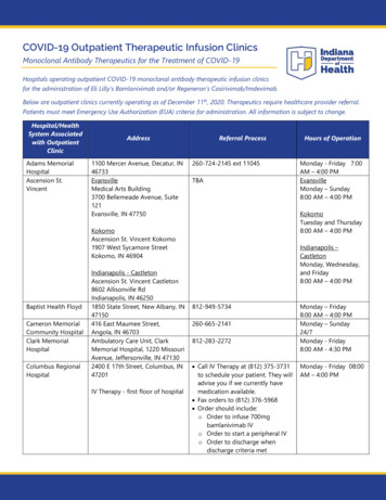
Transcription
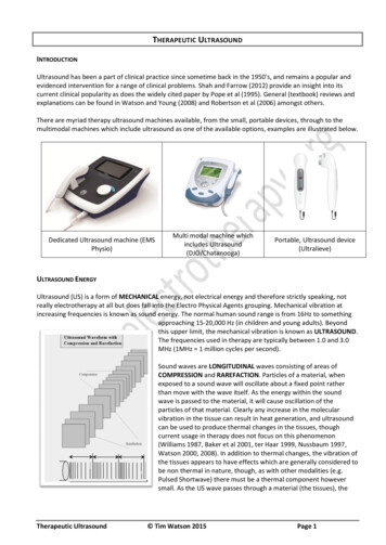
THERAPEUTIC ULTRASOUNDINTRODUCTIONUltrasound has been a part of clinical practice since sometime back in the 1950's, and remains a popular andevidenced intervention for a range of clinical problems. Shah and Farrow (2012) provide an insight into itscurrent clinical popularity as does the widely cited paper by Pope et al (1995). General (textbook) reviews andexplanations can be found in Watson and Young (2008) and Robertson et al (2006) amongst others.There are myriad therapy ultrasound machines available, from the small, portable devices, through to themultimodal machines which include ultrasound as one of the available options, examples are illustrated below.Dedicated Ultrasound machine (EMSPhysio)Multi modal machine whichincludes Ultrasound(DJO/Chatanooga)Portable, Ultrasound device(Ultralieve)ULTRASOUND ENERGYUltrasound (US) is a form of MECHANICAL energy, not electrical energy and therefore strictly speaking, notreally electrotherapy at all but does fall into the Electro Physical Agents grouping. Mechanical vibration atincreasing frequencies is known as sound energy. The normal human sound range is from 16Hz to somethingapproaching 15-20,000 Hz (in children and young adults). Beyondthis upper limit, the mechanical vibration is known as ULTRASOUND.The frequencies used in therapy are typically between 1.0 and 3.0MHz (1MHz 1 million cycles per second).Sound waves are LONGITUDINAL waves consisting of areas ofCOMPRESSION and RAREFACTION. Particles of a material, whenexposed to a sound wave will oscillate about a fixed point ratherthan move with the wave itself. As the energy within the soundwave is passed to the material, it will cause oscillation of theparticles of that material. Clearly any increase in the molecularvibration in the tissue can result in heat generation, and ultrasoundcan be used to produce thermal changes in the tissues, thoughcurrent usage in therapy does not focus on this phenomenon(Williams 1987, Baker et al 2001, ter Haar 1999, Nussbaum 1997,Watson 2000, 2008). In addition to thermal changes, the vibration ofthe tissues appears to have effects which are generally considered tobe non thermal in nature, though, as with other modalities (e.g.Pulsed Shortwave) there must be a thermal component howeversmall. As the US wave passes through a material (the tissues), theTherapeutic Ultrasound Tim Watson 2015Page 1
energy levels within the wave will diminish as energy is transferred to the material. The energy absorption andattenuation characteristics of US waves have been documented for different tissues (see absorption section).ULTRASOUND WAVES :FREQUENCY - the number of times a particle experiences a complete compression/rarefaction cycle in 1second. Typically 1 or 3 MHz (though there are devices which operate in the kHz range - see comments on LowFrequency / Longwave Ultrasound at the end of this paper).WAVELENGTH - the distance between two equivalent points on the waveform in the particular medium. In an‘average tissue’ the wavelength @ 1MHz would be 1.5mm and @ 3 MHz would be 0.5 mm.VELOCITY - the velocity at which the wave (disturbance) travels through the medium. In a saline solution, thevelocity of US is approximately 1500 m sec-1 compared with approximately 350 m sec-1 in air (sound waves cantravel more rapidly in a more dense medium). The velocity of US in most tissues is thought to be similar to thatin saline.These three factors are related, but are not constant for all types of tissue. Average figures are mostcommonly used to represent the passage of US in the tissues. Typical US frequencies from therapeuticequipment are 1 and 3 MHz though some machines produce additional frequencies (e.g. 0.75 and 1.5 MHz)and the ‘Longwave’ ultrasound devices operate at several 10’s of kHz (typically 40-50,000Hz – a much lowerfrequency than ‘traditional US’ but still beyond human hearing range.The mathematical representation of the relationship is V F. where V velocity, F frequency and is the wavelength.ULTRASOUND BEAM, NEAR FIELD, FAR FIELD AND BEAM NON UNIFORMITYThe US beam is not uniform and changes in its nature with distance from the transducer. The US beam nearestthe treatment head is called the NEAR field, the INTERFERENCE field or the Frenzel zone. The behaviour of theUS in this field is far from regular, with areas of significant interference. The US energy in parts of this field canbe many times greater than the output set on themachine (possibly as much as 12 to 15 times greater).The size (length) of the near field can be calculated usingExample of an Ultrasound Beam Plotr2/ where r the radius of the transducer crystal and the US wavelength according to the frequency beingused (0.5mm for 3MHz and 1.5mm for 1.0 MHz).As an example, a 'crystal' with a diameter of 25mmoperating at 1 MHz will have a near field/far fieldboundary at : Boundary 12.5mm2/1.5mm 10cm thusthe near field (with greatest interference) extends forapproximately 10 cm from the treatment head whenusing a large treatment head and 1 MHz US. When usinghigher frequency US, the boundary distance is evengreater. Beyond this boundary lies the Far Field or the Fraunhofer zone. The US beam in this field is moreuniform and gently divergent. The ‘hot spots’ noted in the near field are not significant. For the purposes oftherapeutic applications, the far field is effectively out of reach.One quality indicator for US applicators (transducers) is a value attributed to the Beam Nonuniformity Ratio(BNR). This gives an indication of this near field interference. It describes numerically the ratio of the intensitypeaks to the mean intensity. For most applicators, the BNR would be approximately 4 - 6 (i.e. that the peakTherapeutic Ultrasound Tim Watson 2015Page 2
intensity will be 4 or 6 times greater than the mean intensity). It is considered inappropriate to use a devicewith a BNR value of 8.0 or more. Because of the nature of US, the theoretical best value for the BNR is thoughtto be around 4.0 though some manufacturers claim to have overcome this limit and effectively reduced theBNR of their generators to 1.0.Some recent papers (Alvarenga et al 2010; Gutierrez et al 2010; Straub et al, 2008 and Johns et al 2007) haveconsidered some of the inaccuracies associated with current machines and Pye (1996) presents some worryingdata with regards the calibration of machines in clinical use in the UK.ULTRASOUND TRANSMISSION THROUGH THE TISSUESAll materials (tissues) will present an impedance to the passage of sound waves. The specific impedance of atissue will be determined by its density and elasticity. In order for the maximal transmission of energy fromone medium to another, the impedance of the two medianeeds to be as similar as possible. Clearly in the case of USpassing from the generator to the tissues and thenthrough the different tissue types, this can not actually beachieved. The greater the difference in impedance at aboundary, the greater the reflection that will occur, andtherefore, the smaller the amount of energy that will betransferred. Examples of impedance values can be foundin the literature e.g. Robertson et al 2007, Ward 1986.The difference in impedance is greatest for the steel/airinterface which is the first one that the US has toovercome in order to reach the tissues. To minimise thisdifference, a suitable coupling medium has to be utilised.If even a small air gap exists between the transducer andthe skin the proportion of US that will be reflectedapproaches 99.998% which means that there will be no effective transmission.The coupling media used in this context include water, various oils, creams and gels. Ideally, the couplingmedium should be fluid so as to fill all available spaces, relatively viscous so that it stays in place, have animpedance appropriate to the media it connects, and should allow transmission of US with minimalabsorption, attenuation or disturbance. For a good discussion regarding coupling media, see Casarotto et al2004, Docker et al 1982, Griffin, 1980, Klucinec et al 2000 and Williams 1987. At the present time the gel basedmedia are preferable to the oils and creams. Water is an effective media and can be used as an alternative butclearly it fails to meet the above criteria in terms of its viscosity. There is no realistic (clinical) differencebetween the gels in common clinical use (Poltawski and Watson 2007). The addition of active agents (e.g.anti-inflammatory drugs) to the gel is widely practiced, but remains incompletely researched. We are currentlyevaluating this intervention further.Bacterial Contamination of Ultrasound Treatment Heads and Gel SourcesAs a matter of (clinical) interest, the US treatment should be cleaned with an alcohol based swab (not justwiped with tissue) between treatments (Schabrun et al, 2006) to minimise the potential transmission ofmicrobial agents between patients. Spratt et al (2014) sampled ultrasound treatment heads and gel bottles (inthe USA) reporting over 50% of gel bottles with contamination, some of which were positive for MRSA. Some35% of the ultrasound treatment heads tested also demonstrated contamination, though none with MRSA.The authors report that employment of adequate disinfection techniques was effective in significantlyreducing these levels (they used Protex, Parker Laboratories).Therapeutic Ultrasound Tim Watson 2015Page 3
Ultrasound Application - The Critical AngleIn addition to the reflection that occurs at a boundary due to differences in impedance, there will also be somerefraction if the wave does not strike the boundarysurface at 90 . Essentially, the direction of the US beamthrough the second medium will not be the same as itspath through the original medium - its pathway isangled. The critical angle for US at the skin interfaceappears to be about 15 . If the treatment head is at anangle of 15 or more to the plane of the skin surface,the majority of the US beam will travel through thedermal tissues (i.e. parallel to the skin surface) ratherthan penetrate the tissues as would be expected.ULTRASOUND ABSORPTION AND ATTENUATION :The absorption of US energy follows an exponential pattern - i.e. more energy is absorbed in the superficialtissues than in the deep tissues. In order for energy to have an effect it must be absorbed, and at some pointthis must be considered in relation to the US dosages applied to achieve certain effects (ter Haar, 1999,Watson, 2008, Watson and Young, 2008).Because the absorption (penetration) is exponential, there is (in theory) no point at which all the energy hasbeen absorbed, but there is certainly a point at which the US energy levels are not sufficient to produce atherapeutic effect. As the US beam penetrates further into the tissues, a greater proportion of the energy willhave been absorbed and therefore there is less energy available to achieve therapeutic effects. The half valuedepth is often quoted in relation to US and it represents the depth in the tissues at which half the surfaceenergy is available. These will be different for each tissue and also for different US frequencies. The tablebelow gives some indication of typical (or average) half value depths for therapeutic ultrasound. (afterHoogland 1995)MuscleFatTendon1 MHz9.0 mm50.0 mm6.2 mm3 MHz3.0 mm16.5 mm2.0 mmAs it is difficult, if not impossible to know the thickness of each of these layers in an individual patient, averagehalf value depths are employed for each frequency3 MHz1 MHz2.0 cm4.0 cmThese values (after Low & Reed) are not universallyaccepted (see Ward 1986) and some research (as yetunpublished) suggests that in the clinical environment,they may be significantly lower.Depth (cm)24683 MHz50%25%1 MHz50%25%To achieve a particular US intensity at depth, account must be taken of the proportion of energy which hasbeen absorbed by the tissues in the more superficial layers. The table gives an approximate reduction inenergy levels with typical tissues at two commonly used frequencies, and more detailed information is foundin the dose calculation material and on the web pages (www.electrotherapy.org).Therapeutic Ultrasound Tim Watson 2015Page 4
As the penetration (or transmission) of USis not the same in each tissue type, it isclear that some tissues are capable ofgreater absorption of US than others.Generally, the tissues with the higherprotein content will absorb US to a greaterextent, thus tissues with high watercontent and low protein content absorblittle of the US energy (e.g. blood and fat)whilst those with a lower water contentand a higher protein content will absorb USfar more efficiently. Tissues can be rankedaccording to their relative tissue absorptionand this is critical in terms of clinicaldecision making (Watson, 2008).Although cartilage and bone are at the upper end of this scale, the problems associated with wave reflectionmean that the majority of US energy striking the surface of either of these tissues is likely to be reflected. Thebest absorbing tissues in terms of clinical practice are those with high collagen content – LIGAMENT, TENDON,FASCIA, JOINT CAPSULE, SCAR TISSUE (Watson 2000, 2008, Watson & Young, 2008, ter Haar 1999, Nussbaum1998, Frizzel & Dunn 1982)The application of therapeutic US to tissues with a low energy absorption capacity is less likely to be effectivethan the application of the energy into a more highly absorbing material. Recent evidence of theineffectiveness of such an intervention can be found in Wilkin et al (2004) and Markert et al (2005) whilstapplication in tissue that is a better absorber will,as expected, result in a more effectiveintervention (e.g. Sparrow et al 2005, Leung et al2004).The physiological effects of ultrasound are almostidentical to those of Pulsed Shortwave and Lasertherapy – the key difference however, is thatultrasound energy is preferentially absorbed indifferent tissue to the other modalities – assummarised in the adjacent diagram.PULSED ULTRASOUNDMost machines offer
Ultrasound (US) is a form of MECHANICAL energy, not electrical energy and therefore strictly speaking, not really electrotherapy at all but does fall into the Electro Physical Agents grouping. Mechanical vibration at increasing frequencies is known as sound energy. The normal human sound range is from 16Hz to something approaching 15-20,000 Hz (in children and young adults). Beyond this upper .