Transcription
J Indian Prosthodont Soc (July-Sept 2014) 14(3):305–309DOI 10.1007/s13191-012-0216-8CLINICAL REPORTNarrow Ridge Management with Ridge Splitting with Piezotomefor Implant Placement: Report of 2 CasesDeepak Agrawal Alka Sanjay Gupta Vilas Newaskar Amit Gupta Subhash GargDeshraj Jain Received: 13 July 2012 / Accepted: 11 November 2012 / Published online: 25 November 2012Ó Indian Prosthodontic Society 2012Abstract Narrow dentoalveolar ridges remain a seriouschallenge for the successful placement of endosseousimplants. Several techniques for this procedure may beconsidered, such as guided bone regeneration, bone blockgrafting, and ridge splitting for bone expansion. The ridgesplit procedure provides a quicker method wherein anatrophic ridge can be predictably expanded and graftedwith bone allograft or allograft, eliminating the need forsecond surgical site. Traditionally, osseous surgery hasbeen performed by either manual or motor-driven instruments. Piezosurgery is a relatively new technique forosteotomy and osteoplasty that utilizes ultrasonic vibrationwhich allows clean cutting with precise incisions. This caseseries describes reports of 2 such cases in which narrowD. Agrawal V. NewaskarDepartment of Oral and Maxillofacial Surgery, Govt. Collegeof Dentistry, Indore, Madhya pradesh, Indiae-mail: fordeepak1@yahoo.co.inV. Newaskare-mail: vilasnewaskar@yahoo.comA. S. Gupta (&) D. JainDepartment of Prosthodontics, Govt. College of Dentistry,Indore, Madhya pradesh, Indiae-mail: Dr.alka2000@gmail.comD. Jaine-mail: drdeshrajjain@rediffmail.comA. GuptaDepartment of Orthodontics, Madern Dental College andResearch Centre, Indore, Madhya Pradesh, Indiae-mail: amidont@yahoo.comS. GargDepartment of Periodontics, Govt. College of Dentistry, Indore,Madhya pradesh, Indiae-mail: drsubhasggarg@rediffmail.commandibular ridge splitting was carried by mean of piezotome with immediate placement of implants in the osteotomy site. Five months later, the implants were uncoveredfollowed by impression and restored with impant- supported porcelain-fused-to-metal crowns.KeywordsPiezotomeImplants Narrow ridge Ridge split IntroductionDental implants have become an integral part of comprehensive management of dental patients. Successful implanttherapy often requires sound osseous support. Scipioni et al.[1] suggests that wherever dental implants are placed, aminimum thickness of 1–1.5 mm of bone should remain onboth buccal and lingual/palatal aspects of the implant(s) toensure a successful outcome. For most standard implants, aminimum of 6 mm width is necessary for favourable outcome. A major limitation for successful implant placementremains the problem of inadequate ridge. Several methodshave been described to augment the alveolar crest such asguided bone regeneration, bone block grafting, ridge splitting for bone expansion, and distraction osteogenesis.Ridge splitting creates a sagittal osteotomy of the edentulous ridge using instruments such as chisels between the twocortical plates to expand the ridge width and consequentlyallow for the placement of implants. Ridge splitting approachhas also been referred to as bone spreading, ridge expansion,lateral ridge expansion technique or the osteotome technique.Osborn [2] described this as ‘extension plasty’.Several methods have been accomplished for the osteotomy procedure, manually with osteotomes or motordriven engine. Recently Piezosurgery is being used for123
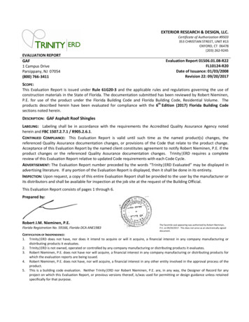
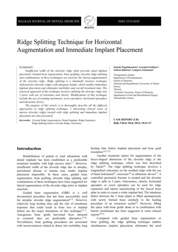
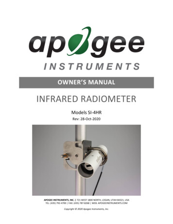
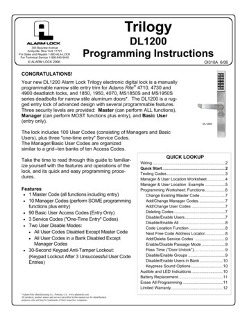
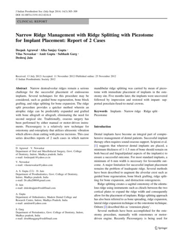
306J Indian Prosthodont Soc (July-Sept 2014) 14(3):305–309osteotomy and osteoplasty that utilizes ultrasonic vibration.This article describes two such cases of narrow ridgemanaged with ridge splitting with the help of piezotome.Case IA 42-year-old woman without medical complications presented for the replacement of a missing mandibular rightfirst, second premolar and first molar (Fig. 1). The teethhad been extracted 4 years before. Clinical examinationrevealed very thin ridge with adequate interarch space. Thepatient was keen for fixed partial prosthesis. So the patientwas planned for placement of implant. A detail dentascanview revealed presence of pointed to 3 mm of ridge widthat first premolar region, the proposed site for placement ofimplant (Fig. 2). Ridge expansion with ridge splittingtechnique was planned. The procedure was performedunder local anaesthesia (Articane 2 %, epinephrine1:100,000). A full thickness mucoperiosteal flap was raisedfrom left canine region to first premolar region on rightside. Osteotomy was performed with piezotome (Ultrasonic bone surgery unit, Resista Verbania, Italy) (Fig. 3). Amidcrestal bone cut along with anterior releasing bony cutwas performed (Fig. 4). The divided ridge was gradualexpanded with increasing size of osteotomes. Implants ofsizes 4.2 9 11.5 mm, 3.75 9 10 mm, and 4.2 9 10 mmwere respectively placed at #28, #29 and #30 region(Fig. 5). Autogenous bone harvested from symphysisregion locoregionally was packed in the expanded crypt(Fig. 6). Rehabilation with full composite splinted crownswas done after 5 months (Fig. 7, 8).Fig. 2 Dentascan distal to #27 revealing ridge crest of 3 mmFig. 3 Piezotome tips used for osteotomy a side cutting, b endcuttingCase IIA 24 years young female approach for replacement ofmissing lower left second premolar and first molar. TheFig. 4 Midcrestal osteotomy with anterior releasing vertical bony cutFig. 1 Missing teeth distal to #27 with narrow ridge crest123teeth have been extracted 3 year ago for grossly cariousnon restorable reason. The patient has been planned forimplant placement. Clinical examination revealed very thinridge width with adequate bone height for implant
J Indian Prosthodont Soc (July-Sept 2014) 14(3):305–309Fig. 5 Implants placed in the splitted ridge in #28, #29 & #30 region307Fig. 8 Post-op orthopentomogram showing stable implants in #28,#29 & #30 region with splinted crownsdistal ends were performed with Piezotome (Fig. 9). Osteotomes of increasing with were used for further expansion of the splitted ridge. Implants of size 4.2 9 13 mmand 5 9 13 mm were placed in #20 and #19 region. Theexpanded crypt was packed with allograft Ossifi (Equniox)Biphasic hydroxyapatite beta tricalcium phosphate. After ahealing period of 5 months the rehabilitation was donewith porcelain fused to metal splinted bridge (Fig. 10).DiscussionFig. 6 Autologous graft harvested from symphysis regionThe ridge split procedure represents one form of agumentation technique for narrow rigdes. The procedure can beconsidered for simultaneous placement of implants inaddition to ridge expansion, thus reducing the overall timefor implant therapy. Ridge splitting for root-form implantplacement was developed in the 1970s by Dr. Hilt Tatum[3]. Tatum developed specific instruments includingFig. 7 Implant retained composite splinted crown in #28, #29 & #30regionplacement. So ridge splitting was planned under localanaesthesia. A full thickness mucoperiosteal flap was elevated from first premolar to second molar. Midcreastalosteotomy along with vertical bony cuts on mesial andFig. 9 Ridge splitting with midcrestal incision and mesial and distalvertical osteotomy cuts123
308Fig. 10 Post-op orthopentogram showing stable implanted withsplinted PFM crowns n #19 & #20 regiontapered channel formers and D-shaped osteotomes toexpand the resorbed residual ridge. Summers [4] later revivedthe interest in this technique; he reported that in 143maxillary implants placed using the osteotome technique,only 5 failed (96 % implant survival). In 1992, Simionet al. [5] introduced a split-crest bone-manipulation technique by means of provoking a longitudinal greenstickfracture at the top of the bone that would split the atrophiccrest in two parts. In 1994, Scipioni et al. [6] presented theclinical results of an edentulous ridge expansion technique.They placed 329 implants in 170 patients with success rateof 98.8 %. Sethi and Kaus [7] placed 449 implants in 150patients in thin maxillary ridges of adequate height andcomprising 2 separate cortical plates with interveningcancellous bone and observed them for a period of up to93 months. A 97 % implant survival rate after a 5-yearobservation period was found.Ridge split augumentation aims at the creation of a newimplant bed by longitudinal osteotomy of the alveolar. Tostart, adequate bone height for implant placement shouldbe present because the splitting of the crest will notincrease bone volume vertically. A minimum of 3 mm ofbone width, including at least 1 mm of cancellous bone, isdesired to insert a chisel between cortical plates and consequently expand the cortical bones.Ridge splitting is more applicable to the maxilla than themandible. The thinner cortical plates and softer medullarybone make the maxillary ridge easier to expand.Favorable conditions for the posterior mandible includea long edentulous span (missing molar and premolar teeth),abundant bone height superior to the mandibular canal([12 mm), and the presence of some cancellous bonebetween the dense outer cortical plates. These considerations for ridge splitting were favourable for both of ourcases.Several authors have suggested the use of a partialthickness flap to help immobilize the displaced buccal123J Indian Prosthodont Soc (July-Sept 2014) 14(3):305–309cortical plate. In cases where there is thin connective tissue,the partial-thickness flap procedure becomes extremelydifficult, and the remaining tissue over the alveolar bone istoo thin to protect the bone adequately. In the presentedcases, the use of a full-thickness flap helped to avoidexcessive bleeding, resulting in better visualization of theoperating sites and better handling of the surgical steps.Number of surgical techniques has been employed forridge splitting. The initial osteotomy can be performed onmidcrestal bone using a No. 15 blade or a carbide tungstenbur, Flat-end Fissure 701 (Brasseler USA, Savannah, GA,USA). Suh et al. [8] describe the microsaw techniquewhich provides better control when preparing a cut along anarrow alveolar ridge and appears less traumatic to thebone. Additionally, less bone is lost because the microsawcreates much thinner cuts compared to conventional burs.In the present clinical report, a piezoelectric surgery systemwas used to create the vertical and horizontal corticotomies. Advantages of piezoelectric ultrasonic surgicalinstruments are1.2.3.micrometric bone cutselective cutclear surgical field.Microvibrations cut hard tissue with precision andaccuracy. The Piezotome device operates with modulatedultrasound micromovements and with an oscillating frequency of 29 and 32 kHz, making it specifically suitablefor osteotomies but not soft tissue cutting. The accidentalslipping of the titanium tips onto surrounding soft tissuedoes not cause any injuries if only a very small degree ofpressure is adopted, as recommended by the manufacturer.Unlike rotary bur, ultrasonic device can be used in ridgesplitting of as narrow as 2 mm ridge without bony perforation. Thanks to cavitation effect of the sterile saline ascoolant, maximum surgical visibility is allowed duringosteotomy. Insignificant noise and vibrations compared tothe ones by rotary bur or saw provide better comfort topatients.The mid crestal osteotomy should come within to 2 mmof the adjacent teeth but not closer. Because of the thickness of the cortical bone in the mandible, one or two vertical osteotomy cuts are required, extending from the edgesof the initial midcrestal osteotomy. Typically the length ofthe bony cut will be 3 mm shorter than the final length ofthe implant. The length of the osteotomy along the edentulous span should extend well beyond the planned implantsites. This extended length will allow the plates to expandor bow during preparation of the osteotomy and implantinsertion (Case I).Chisels or Osteotomes of increasing width and a malletwere used to further enlarge the osteotomy to a point 3 mmshorter than the final length of the implants to be placed.
J Indian Prosthodont Soc (July-Sept 2014) 14(3):305–309The osteotomes are gently malleted to expand the bone.The force should be directed over the long axis of theosteotome, and periodic pauses allow the viscoelastic boneto adapt to the expansion. The osteotome should be rotatedwith a gentle pulling force to allow atraumatic removal ofthe instrument.To prevent a fracture of the buccal plate during theexpansion process, Suh et al. [8] recommended an additionalapical osteotomy connecting the apical ends of the two‘‘bony verticals’’ cuts in cases of more cortical mandibularbone. Enislidis et al. [9] and Elian et al. [10] recommended astaged approach to avoid postoperative complications frommalfracture of the buccal segment in which midcrestalosteotomy was carried out in stage one followed by ridgeexpansion and implant placement in stage two.The segmental ridge split procedure creates a crypt surrounded by bone and periosteum into which implants andbone graft material can be introduced with reasonableconfidence that new bone can be constructed and this newbone will produce a solid bone for dental implants. Different combinations of autogenous and alloplastic graftmaterials have been employed. The site is then covered witha bioresorbable membrane. Scipioni et al. [11] have foundthat a bone graft is usually unnecessary. In general, interpositional grafts have an improved prognosis because theyhave an enhanced vascular bed in an osteogenic environment and are protected from masticatory function. In ourstudy, in case I, autogenous bone harvested from symphysisregion have been employed while in case II allograft Ossifi(Equinox) Biphasic hydroxyapatite beta tricalcium phosphate molecule have been used as interpositional material.Wound healing in these cases is similar to the fracturerepair of bone. The gap fills with a blood clot that organizesand is replaced with woven bone. This immature osseoustissue develops into load-bearing lamellar bone at theimplant interface.In the present clinical study, we used a tapered screwtype implant to increase the initial stability and preventbuccal bone segment fracture. Brunski [12] reported thatscrew-shaped implants provided the strongest retentionimmediately after implant placement. Kan et al. [13]reported a notably greater implant survival rate for threaded implants (titanium 94.9 %; HA-coated 96.0 %) thanfor nonthreaded implants (HA-coated 75.4 %).Second-stage surgery was performed 5 months later,healing abutments were placed, and the soft tissue wereallowed to heal for an additional 2 weeks. Full compositesplinted crowns (Fig. 8) and splinted porcelain-fused-tometal (PFM) crowns (Fig. 10) supported by abutments thenwere delivered in case I and case II respectively. Thus, theoverall treatment time has been shorten since this procedure allowed simultaneous placement of implants. Contrary, onlay bone grafting procedures require second309surgical site, which typically exhibit post-operative morbidity associated with bone harvesting. Additionally, onlaybone graft require a healing period of 6 months to a yearbefore dental implants can be placed, and the onlay graftsometimes fails to fuse to the augmented site.The disadvantage of ridge splitting is that if complications arise and bone loss occurs, then the patient is usuallyleft with an even greater bone defect than before treatment.Mitigating factors that may produce less favourable resultsin ridge splitting include infections, implant loosening,sloughing of the implant edges, and loss of graft material.ConclusionWhen judiously used ridge splitting is helpful technique inmanaging narrow ridges. Since this approach allowssimultaneous placement of implants, shortening the overalltreatment time. The implementation of newer cutting toolPiezotome allows precise osteotomy.References1. Scipioni A, Bruschi GB, Calesini G (1994) The edentulous ridgeexpansion technique: a five-year study. Int J PeriodonticsRestorative Dent 14(5):451–4592. Osborn JF (1985) Die alveolar-expansions-plastik. Quintessenz39:9–143. Tatum H (1986) Maxillary and sinus implant reconstructions.Dent Clin N Am 30:207–2294. Summers RB (1994) A new concept in maxillary implant surgery:the osteotome technique. Compend Contin Educ Dent 15:152–1605. Simion M, Saldoni M, Zaffe D (1992) Jawbone enlargementusing immediate implant placement associated with a split cresttechnique and guided tissue regeneration. Int J PeriodonticsRestorative Dent 2:4626. Scipioni A, Bruschi GB, Calesini G (1994) The edentulous ridgeexpansion technique: a five-year study. Int J PeriodonticsRestorative Dent 14:4517. Sethi A, Kaus T (2000) Maxillary ridge expansion with simultaneous implant placement: 5-Year results of an ongoing clinicalstudy. Int J Oral Maxillofac Implants 15:4918. Suh JJ, Shelemay A, Choi SH, Chai JK (2005) Alveolar ridgesplitting: a new microsaw technique. Int J Periodontics Restorative Dent 25(2):165–1719. Enislidis G, Wittwer G, Ewers R (2006) Preliminary report on astaged ridge splitting technique for implant placement in themandible: a technical note. Int J Oral Maxillofac Implants 21:44510. Elian N, Jalbout Z, Ehrlich B et al (2008) A two-stage full-archridge expansion technique: review of the literature and clinicalguidelines. Implant Dent 17:1611. Scipioni A, Bruschi GB, Giargia M et al (1997) Healing atimplants with and without primary bone contact. Clin OralImplants Res 8:39 (14:451, 1994)12. Brunski JB (1992) Biomechanical factors affecting the bonedental implant interface. Clin Mater 10:15313. Kan JY, Rungcharassaeng K, Kim J et al (2002) Factors affectingthe survival of implants placed in grafted maxillary sinuses: aclinical report. J Prosthet Dent 87:485123
Ridge splitting for root-form implant placement was developed in the 1970s by Dr. Hilt Tatum [3]. Tatum developed specific instruments including Fig. 5 Implants placed in the splitted ridge in #28, #29 & #30 region Fig. 6 Autologous graft harvested from symphysis region Fig. 7 Implant retained composite splinted crown in #28, #29 & #30 region