Transcription
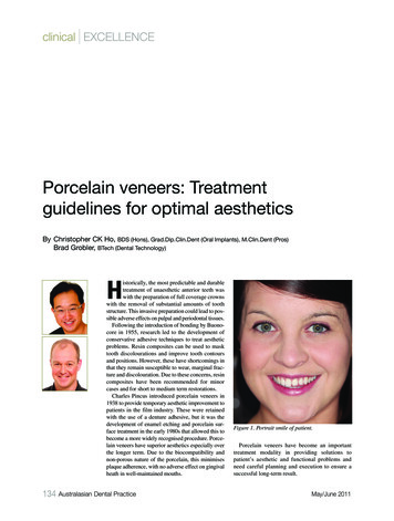
ADP May-Jun 2011Quark9 Web Layout 1 30/08/11 10:21 PM Page 134clinical EXCELLENCEPorcelain veneers: Treatmentguidelines for optimal aestheticsBy Christopher CK Ho, BDS (Hons), Grad.Dip.Clin.Dent (Oral Implants), M.Clin.Dent (Pros)Brad Grobler, BTech (Dental Technology)Historically, the most predictable and durabletreatment of unaesthetic anterior teeth waswith the preparation of full coverage crownswith the removal of substantial amounts of toothstructure. This invasive preparation could lead to possible adverse effects on pulpal and periodontal tissues.Following the introduction of bonding by Buonocore in 1955, research led to the development ofconservative adhesive techniques to treat aestheticproblems. Resin composites can be used to masktooth discolourations and improve tooth contoursand positions. However, these have shortcomings inthat they remain susceptible to wear, marginal fracture and discolouration. Due to these concerns, resincomposites have been recommended for minorcases and for short to medium term restorations.Charles Pincus introduced porcelain veneers in1938 to provide temporary aesthetic improvement topatients in the film industry. These were retainedwith the use of a denture adhesive, but it was thedevelopment of enamel etching and porcelain surface treatment in the early 1980s that allowed this tobecome a more widely recognised procedure. Porcelain veneers have superior aesthetics especially overthe longer term. Due to the biocompatibility andnon-porous nature of the porcelain, this minimisesplaque adherence, with no adverse effect on gingivalheath in well-maintained mouths.134 Australasian Dental PracticeFigure 1. Portrait smile of patient.Porcelain veneers have become an importanttreatment modality in providing solutions topatient’s aesthetic and functional problems andneed careful planning and execution to ensure asuccessful long-term result.May/June 2011
ADP May-Jun 2011Quark9 Web Layout 1 30/08/11 10:21 PM Page 135clinical EXCELLENCEFigure 2. Frontal smile.Figure 3. Frontal retracted photos.Figure 4. Diagnostic wax-up.Figure 5. Silicone reduction indexes in use.Figure 6. Completed preparation.Figure 7. Bis-acryl resin temporaries being constructed.Figure 8. Provisionals straight after removal of putty key.Figure 9. Spot-etch temporisation.Informed consentmust be mentioned and a discussion on the procedures involvedwith the steps from start to completion. The patient mustbe educated on the care and maintenance of the veneersand mention made of the longevity of the veneers and theireventual replacement.Porcelain veneers are often an aesthetic and elective procedureand as such require a full discussion on the benefits and riskswith the functional and aesthetic objectives defined withinthis process. Alternative means of achieving the patient’s goalsMay/June 2011Australasian Dental Practice135
ADP May-Jun 2011Quark9 Web Layout 1 30/08/11 10:21 PM Page 136clinical EXCELLENCEFigure 10. Scan of the wax-up.Figure 11. The wax-up scan is overlaidon the working model and made transparent, it will be used to position theinitial shapes.Figure 12. Initial Positioning oftooth shapes.Figure 13. Precise control of the cementgap and fit.Figure 14. Exact duplication of the waxup into veneers.Figure 15. Adaption of occlusal contactsand interproximal contacts.Figure 16. Freeform Modifications andartistic improvements.Figure 17. Veneers ready for milling ine.max CAD Lithium Disilicate.Figure 18. Lab-Dentist communicationto visualize preps and ceramic thickness.Shade selectionPreparationThe selection of a shade and masking with veneer restorations canbe a challenging procedure due to the thin nature and translucency of the restoration. The final colour that is attained is a resultof the underlying tooth colour, the resin cement that is selectedand the opacity/translucency of the porcelain used.The underlying tooth structure may be discoloured due to previous excellencetics, old restorations, age, trauma and tetracyclinecases. The colour of the existing teeth should be communicated tothe ceramist (stump shade), as this allows the ceramist to determine the final opacity and translucency needed of the restoration.The influence of the substrate determines the final shade.A mental picture should be developed of the requirements of thetooth preparation needed from the amount of depth reduction, tothe preparation margins to attain the final outcomes.The aims of tooth preparation are to: Provide enough thickness for the porcelain for sufficient fracture resistance and to not over contour the final restoration; Provide a margin, so that the ceramist has a definite finishingline, allowing normal emergence of the veneer from the gingival margin; Maintain the preparation within enamel wherever possible; Provide a finished preparation, which is smooth and free of anysharp internal line-angles, which may cause stress concentration within the ceramic; and Provide definite seating landmarks making proper seating ofthe veneer.Diagnostic wax-up/mock-upIt is critical to have a clear plan of where treatment is heading andthe utilisation of a wax-up can assist the planning of the desiredaesthetic appearance. This should incorporate the patient’s wantsthat were expressed in the initial treatment planning discussions.This wax-up (Figure 4) also allows the manufacture of puttykeys for provisionalisation and reduction guides (Figure 5) for thepreparation process. The contours and form of the final teeth canbe transferred from the desired wax up to the provisionalsallowing patients to have a preview of their desired appearance andreconfirm that they are happy with the changes that are planned.136 Australasian Dental PracticeLabial preparationThe preparation of the buccal plane of the incisors (which areconvex) needs to be addressed in three planes with incisal, middlethird and cervical planes.A careful labial reduction of tooth structures is carried out toprovide a minimum of 0.3mm (feldspathic porcelain) or 0.6mm(Empress esthetic, e.max) preparation.May/June 2011
ADP May-Jun 2011Quark9 Web Layout 1 30/08/11 10:21 PM Page 138clinical EXCELLENCEFigure 19. Pumice slurry used to clean preparations. If a spotetch provisional technique has been used this area is carefullyprepared with a diamond bur to remove any resin tags.Figure 20. Interproximal margins cleaned with finishing stripsprior to bonding.Figure 21. Full arch rubber dam (Roeko Flexi Dam, Coltene)applied with knitted retraction cord (Ultrapak 0, Ultradent).Figure 22. The use of a total etch technique, etching dentine for15 seconds maximum.There are several methods of attaining the reduction requiredwith the preparation:1. Freehand;2. Use of depth cuts/grooves. The use of depth cutters or grooves anddimples has been recommended to control tooth preparation, as theuse of standardised objects allows accurate judgement of depth; or3. Use of silicone putty index.Using a silicone index may help in assessing the amount oftissue reduction and can be prepared by the dental ceramist fromthe initial wax-up model. When viewed from the occlusal view,this can be cut in horizontal slices which can be peeled back toassess different vertical positions of the reduced teeth. Utilisationof a silicone index derived from the wax-up allows a visualisationof the reduction required to achieve the form and contours of thepre-planned shape and length of the final veneers.tage of simple tooth preparation and the aesthetic characteristicsare easier to fabricate with the ceramist, as it is possible to developincisal translucency. The margin is not in a position where it willbe subjected to protrusive forces and the preparation is reported toreduce stress concentration within the veneer by distributing theocclusal load over a wider surface (Highton et al, 1987).Proximal preparationThis preparation in the interproximal region can be made eitherby stopping short of breaking the contact, or by preparing throughthe contact point.Breaking the contact (sometimes called the “slice preparation”)may be necessary to clear the contact in certain situations, such aschanging the shape or position of teeth. With the additional spaceinter-proximally, this allows the ceramist freedom to adjust thecontours and position of the teeth.Incisal edge reductionDifferent preparation designs have been advocated from featherand window preparations that involve no reduction of the incisaledge or preparation of the lingual surfaces, to other preparationsthat involve a reduction of the incisal edges. The author prefers anincisal preparation that is carried over the incisal edge from buccalto palatal, with up to 1.5 mm of incisal reduction. According toCalamia, a tooth preparation that incorporates incisal overlap ispreferable, because the veneer is stronger and provides a positiveseat during cementation. This preparation design has the advan-138 Australasian Dental PracticeCervical marginThe cervical preparation for a veneer is recommended to be achamfer design with a maximum depth of 0.4mm. This allows theveneer to reproduce natural tooth contours and not be over-contoured. Additionally, it allows simple seating of the veneer andminimises stresses, enhancing the future fracture resistance of theveneer. Unlike crown margins that are often buried in the sulcus,the use of the thin translucent porcelain allows often a “contactlens” effect where the margins are blended with no discernibleMay/June 2011
ADP May-Jun 2011Quark9 Web Layout 1 30/08/11 10:21 PM Page 140clinical EXCELLENCEFigure 23. The use of a total etch technique with a fifth generation bonding agent Optibond Solo Plus (sds Kerr).Figure 24. The adhesive luting of the veneer with light cure resincement (Nexus 3; Kerr).Figure 25. The use of a tacking tip (Demi LED KERR) which istypically 2-4mm in diameter spot tacks the veneer, stabilising itin the correct position. Cleanup of the excess cement is undertaken followed by final curing.Figure 26. The use of a #12 blade to carefully removeexcess cement.demarcation. This enables margins to be either equi-gingival orsupra-gingival. Additionally there is a greater possibility that themargins will be in enamel. This ”contact lens effect” may extendthe longevity of the veneer, with the apical migration of gingivalmargins not revealing a distinct line of demarcation between theveneer and the natural tooth.A supra-gingival margin has many advantages with less risk ofexposing dentine and less chance of injury to the soft tissuesduring preparation. Impression taking is also easier with no needfor retraction and the likelihood of a clear margin captured. Dueto the likelihood of the margin being in enamel, there is lesschance of microleakage associated with enamel bonding. Subgingival margins may be required when there are caries orprevious restorations extending sub-gingivally.ProvisionalisationFacebow transfer and bite registrationTo communicate clearly to the ceramist the correct final orientation of the incisal plane of the planned veneers, it is important thatthey receive a “stick bite” or “symmetry bite”. This can be assimple as two sticks within the bite registration to register themidline and the interpupillary line to the teeth.140 Australasian Dental PracticeSome clinicians that feel that provisionalisation is not necessarywith veneers due to the minimal tooth reduction required. On thecontrary, provisionalisation is an integral part of the treatmentprocess with the ability to communicate with the patient and laboratory what enhances a patient’s smile. The provisionals areconstructed from the diagnostic wax-up with many patients notable to determine their desires or visualise the final result by justdiscussing the changes verbally.Utilising provisionals gives the patient a “test run” or previewof the final result planned, which is strongly recommended(Figures 7-9).Material selectionA lithium disilicate (e.max, Ivoclar Vivadent) material wasselected for its optical properties and excellent flexural strengthand fracture toughness.It is also available in different translucencies and opacitiesallowing the ceramist better colour masking if required. The procedure for manufacture of this material can be from either hotpressing or CAD/CAM.May/June 2011
ADP May-Jun 2011Quark9 Web Layout 1 30/08/11 10:21 PM Page 142clinical EXCELLENCEFigure 28. Frontal smile.Figure 27. Final portrait smile.Laboratory procedures by Brad GroblerCreating lifelike ceramic veneers has been a skill reserved for greatceramists using feldspathic porcelain or meticulous waxers usingthe hot press technique, but now CAD/CAM is giving techniciansthe ability to easily create veneers with a few clicks of a mouse.The diagnostic wax-up, which has been approved by the patientand who is now wearing the design as temporaries, can bescanned into the system to be used as the blueprint for the finalveneers (Figure 10). At this stage in the dental veneer process, thejob of the technician is to exactly reproduce the design that thepatient and clinician have approved. Any deviation from thisdesign can result in numerous problems with acceptance, aesthetics, phonetics and function. However, small details andsurface characteristics can be improved and minor design changescan be made to improve the artistic and aesthetic outcome.The wax-up scan is overlaid on the working model and madetransparent (Figure 11). It will be used to position the initialshapes. The fit of the restorations can be precisely controlled sothat the individual preferences of clinicians and variations inbonding materials can be accommodated (Figure 13).The ZFX System has the ability to adapt your design to fit orduplicate the wax-up scan exactly. With conventional techniques,this duplication was difficult to achieve; wax injection into silicone putties made from the diagnostic wax-up can create exact142 Australasian Dental PracticeFigure 29. Completed veneers - retracted view.duplication but this is still time consuming and technique sensitive. Now with the click of the mouse, duplication is achieved(Figure 14).The shapes are minimally altered to ensure proper contacts,embrasures and interdental spaces (Figure 16). Surface irregularities and final aesthetic alterations are made to ensure you get agreat milling result. The design is then finalized (merged) and themilling preview is generated (Figure 17).The designs are milled from e.max CAD HT, shade B1 in a 5axis ultrasonic milling machine at ZFX Australia. The millings arereturned to the laboratory, where the small milling tags are removedand the blue (pre-crystalized) restorations are placed on the model,with no or very minimal adjustment needed to fit and contacts.Final finishing requires all the usual ceramics techniques ofshape, morphology and surface texture. The majority of this shouldbe done in the blue (pre-crystalized ) phase of the e.max CADmaterials due to the surface hardness characteristics. Lithium disilicate in the “blue” phase has a surface hardness similar to layeredglass ceramics but once crystalized (in the correct crystallizationMay/June 2011
ADP May-Jun 2011Quark9 Web Layout 1 30/08/11 10:21 PM Page 144clinical EXCELLENCEfiring cycle from Ivoclar Vivadent), the material displays its fullflexural strength but also a very high surface hardness which makesshaping and texturing with rotary burs very difficult.Cut back and layering can done if adequate space is available,a small incisal cut back can be made and e.max ceram layeringceramic can be fired onto the cutback to increase the incisaltranslucency and internal characteristics.Another valuable feature of the ZFX CAD/CAM system is theability to pick up potential problems with aspects of the casebefore anything is actually made. Checking for adequate reduction, even thickness and proper interdental margin placement forcontact creation (Figure 18).Try-in of veneersThe tooth surface should be cleaned of any residual resin cementor provisional material, to ensure perfect adaptation of theveneers (Figures 19-21).Each veneer should be tried individually to assess fit. This isbest done dry (without water or try-in gels) at this stage as complete seating and marginal adaptation can be better visualised.Incomplete seating is normally due to remaining provisional material, luting resin that has not been removed or tight contact points.Once the patient is happy and has approved the final aesthetics,the restorations are prepared for cementation. The veneers (beingsilica-based restorations) must be etched with hydrofluoric acid,which allows a micro-mechanical bond when adhesively bonded.The intaglio surface is etched with 9.5% hydrofluoric acid for 20seconds with lithium disilicate (e.max) or 60 seconds for othersilica based ceramics. The use of hydrofluoric acid dissolves theglassy matrix surrounding the crystalline phase within the porcelain, leaving retentive areas between the acid resistant crystals.The acid should be thoroughly cleansed with air-water sprayand the porcelain should then be placed into a container of distilled water (or 95% alcohol or acetone) and put into an ultrasonicbath for 4 minutes to remove any residues remaining on the surface. Restorations are removed, dried and silane primer is appliedto the fitting surface which helps provide a chemical covalentbond to the ceramic. This is allowed to remain on the veneer for 1minute and after that the veneer should be gently blown with airto evaporate any remaining solvent.Bonding veneersThe application of rubber dam is recommended to achieveadequate isolation, which helps to provide a clean, dry environment and minimises contamination from saliva and blood(Figures 22-25).Light curing composite resin is preferred for cementation of theveneers as they have a longer working time than dual cure orchemically cured composites. This allows sufficient time toremove excess composite prior to curing and thus reduces the finishing procedures. The colour stability of light curing resincements are much better compared to dual or chemical cure composites. Dual cure resin cements contain tertiary amines whichmay undergo long term colour change (“amine discolouration”)with overall darkening and thus are normally contraindicated withveneers due to their thin nature and translucency.The use of various coloured resin cements has an influence onthe final result attained and is often used to fine tune the final144 Australasian Dental Practiceshade of the restoration to attain the desired outcome. Itsinfluence is minimal, contributing less than 10% of the finalcolour of the restoration.Finishing and polishingIf the bonding procedure was completed smoothly with a well fitting veneer, there should be very little cement to clean up fromaround the margins. It is preferred to not use a rotary instrumentto finish the margins, as this may remove the glaze layer,increasing the roughness of the porcelain and causing increasedplaque retention. The use of a #12 blade (Figure 21) to carefullyremove excess cement is a preferred technique, however at timesif necessary a fine diamond can be used to adjust the porcelainand then carefully polished.Occlusion is carefully checked initially with centric occlusionfollowed by other excursive movements. The use of rugby-shapeddiamonds with water spray can be used to adjust the porcelain.Any adjustments must be further polished.The long term clinical success of porcelain veneers dependson a careful case selection and diagnostic approach, as wellas accurate and appropriate tooth preparation and adhesivebonding procedures.References1. Calamia JR. The Etched Porcelain Veneer Technique. NY State Dent J.1988:54:48- 50.2. Magne P, Belser U. Bonded porcelain restorations in the anterior dentition: A Biomimetic Approach. Germany: Quintessence. 2003.3. Calamia JR, Calamia CS. Porcelain laminate veneers: reasons for 25 years of success. Dent Clin North Am. 2007;51(2):399-4174. Highton R, Caputo AA, Maytas JA. A photoelastic study of stress on porcelainlaminate preparations. J Prosthetic Dent. 1987;58:157-161.About the authorsDr Christopher Ho received his Bachelor of Dental Surgery withFirst Class Honours at the University of Sydney. He has completed postgraduate studies in the Graduate Diploma in ClinicalDentistry in Oral Implants at the University of Sydney and Masters of Clinical Dentistry in Prosthodontics with Distinction fromKings College, London. Dr Ho is a lecturer on aesthetic andimplant dentistry locally and internationally and is involved withthe evaluation and development of new dental products and materials. He is a faculty member with the UCLA/Global Institute forDental Education teaching in the one year Master programs inEsthetic Dentistry and Implant Dentistry. Dr Ho’s research interests are in immediate placement and loading of dental implants.He has a referral-based private practice in prosthodontic andimplant dentistry in Sydney, Australia.Mr Brad Grobler received his Degree in Dental Technology fromTechnikon Kwazulu Natal, Durban. He is a Dental Ceramist whospecializes in Aesthetic and Cosmetic Dentistry, CAD/CAM andImplant Restorations Fixed and Removable. Mr Grobler alsolectures locally and abroad and is an opinion leader for IvoclarVivadent and ZFX. He actively searches out the best continouseducation and latest techniques, has attended workshopswith numerous Master Ceramists and is a Kois Center Student.He is a Sustaining Member of the AACD and an ExecutiveMember of the NZACD.May/June 2011
Charles Pincus introduced porcelain veneers in 1938 to provide temporary aesthetic improvement to patients in the film industry. These were retained with the use of a denture adhesive, but it was the development of enamel etching and porcelain sur-face treatment in the early 1980s that allowed this to become a more widely recognised procedure .