Transcription
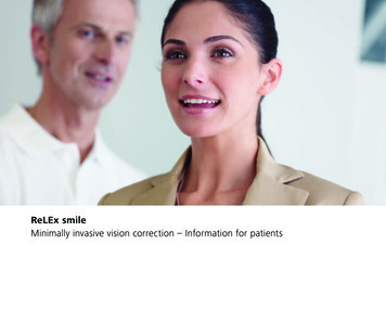
ReLEx smileMinimally invasive vision correction – Information for patients
Seeing is livingOur eyes are our most important sensory organ. The human brain obtains over 80 % ofits information via the sense of sight. Our eyes are our windows to the world. Seeing isrecognizing. Seeing is experiencing. Seeing is independence and freedom. Seeing is living.Well over half the world’s population relies on glasses or contact lenses to see well. Butmany find that being dependent upon optical appliances interferes with their professionallives and leisure time.Simply being able to see. Without glasses or contact lenses. Completely free of anyoptical appliances. This is now possible in increasing numbers of cases thanks to ongoingdevelopments in medicine and technology. Refractive visual correction techniques havebeen scientifically recognized and clinically tested over the last few decades. They havecome to represent an important alternative to traditional correction methods such asglasses and contact lenses.ReLEx smile is the new laser technique by Carl Zeiss for the gentle correction of visiondefects. It is a minimally invasive treatment method which combines the extensiveexperience and superior safety of traditional vision correction techniques with numerousinnovative benefits, high precision levels and perceptibly greater comfort during thetreatment itself.23
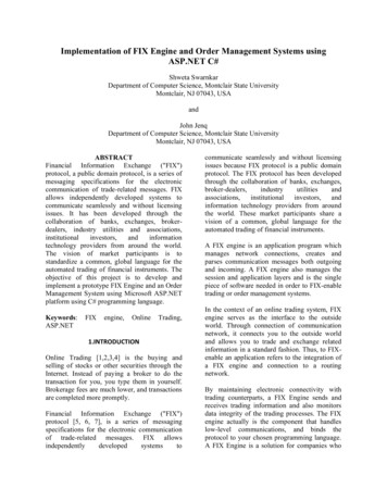
LensIn and out of focus –Types of vision defectRetinaNormal vision (emmetropia):Light rays are refracted by the cornea and thelens in such a way that the focal point is directlyon the retina. Objects both near and far appearin sharp focus.PupilCorneaThe physical/optical principles behind the human eye areIn farsighted people, the eye is too short in relation tosimilar to those in a camera. The cornea and lens assumethe refractive power. Light rays are refracted by thethe role of the camera lens. They bundle the parallelcornea and the lens in such a way that the focal point isincidental light rays and determine the focal distance. Inbehind the retina. A blurred image is then created onsuch a way that the focal point is in front of the retina.an eye with normal vision, the light rays are focused sothe retina because the rays are not yet focused whenDistant objects appear out of focus. Depending on thethat the focal point is on the retina itself. The result is athey hit it. Up to a certain age this lack of refractivedegree of myopia, near objects appear in sharp focus.sharply focused image. This is then transmitted via thepower can be compensated by changing the shape ofoptic nerve to the brain.the lens (accommodation). Depending on the extent ofNearsightedness (myopia):Light rays are refracted by the cornea and the lens inImage in front of retinathe farsightedness, objects which are close, and evenNearsightedness is the most common vision defect world-distant ones in some cases, are no longer in sharp focus.wide. Almost half the global population is – to varyingOptic nervFarsightedness (hyperopia):Light rays are refracted by the cornea and the lens indegrees – affected by it. In nearsighted people, the eye isIn people with astigmatism, the curvature of the corneasuch a way that the focal point is behind the retina.too long in relation to its refractive power. Light rays areis uneven. The resulting refraction causes multiple focalDepending on the extent of the farsightedness, objectsrefracted by the cornea and the lens in such a way thatpoints to be created. Objects both near and far appearwhich are close, and even distant ones in some cases,the focal point lies in front of the retina. By the time theskewed or distorted. Astigmatism can occur independentlyappear out of focus.rays hit the retina itself, they are already drifting apart.or be accompanied by farsightedness or nearsightedness.Image behind retinaThe result is a retinal image which is out of focus. Distantobjects appear blurred. Depending on the degree of visionAstigmatismdefect, near objects are in sharp focus.The irregular curvature of the cornea causes thelight rays to be refracted into multiple focal pointsand not just one. Depending on the extent of theastigmatism, objects both near and far appearskewed and distorted.Image in front of and behind retina45
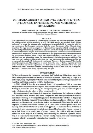
A life without glasses –The different laser treatment optionsThe treatment stepsLaser surgery techniques for refractive vision correctionIt then adheres in the original place over the comingwere well established, both scientifically and clinically, byweeks and months. The disadvantage is that the flap canthe mid-80s. The basic principle behind all the methods isshift or even tear off (flap displacement) as the result ofusing a laser to model the outer corneal layer so that therubbing or other physical contact after the operation.ReLEx smilefocal point of the bundled incidental light is on the surface of the retina itself and not in front of or behind it.The ReLEx smile principle: The high-precision VisuMax femtosecond laser creates a small lens (lenticule) insideThe original treatment methods, PRK and LASEK, manu-the intact cornea, the volume and form of which areally removed the outer protective layer (epithelium) ofdetermined by the degree of vision defect to be corrected.Step 1:Step 2:Step 3:the cornea before the laser surgery. The consequencesThis lenticule is then removed in a minimally invasiveIn a single step the VisuMax femtosecondThe surgeon removes the lenticule throughThe minimally invasive removal of theoften included post-operative pain and very slow recov-procedure from the interior of the cornea through alaser creates a thin lenticule and a smallthe small access. There is minimallenticule changes the shape of the cornea,ery of visual acuity. As a consequence, laser surgery tech-small access measuring just a few millimetres. In contrastaccess measuring less than 4 mm in thedisruption to the biomechanics of thecorrecting the refractive error of the eye.niques which involved preparing a corneal flap (LASIKto earlier techniques, it is no longer necessary to foldintact cornea.cornea. No flap needs to be cut.and Femto-LASIK) were then developed. Here, a mechani back the cornea. There is no flap incision, the area ofcal cutting tool (microkeratome) or a femtosecond laserthe incision is reduced to a minimum and the outeris used to create a thin flap of cornea which is justcorneal layers remain more or less intact. All of which120 micrometers thick. Almost a complete circle (270 )is made possible by a sensitive, precise and convenientis cut in the upper layers of the cornea. The flap istreatment method.Femto-LASIKthen folded back like the cover of a book. Subsequently,the exposed corneal tissue is then ablated using anexcimer laser in a separate stage of treatment. Thegreater the level of vision defect, the more tissue needsStep 1:Step 2:Step 3:Step 4:Step 5:The femtosecond laserThe patient is moved toThe surgeon opens the flapThe excimer laser ablatesAfter the laser surgerycreates a corneal flap.the excimer laser.and folds it back to exposethe pre-calculated amountis complete, the flap isto be removed. In the final stage of treatment, thethe lower corneal layerof corneal tissue, pointreturned to its original posi-corneal flap is then folded back into its original position.(stroma) beneath.by point.tion. It then adheres in theoriginal place over time.67
Minimally invasive – with no flap incisionThe benefits of ReLEx smilePrecise and sensitive treatmentVisuMax femtosecond laserIn recent years minimally invasive diagnostic and treat Entire treatment carried out usingThe specially developed laser system used for the ReLEx procedure is the Carl Zeissment methods have set new standards in modernfemtosecond laser (all-femto)VisuMax femtosecond laser. In Femto-LASIK surgery it has already impressed patientsmedicine. This is because minimally invasive very often Use of high-precision femtosecond technologyand physicians alike with its sophisticated technology, its precision and its reliability.means minimum discomfort for the patient. In its O utstanding predictability of results, even in casesReLEx using the VisuMax now permits accuracy down to nearest micrometer precisionReLEx smile method Carl Zeiss has developed a new technique for the minimally invasive laser correction ofvision defects. This innovative treatment techniqueyields a whole range of benefits:of severe myopia ( -7.00 D)in the preparation of the lenticule within the intact cornea. A natomically shaped contact glass avoids unnecessarycompression of the cornea No loss of vision during treatmentThe high-precision femtosecond laser provides targeted correction of the vision defectwhile leaving the surrounding corneal tissue virtually unaffected. Noiseless and odorless treatmentFlapless treatment Stabilization of visual acuity typically within 14 daysA contact glass specially designed to match the individual anatomy of the corneapermits customized treatment in which the corneal tissue does not need to be No folding back of a corneal flap Lenticule preparation in the intact corneaSingle-step treatmentunnecessarily compressed. This helps avoid temporary loss of vision caused by Minimally invasive access measuring just a few Entire laser correction in a single treatment stepexcessive intraocular pressure.millimeters (80 % less lateral incision than in No device change during the operationLASIK and Femto-LASIK) Short procedure A lmost complete preservation of protective cornealAn ergonomically designed and functionally versatile patient supporting systemprovides extra comfort and relaxation during treatment. The position of the patientlayer (epithelium) and stabilizing outer corneal layersis continuously monitored during the laser procedure and automatically adjusted(Bowman’s membrane)and corrected, if necessary. Maximum number of corneal nerves responsible fortear regulation remain intact Practically painless during and after the procedure89
Other things you might like to know about ReLExWhat makes ReLEx smile so special?and the stabilization of visual acuity takefirst patients have already undergone 5 yearsAfter ReLEx, how long will it be beforeUnlike in all other laser eye surgery proce-a relatively long time. PRK and LASEKof post-treatment monitoring. ReLEx smile isI can see properly without glasses ordures, the corneal tissue is not ablated usingare not recommended for the treatment ofnow being deployed in many countriescontact lenses and return to my normalan excimer laser. In ReLEx, proven femtosec-severe nearsightedness. The risk of scarringworldwide as a standard treatment method.routine?ond laser technology is the only technology(haze) is significantly higher. ReLEx smileused. A small lenticule is prepared inside thecombines the benefits of PRK and LASEKHow can I find out if I am suitablecases visual acuity is very good one or twointact cornea using the femtosecond laser.(no flap incision) with those of LASIK andfor ReLEx?days after the operation and stabilizes withinThis lenticule is removed through an accessFemto-LASIK (minimum discomfort andAs with all other vision correction methods,two to three weeks. Following PRK or LASEK,point just a few millimeters across. In fact,rapid restoration of visual acuity) – butyou will have to undergo a detailed eyeby comparison, full recovery can take up tothe cornea does not have to be opened atwithout any of their disadvantages.examination. The nature and degree ofthree months. After ReLEx, you will generallyametropia, the curvature and thickness ofbe able to drive, work and do sports withoutHow much experience has gonethe cornea, as well as many other factorsglasses or contact lenses just a few daysinto ReLEx?play a role. Your ophthalmologist will advisefollowing treatment.The name Carl Zeiss has stood for qualityyou personally after conducting a detailedand precision in optics since 1846. In 1986examination.all – unlike with LASIK and Femto-LASIK. Thetechnique is made possible by the uniqueprecision and performance of the VisuMax femtosecond laser made by Carl Zeiss.Every healing process is different. In mostWhat are the risks?There is no flap in PRK or LASEK either.Carl Zeiss unveiled the first excimer laser forLike all medical techniques, ReLEx smile isHow do these procedures differ fromrefractive correction of the eye. Over thenot without side effects. Only your physicianReLEx?last 25 years the company has been at thecan explain the individual risks and possibleIn PRK and LASEK procedures, the top layer offorefront of advances in the laser correctionside effects to you and decide whetherthe cornea (epithelium) is removed manually.of vision defects. Femtosecond technologytreatment with ReLEx smile is the rightThe exposed deeper layers of the cornea arehas been extensively trialed and has provenoption for you.then ablated using an excimer laser. The dis-its clinical value over many years. Refractiveadvantages: there is significantly greater painlenticule extraction has been performed inafter the operation, and the healing processcontrolled clinical studies since 2006. The1011
CZ-VII/2012Printed in GermanyPublication No: 000000-1964-124Carl Zeiss Meditec AGGoeschwitzer Strasse 51–5207745 JenaGermanywww.meditec.zeiss.com/ReLEx
all - unlike with LASIK and Femto-LASIK. The technique is made possible by the unique precision and performance of the VisuMax femtosecond laser made by Carl Zeiss. There is no flap in PRK or LASEK either. How do these procedures differ from ReLEx? In PRK and LASEK procedures, the top layer of the cornea (epithelium) is removed manually.