Transcription
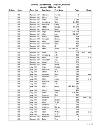
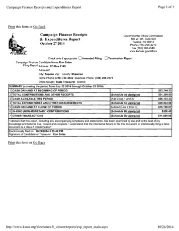
NEWSLETTERJanuary 2018Bright Field-STEM image of porosity in a beta-zeolite (FEI Titan Themis G2 300image courtesy of James Cattle, PhD Student, University of Leeds. Sampleprovided by Dr Stig Helveg, Haldor Topsoe A/S).See http://emag.iop.org for further details
EMAG Group newsletterJan 2018CONTENTSEMAG COMMITTEE3LETTER FROM THE CHAIR4FORTHCOMING EVENTS5EMAG 2018 CALL FOR PAPERSEMAG ANNUAL GENERAL MEETING 2018NEWS567PROF. PRATIBHA GAI DBEUNIVERSITY OF GLASGOW PLASMA-FIBELECTRON MICROSCOPY AT DARESBURY LABSMEETING REPORTS781011MSM-XX, OXFORDVISION AND OPHTHALMOLOGY, BALTIMOREMMC17, MANCHESTERM&M, ST LOUIS11121415APPLY FOR IOP RESEARCH STUDENTS CONFERENCE FUND15EMS MEMBERSHIP16ADDITIONAL FUTURE MEETINGS OF INTEREST172
EMAG Group newsletterJan 2018EMAG COMMITTEEChairDr Sarah HaighSchool of Materials, University of Manchester, Manchester, M13 9PLTel: 0161 306 3618 Email: sarah.haigh@manchester.ac.ukSecretary & Honorary TreasurerDr Andy BrownSchool of Chemical and Process Engineering, University of Leeds, Leeds, LS2 9JTTel: 0113 343 2382 Email: a.p.brown@leeds.ac.ukOrdinary MembersDr ZiYou Li, University of Birmingham, ziyouli@nprl.ph.bham.ac.ukDr Ana Sanchez, University of Warwick, a.m.sanchez@warwick.ac.ukDr Cornelia Rodenburg, University of Sheffield, c.rodenburg@sheffield.ac.ukDr Larry Stoter, Retired, larry.stoter@gmail.comMr Michael Dixon, Hitachi Europe, Michael.Dixon@hht-eu.comProf Jun Yuan, University of York, jun.yuan@york.ac.ukDr Donald MacLaren, University of Glasgow, donald.maclaren@glasgow.ac.ukDr Sarah Karimi, JEOL Ltd, sarah.karimi@outlook.comCo-optedDr. Caterina Ducati, University of Cambridge, cd251@cam.ac.ukDr Thomas Slater, University of Manchester, thomas.slater-2@manchester.ac.uk3
EMAG Group newsletterJan 2018LETTER FROM THE CHAIRDear Friends and Colleagues,My main task here is to thank the many people who have made EMAG such asuccess this year. I’d like to start by thanking the many people who volunteeredto be part of the EMAG committee last summer and to everyone who voted. I’mdelighted to welcome our new members, Cornelia Rodenburg and Larry Stoter.Larry brings to the committee vital industrial insights while Cornelia brings newexpertise and experience in scanning electron microscopy of soft matter. Wealso welcome back Jun Yuan and Michael Dixon who have done a great joband been re-elected for a further 4 years. Special thanks are also due to ouroutgoing committee members, Budhika Mendis, Caterina Ducati and DavidBeamer for their fantastic contributions to EMAG over many years. We furtherdecided to co-opt from our nominated candidates, Thomas Slater, who is apostdoctoral research. Tom will take on the new role to represent the interestsof our younger members (so please do get in touch with him if you haveideas/suggestions!).I would also like to thank the team at the Royal Microscopy Society for all theirsupport in the organisation of our full 3.5 day biennial EMAG conference whichwas again held as part of the MMC conference in Manchester from 3 rd-6th July2017 (www.mmc2017.org.uk). I really enjoyed the meeting and the feedbackwe have had from attendees has been great. We had over 200 abstractssubmitted to EMAG alone, 1340 attendees at the MMC conference overall and 70 companies represented at the exhibition. The pre-meeting schools wereoversubscribed and all the poster and oral presentations were exceptionallyhigh quality showing the excellence and diversity of the EMAG community. Forexample, you may be interested to know that of the 66 EMAG speakers 20 werefemale and 35 international. The flash presentations worked really well forincreasing the visibility of posters and encouraging students to present. I’d liketo encourage students to consider requesting an oral contribution whensubmitting to our exciting EMAG 2018 workshop which focusses on applicationsof electron microscopy to beam sensitive materials (page 5).Finally I’d like to thank Andy Brown for putting together this excellent newsletter– we welcome your contributions for future issues!Best wishes,Sarah Haigh, University of Manchester, EMAG Chair4
EMAG Group newsletterJan 2018FORTHCOMING EVENTSEMAG 2018: Applications of Electron Microscopy toBeam Sensitive MaterialsCall for PapersAbstract submission deadline 7 April 2018.Submit through http://emag2018.iopconfs.org/homeThe EMAG conference in 2018 will be held at the University of Warwick on 4-6July 2018. This single session format conference will focus on Applications ofElectron Microscopy to Beam Sensitive Materials. We have a fantastic group ofinvited speakers (see below) who will present alongside contributed oralpresentations as well as poster sessions. Prizes will be awarded to the beststudent contributions. A table-top trade exhibition will be held for delegates toexplore the latest advances in instrumentation. Conference delegates will beable to enjoy a dinner in the historic Warwick Castle.There will also be a set of company workshops with experimental and softwaredemonstrations running from 1 pm on Wed 4th July – check the conferencewebsites for more detail on these as they develop Invited Speakers Professor Elena Besley, University of Nottingham, UK Dr Stuart Boden, University of Southampton, UK Professor Nigel Browning, University of Liverpool, UK Professor Ray Egerton, University of Alberta, Canada Dr Lewys Jones, Trinity College Dublin, Ireland Professor Nico Sommerdijk, Technische Universiteit Eindhoven,Holland Professor Kazu Suenaga, National Institute of AIST, JapanAbstract submission closes 7 April 2018.5
EMAG Group newsletterJan 2018EMAG Annual General Meeting 2018The AGM of the EMAG group will be held as part of EMAG 2018 in Warwick (4- 6th of July 2018).http://emag2018.iopconfs.org/homeNo fee is charged to attend the Annual General Meeting. Agenda and moredetails will be circulated nearer the time. If you cannot attend the AGM but haveany issues you would like to be raised at the meeting, please contact thehonorary secretary (a.p.brown@leeds.ac.uk).6
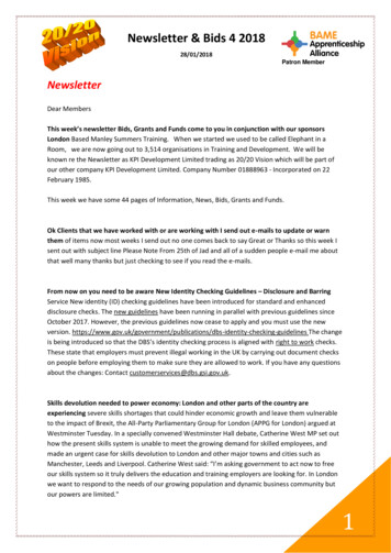
EMAG Group newsletterJan 2018NEWSCongratulations to Prof. Pratibha Gai, FREng, FRS, FRMS andfounding director of the York-JEOL Nanocentre, on being made a DameCommander of the Order of the British Empire (DBE) for services to ChemicalSciences and Technology in the Queen’s New Year Honour List.Prof. Pratibha Gai is an early pioneer of research into atomic processes importantto the chemical industry, particularly using in-situ electron microscopytechniques. With Prof. Edward D. Boyes, they custom modified the pole piecesof a conventional high-resolution analytical electron microscope in 1995 to enableinteraction between gas molecules and catalysis to be probed directly in realtime, at the atomic scale, under a controlled environment. Their innovation hasnow been commercialized as Environmental Transmission Electron Microscopesby several major electron microscope manufacturers. After many years workingin DuPont, USA, Profs Gai and Boyes joined the University of York to found theYork-JEOL Nanocentre in 2006, bringing their innovative approach to newlydeveloped aberration corrected electron microscopy. They have converted aJEOL double-aberration corrected 2200FS (S)TEM into the world’s onlyenvironmental electron microscope capable of both aberration corrected highresolution transmission electron microscopy and high resolution scanningtransmission electron microscopy, including high angle annular dark field imagingcapabilities with single atom sensitivity. Her work has already been recognizedby the IoP’s award of the Gabor medal and prizes. She has also been namedas L’Oréal-UNESCO Women in Science European Laureate for 2013 and nowfittingly, has royal recognition.In-situ electron microscopy at the atomic scale has become an indispensablecharacterization tool, thanks to pioneering work such as those by Prof Gai andothers. We will expect to see more fundamental understanding of the dynamicalprocesses in advanced materials reported using such approaches.Prof Jun Yuan, University of York7
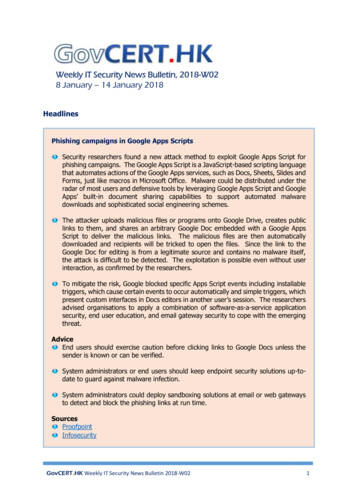
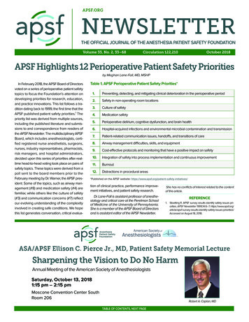
EMAG Group newsletterJan 2018Glasgow Plasma-FIB Now Open For New wing a successful inauguration event last summer, the new Xenon PlasmaFocused Ion Beam Microscope (PFIB) at the University of Glasgow is now fullyoperational. The system, a Helios DualBeam microscope supplied by ThermoFischer Scientific, has a unique configuration that enables the preparation,manipulation and analysis of materials in three dimensions and across micron tonanometre length scales. Funded by the EPSRC, the system is open to externalusers through a simple access scheme that will be free over the coming year. Weare keen to facilitate as wide a variety of nanoscience as possible.The PFIB's primary role is in the fabrication of large-area, low-damage crosssections suitable for electron microscopy analysis. The instrument uses a noblegas beam in place of established Gallium (Ga) ion-beam technology, therebyreducing artefacts and damage in specimens. A 50-100 times increase in beamcurrent also enables the site-specific preparation of far larger areas and volumesthan were previously feasible. It has already been exploited in studies ofmicroelectronic devices, thermoelectric materials and meteorites and is expectedto be of tremendous benefit to the study of a far wider range of materials.Coupled with detectors for electron backscattered diffraction (EBSD) and energydispersive spectroscopy (EDS), the new instrument facilitates three-dimensionalchemical and structural analysis of specimens. Tomography is enabled usingserial sectioning, to create a stack of images, elemental maps and diffractionpatterns that can be processed to produce a 3D representation of the sample.Perhaps the most exciting capability - which is unique to the UK - is lithographicdeposition of magnetic nanostructures. The PFIB is equipped with multiple gasinjector systems that enable the localised deposition of C, Pt, Co and Fe. Usingthe electron beam induced deposition (EBID) technique, it is now possible topattern arbitrary metallic structures in three dimensions, enabling a variety of newmagnetic studies.If you are interested in making use of this new technology, please contact phasknc@glasgow.ac.uk for further details or download an application form directlyfrom ups/mcmp/facilities/ ,following the link to the PFIB.Dr Donald MacLaren, University of Glasgow8
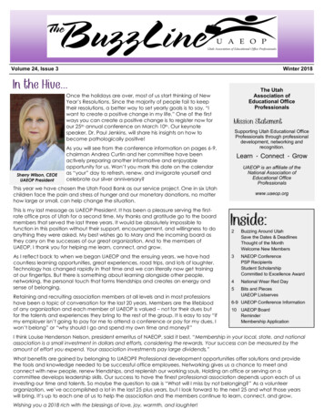
EMAG Group newsletterJan 2018(a) The Glasgow PFIB; (b) a 3D section of a geological specimen; (c) wide-areacross-section preparation; (d) 3D EBID patterning of free-standing magneticwires and structures; (d) EBSD mapping of a polycrystalline heusler alloy.9
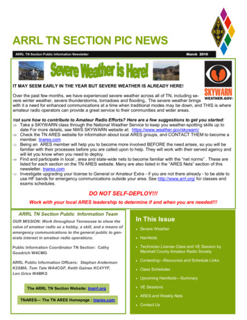
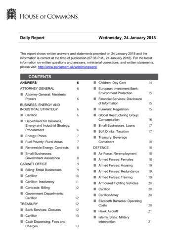
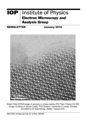
EMAG Group newsletterJan 2018A boost for electron microscopy at 7th January saw the official opening of Hitachi’s new “collaboration lab” at theDaresbury Science and Innovation Campus in Cheshire.The event was attended by over 100 microscopists from across the UK, withinvited speakers including Prof Quentin Ramasse (SuperSTEM / University ofLeeds) and Prof Raynald Gauvin (McGill University, Montreal) as well aspresentations from Hitachi’s designers and global managers. Attendees werealso given the opportunity to see the facility which houses advanced FESEM,STEM, correlative EM-SPM, plus ion milling and other advanced samplepreparation tools, with the opportunity for further instrumentation to locate thereaccording to scientific need.The new facility makes Daresbury a significant hub for electron microscopy in theUK, complementing the existing EPSRC National Facility for AberrationCorrected STEM (SuperSTEM) which also resides on the campus.The “collaboration lab” concept is designed to offer easy access in order to trialtechniques or to engage with Hitachi in method and instrument development.With proximity to many academic and industrial scientists in the region, and easyaccessibility from across the UK, it is anticipated that the new facility will bringnew microscopy applications to Daresbury, as well as play a significant role in theEMAG community.Mike Dixon, Hitachi High-Technologies EuropeLHS; Official lab opening by (from left to right) Yasukuni Koga (EuropeanPresident), Mike Dixon (UK manager), Toshiyuku Ikeda (Senior Vice President),Mats Eriksson (EMEA manager), Sukehiro Ito (Design and Production Director),Hiroshi Kato (Global EM business manager)RHS; Lab tours with Yukari Dan (UK Applications Manager)10
EMAG Group newsletterJan 2018MEETING REPORTSMicroscopy of Semiconducting Materials, MSM-XX 2017OxfordVeerendra C Angadi, PhD student, University of SheffieldI had an opportunity to attend the 20th Microscopy of Semiconducting Materials2017 (MSM-XX 2017) held in Oxford from 9 - 13th April 2017 which is about a 2hour drive from Sheffield. The conference included multiple sessions, in whicheach session is focused on various aspects of semiconductor microscopy likeanalytical TEM, cathodoluminescence, lattice defects, nanowires, strained layersand quantum wells etc.Lady Margaret Hall of University of Oxford was chosen as the venue. The venuehas a very historic significance as it is the first women’s college in Oxford foundedin 1878. The number of attendees for the conference were relatively low as it wasvery specific to microscopy of semiconductors. As a PhD student I had to presentmy poster on the topic “Determination of bandgap onset by blind deconvolutionof electron energy-loss spectra (EELS): 1D EELS vs 2D EEL spectrum images”.My session was on 10th April, Monday 17:00 - 19:00 in Symposium A: AnalyticalTEM poster session. It was a great opportunity for me to present among fellowresearchers. The discussions and questions posed by the attendees were reallyenlightening. The interactions and suggestions gave a lot of opportunity toimprove my work. In the mean time I attended many talks by the invited speakersand the industrial exhibitors. The one that I found very interesting was a talk byDr Peng Wang on the topic of 2D and 3D electron ptychographic reconstruction.On Wednesday evening the congress banquet was arranged in the dining hall ofLady Margaret Hall, which was right in front of the conference venue. At thebanquet, a MSM traditional pub quiz was held.Overall the conference was a fantastic learning experience for me and I am sureeveryone felt the same. I would like to thank IoP for the bursary support and anopportunity to write a report on my experience of the conference.11
EMAG Group newsletterJan 2018Association for Research in Vision and OphthalmologyAnnual Meeting 2017, BaltimoreMatthew G. Pilgrim, PhD student, University College LondonThe Association for Research in Vision and Ophthalmology annual meeting(ARVO) is the largest and most significant conference for any researcher workingin the field of ophthalmology. This year the conference took place from the 7-11thMay 2017 in the city of Baltimore, United States of America. By attending thisyear’s ARVO annual meeting I was able to disseminate my current research toboth researchers and clinicians working in the field of ophthalmology.Currently, my research is focused on determining the composition of sub-retinalpigment epithelial deposits, the earliest pathological lesion of the blinding diseaseage-related macular degeneration (AMD). To understand the elemental andmolecular composition of these lesions I am utilizing a number of high-resolutionimaging modalities and molecular analysis techniques including scanningelectron microscopy (SEM), density dependent colour SEM, SEM coupled energydispersive X-ray analysis, secondary ion mass spectrometry, synchrotron microfocus X-ray fluorescence and transmission electron microscopy coupled withselected area electron diffraction. This novel approach to answering a biologicalquestion in the field of vision and ophthalmology has proven highly successful.The conference provided the perfect opportunity to showcase thesemethodologies and demonstrate how they can assist ophthalmic research. Ipresented this study in the form of a poster that was entitled “A mineralomic studyof the retinal pigment epithelium – Bruch’s membrane complex in human eyeswith age-related macular degeneration”.As a PhD student, attendance to ARVO 2017 was extremely beneficial. Itprovided a rare opportunity to discuss my research with the internationalcommunity of AMD researchers. These discussions were extremely useful as Iwas able to discuss my results in the context of other areas of AMD research.This was invaluable as it highlighted additional lines of enquiry, in my caseanother inorganic compound. The poster session also provided the chance todiscuss the recent publication “Sub retinal pigment epithelial deposition of drusencomponents including hydroxyapatite in a primary cell culture model”. This studywas completed and co-authored with Professor Christine Curcio of the Universityof Alabama at Birmingham, who I met at ARVO 2015, and my research group atthe UCL Institute of Ophthalmology. Receiving feedback about our model systemas well as discussing potential experiments was extremely insightful andmotivating.One of the highlights of the conference was a presentation by Dr Ruchira Singh12
EMAG Group newsletterJan 2018from the University of Rochester Medical Center, United States of America. DrSingh gave a 15-minute presentation on her current research investigating therole of the retinal pigment epithelium in macular dystrophies, including AMD. Tounderstand the role of this cell type in disorders like AMD and Sorsby’s fundusdystrophy, Dr Singh and colleagues utilized human induced pluripotent stem cells(hiPSC), these are of great interest in ophthalmic research as they have beenshown to differentiate into retinal pigment epithelial cells, the support cells of thelight sensitive photoreceptor cells of the retina. Dr Singh and her group showedthat hiPSCs in culture were sufficient to promote drusen formation. Cells culturedfor 90 days produced sub-RPE deposits containing lipids and known drusenmarkers like APO-E. This study was particularly interesting as Dr Singh usedTranswell inserts identical to the ones used in the aforementioned cell culturemodel. In our model system we found that primary RPE cells produced depositscontaining both lipid and APO-E, like Dr Singh’s model, as well as hydroxyapatite.Whilst Dr Singh didn’t show data or appear to investigate whether hydroxyapatitewas present in her model system, this study was of great interest as hiPSCs havebeen used in a recent clinical trial aiming to restore or prevent further vision loss.However, if these cells can still produce deposits containing the key molecularcomponents of drusen, including inorganic hydroxyapatite, then perhaps this isnot an optimal treatment.Overall, the annual meeting for the Association for Research in Vision andOphthalmology was a huge success. Through discussions with the internationalcommunity of AMD researchers I was able to identify possible new lines ofresearch. Furthermore, the various talks and posters that were available eachday provided a rich source of knowledge on the biological question I am currentlyinvestigating as well as providing technical information.13
EMAG Group newsletterJan 2018Microscience and Microscopy Congress 2017, ManchesterJulie. A. Watts, PhD, student, University of NottinghamThe conference chairs, Peter O’Toole and Rik Brydson, set the tone for a relaxed,welcoming, and informative international congress, which was the biggest yetwith more attendees than previous events, 6 plenary lectures, 6 parallel sessionsand a large exhibition hall packed with demonstration kit alongsideknowledgeable technical experts. Three words summed up the event for me:aspirational, fast-paced, and inspirational.Aspirational because the Plenary lectures from world leaders in their variedsubject areas were packed with information and brilliant concepts, yet wereunderstandable and entertaining. I particularly enjoyed the historical perspectivesand scientific breakthroughs discussed by the speakers, and the unexpectedinteractivity: I can see where my individual research findings fit into the biggerpicture.Fast-paced, since with six parallel sessions running, not only was there scope toattend lectures highly relevant to my topic, but also to find out about differentresearch areas and techniques. The exhibition was the place to go for advice andtips from an array of companies, and included a learning zone and workshopareas which were good places to hear about the latest microscopes, equipmentand detectors.Inspirational by virtue of the variety and number of opportunities to connect withother individuals from around the world, informally at drinks receptions, the EMAGdinner or at the congress banquet, or more formally during one of the two postersessions. The breadth of research on display was astounding. The EMAG lecturefrom Professor Angus Kirkland, 2017 winner of the Alan Agar Medal for ElectronMicroscopy, explaining how reconstruction of exit wave function phase dataprojected onto image data results in super-resolution was definitely inspirational.Attending the Microscience Microscopy Congress 2017 has benefitted me inpractical ways thanks to helpful discussions with researchers at all levels whokindly participated in the poster sessions, and the pre-congress workshop(ImageJ/FIJI) that has enabled me to process data with vastly improvedefficiency. I am very grateful to have been given the opportunity to attend thisvery successful four-day congress, held at Manchester Central, to the EPSRC forfunding my research and to the IOP for providing a bursary towards the cost ofattending the event.14
EMAG Group newsletterJan 2018Microscopy and Microanalysis 2017, St LouisAnuj Pokle, PhD student, Trinity College DublinThe Microscopy and Microanalysis conference is an annual internationalconference held in US in the field of electron microscopy. This year it was in St.Louis, Missouri (6-10th August 2017). Fortunately, this time I got the opportunityto give two platform presentations. It was very exciting but at the same time hecticto prepare yourself for two talks. The best plenary talk that I enjoyed was on‘Detecting Massive Black Holes via Attometry’ by Keith Riles from University ofMichigan. Prior to the conference there were pre-meetings scheduled for studentsand scientists across the fields followed by a welcome reception on Sundayevening (6th Aug). It was an interesting informal meeting where you could get intouch with the people and have a chat over a beer and good food!There were numerous microscopy sessions organized over cellular, food,pharmaceuticals semiconductors and energy related materials. Variouscharacterization techniques within the microscopy world were presentedincluding In-situ experiments, FIB (Focussed Ion Beam), Cryo, Tomography,Aberration-corrector, machine learning and implementing advanced computationin microscopy.The parallel sessions going on made it pretty tough to choose the talks to attend.My prime focus of work is on aberration corrected STEM (Scanning TransmissionElectron Microscopy), EELS (Electron Energy Loss Spectroscopy, EDX (Energydispersive X-ray Spectroscopy) over two –dimensional materials. Presentationson low loss vibrational spectroscopy (equivalent of doing optical spectroscopy athigh spatial resolution) and quantified imaging which allows you to count thenumber atoms in a nanoparticle were very interesting. These are pretty recentdevelopments in the field of electron microscopy. Poster presentations weremuch helpful to discuss the theoretical and experimental aspects in more details.The Microscopy Society of America (MSA) and the Student council organizedmeetings to update on the current society situation. The student council electedtheir new board members and put across plans of providing further travel grantsfor students from next year. The MSA had also organized a lunch meetingespecially for early stage researchers to get proper guidance from mentors andto share their views.Thanks to the IOP Research funds, I could take care of some of my travelexpenses. This was really helpful. I would strongly recommend the M&M 2017Conference not only for electron microscopists but people from other fieldsincluding materials, biology etc. It’s a perfect way to learn new things in a friendlyenvironment, meet the experts and networking for future collaborations andcareer planning.15
EMAG Group newsletterJan 2018APPLY FOR IoP RESEARCH STUDENTSCONFERENCE FUNDIf you are a student member and are looking for funding to attend a meeting orconference, please apply for an RSCF bursary, which may give you up to 300towards your costs. We have several of these bursaries to give away eachyear. Check eligibility criteria and download the form athttp://www.iop.org/about/grants/research student/page 38808.htmlEMS MEMBERSHIPEMAG members are reminded that they are all automatically members of theEuropean Microscopy Society, at no cost to themselves. However, in order toreceive information from the EMS, it is essential to send your e-mail address tothe EMS secretary - this cannot be sent by the IOP due to the Data ProtectionAct. This is important, since almost all communications from the EMS are sentby e-mail, including information for voting for the next Executive Board.Send your e-mail address (and preferably your other details, postal address,phone & fax numbers) to: secr@eurmicsoc.org and indicate whether you agreeto include this information in the EMS Yearbook. If you do NOT wish to appearin the Yearbook, your e-mail address will be used solely for the dispatch ofinformation by the EMS secretary (virginie.serin@cemes.fr).The EMS web page can be viewed at: http://www.eurmicsoc.org/EMAG members are also reminded of the availability of EMS Bursaries. Formore details, see http://www.eurmicsoc.org/scholarships.htm16
EMAG Group newsletterJan 2018ADDITIONAL FUTURE MEETINGS OF INTERESTMicroscopy characterization of organic-inorganic Interfaces22-23rd February 2018, tron Back Scatter Diffraction Meeting 201810-11th April 2018, -calendar/ebsd-2018.html19th International Microscopy Congress (IMC19)9-14th September 201, Sydney, Australiahttp://imc19.com/mmc2019: Microscience Microscopy Congress 20191-4th July 2019, ess-2019.htmlFor more microscopy events alendar/17
EMAG Group newsletterJan 2018EMAG contact pointsIOP:Institute of Physics, 80 Portland Place, London, W1B 1NT, UKTel: 44 (0)20 7470 4800, Fax: 44 (0) 20 7470 4900For conferences:Email: tific/conferences/index.htmlGroup matters: Science Support OfficerEmail: groups@iop.orgEMS:European Microscopy SocietyEmail: tmlMRS:Materials Research Society, 9800 McKnight Road, Pittsburgh,PA 15237, USA.Tel: 1 412 779 3003, Fax: 1 412 779 8313http://www.mrs.org/meetings/MSA:Microscopy Society of America, 12100 Sunset Hills Rd., Suite 130,Reston, VA 20190, USA.Tel: 1 703 234 4115, Fax: 1 703 435 4390http://www.microscopy.org/RMS:Royal Microscopical Society, 37/38 St. Clements, Oxford, OX4 1AJ.Tel: 44 1865 248 768, Fax: 44 1865 791 237Email: is newsletter is also available on the web and in larger print sizesThe contents of this newsletter do not necessarily represent the views orpolicies of the Institute of Physics, except where explicitly stated.The Institute of Physics, 80 Portland Place, W1B 1NT, UK.Tel: 020 7470 4800Fax: 020 7470 484818
School of Chemical and Process Engineering, University of Leeds, Leeds, LS2 9JT Tel: 0113 343 2382 Email: a.p.brown@leeds.ac.uk . now been commercialized as Environmental Transmission Electron Microscopes by several major electron microscope manufacturers. After many years working in DuPont, USA, Profs Gai and Boyes joined the University of .