Transcription
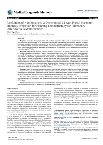
Higashihara, J Med Diagn Meth 2014, cal Diagnostic MethodsResearchArticleResearch ArticleOpenOpenAccessAccessUsefulness of Non-Enhanced 3-Dementional CT with Partial MaximumIntensity Projection for Planning Embolotherapy for PulmonaryArteriovenous MalformationsHiroki Higashihara*Department of Radiology, Osaka University Graduate School of Medicine, Osaka, JapanAbstractPurpose: Computed Tomography (CT) with contrast material is often used for preoperative assessmentand planning of embolotherapy in the treatment of Pulmonary Arteriovenous Malformations (PAVMs). However,pulmonary vasculature is well demonstrated in the lung window setting without the need for contrast material; thus,risks of adverse effects of contrast material itself or paradoxical emboli through PAVMs can be avoided. The purposeof this study was to determine the usefulness of non-enhanced 3-Dimentional (3D)-CT angiography for planning ofembolotherapy for PAVMs.Materials and methods: Between February 2004 and October 2011, 20 patients (nine males, 11 females) with41 PAVMs underwent non-contrast CT using a multi-detector-row CT prior to coil embolotherapy. A high-resolutiontailored 3D-CT angiogram was reconstructed on a workstation with partial Maximum Intensity Projection (MIP) in thelung-window to assess the angioarchitecture of the lesions for preoperative planning of embolotherapy. For eachlesion, location, number, and diameter of feeding arteries and draining veins were measured. It was determinedwhether there was any side branch close enough to the sac to anchor the first coil to prevent coil migration. Thedifference between the diameter of the first coil and the feeding artery was measured. Based on findings, diagnosticpulmonary angiography and coil embolotherapy were performed. The depiction of the side branch of the feedingartery close to sac between preoperative partial MIP 3D-CT images and selective pulmonary angiography wasevaluated by using the un-weighted κstatistical analysis.Results: A total of 49 feeding arteries were embolized. Mean diameter of the feeding arteries and drainage veins were3.6 mm and 4.6 mm, respectively. Sixteen and eighteen feeding arteries with a side branch to anchor were depicted on CTand angiography, respectively. In the depiction of the side branch of the feeding artery close to sac between preoperativepartial MIP images and selective pulmonary angiography, excellent agreement was obtained (κ 0.91).The mean difference between diameter of the first coil and the feeding artery was 1.5 1.47 mm. All 41 lesionscould be identified on angiography, and embolization procedures could be executed as planned based on partialMIP images.Conclusion: Non-enhanced tailored partial MIP 3D-CT in the lung window appears to be a feasible and usefulvascular imaging technique for planning coil embolotherapy of PAVMs.IntroductionPulmonary Arteriovenous Malformations (PAVMs) are anomalousarteriovenous communications without intervening capillary beds andconsist of a dilated, tortuous feeding artery, an aneurysmal sac, and adrainage vein. Symptoms are mainly caused by right-to-left shunting orrupture and include dyspnea, cyanosis, polycythemia, and hemoptysis.In addition, more serious neurological complications such as cerebralstroke and brain abscess can be caused by paradoxical embolization,the risk of which is significantly higher for PAVMs with feeding arteriesthat are 3 mm or more in diameter [1]. This is the threshold size forwhich occlusion is required. For symptomatic PAVMs or lesions atrisk, transcatheter embolization is the treatment of choice to controlsymptoms or reduce risk [2-6].The goal of imaging studies, especially Computed Tomography(CT), is to identify all PAVMs with feeding arteries that are 3 mm ormore in diameter. In addition, 3-Dimensional (3D)-CT images arehelpful to understand the angioarchitecture of the individual lesion andaid in the planning of embolotherapy. Because a PAVM is a “vascularlesion,” Contrast-Enhanced CT (CECT) for the mediastinal windowview is often preferred to identify the anomalous vessel components forpre- and/or post-treatment evaluation [7,8].Although infrequent, intravenous administration of contrastmaterial carries the risk of paradoxical embolization due to possibleJ Med Diagn MethISSN: 2168-9784 JMDM, an open access journalcontamination of air bubbles. Although we are unable to quantify therisk of paradoxical embolization during CECT, a complication such ascerebral infarction could be serious if paradoxical embolization occurs.In addition, contrast enhancement does not enhance the lung-windowdisplay. On the other hand, PAVMs can be clearly identified on nonenhanced 3D-CT images in the lung window.The 2009 International Guidelines for the Diagnosis andManagement of Hereditary Hemorrhagic Telangiectasia also state thatunenhanced CT should follow a positive contrast echocardiogram [9].*Corresponding author: Hiroki Higashihara, Department of Radiology,Osaka University Graduate School of Medicine,2-2, Yamada-oka, Suita,Osaka, Japan, Tel: 81-6-6879-3434; Fax: 81-6-6879-3439; ved September 06, 2013; Accepted December 23, 2013; PublishedJanuary 01, 2014Citation: Higashihara H (2014) Usefulness of Non-Enhanced 3-DementionalCT with Partial Maximum Intensity Projection for Planning Embolotherapyfor Pulmonary Arteriovenous Malformations. J Med Diagn Meth 2: 148. doi:10.4172/2168-9784.1000148Copyright: 2014 Higashihara H. This is an open-access article distributed underthe terms of the Creative Commons Attribution License, which permits unrestricteduse, distribution, and reproduction in any medium, provided the original author andsource are credited.Volume 2 Issue 6 1000148
Citation: Higashihara H (2014) Usefulness of Non-Enhanced 3-Dementional CT with Partial Maximum Intensity Projection for Planning Embolotherapyfor Pulmonary Arteriovenous Malformations. J Med Diagn Meth 2: 148. doi: 10.4172/2168-9784.1000148Page 2 of 5We demonstrate here the usefulness of non-enhanced 3D-CT forplanning treatment of PAVMs using embolotherapy.distance for the coil placement, we were planning to place the coils usingSac embolization technique described later. We defined the followingfour techniques for coil placement: “Push technique” indicated thatthe first coil was placed without anchoring the side branch; “Anchortechnique” indicated that the tip of the first coil was placed into theside branch to avoid coil migration; “Scaffold technique” indicated thatthe large first 2 or 3 coils were placed in the feeding artery for scaffoldformation to avoid coil migration; and “Sac embolization technique”indicated that the coil was placed in only the aneurysmal sac becausethe distance to the feeding artery was too short to be embolized.Materials and MethodsPatient population and back groundOur institution review board does approve this study asretrospective study. Informed consent was obtained from all patientsbefore procedures. Between February 2004 and April 2011, 20patients (9 men and 11 women) with a total of 41 PAVMs underwentcoil embolotherapy. Fifteen patients were suspected as HereditaryHemorrhagic Telangiectasia. In the remaining 5 patients, 3 patientswere incidentally suspected by the chest X-ray. The one patient washypoxemia and the other was post cerebral infarction. The mean patientage was 40 years (range, 13-72 years). For preoperative planning, allpatients were evaluated with non-enhanced CT.Embolization procedureBefore embolotherapy, all patients underwent pulmonaryangiography with a 4-F pig tail catheter and a 6-F multipurpose catheterthrough the right common femoral vein. First, the main pulmonaryangiogram was obtained to detect the lesions of PAVMs, followed byselective catheterization of the feeding artery with either a 4-Fr endhole or a 2.3-Fr microcatheter (Rapidtransit, Cordis Corporation,Miami Lakes, FL), depending on the partial MIP image obtained forpre-therapeutic planning. The embolic materials used in this study werehigh radial force Inconel coils or soft platinum coils. The success of theprocedure was determined by whether PAVMs were embolized or notin accordance with the preoperative planning based on partial MIPimages as described earlier.CT techniqueCT angiography equipment and CT scanners changed during thestudy. An 8-detector CT scanner (Lightspeed Ultra; General ElectricMedical Systems, Milwaukee, WI) was used for two patients fromFebruary 2004 to March 2005 and a 64-detector CT scanner (LightSpeedVCT; General Electric Medical Systems) was used for eighteen patientsfrom April 2005 to April 2011. The imaging parameters for the8-detector CT scanner were as follows: collimation, 1.25 mm; and pitch,1.375. The imaging parameters for the 64-detector CT scanner were asfollows: collimation, 0.625; and pitch, 1.375.For each patient, we investigated the size of the first coil, the numberof coils placed, and which technique was used to place the coils. We alsoevaluated whether the side branch to anchor the first coil was depictedon the angiographic image.None of the patients was administered nonionic contrast material.The scan region ranged from the apical portion of the lung to thebottom of the diaphragm. A high-resolution tailored 3D-CT angiogramwas reconstructed with partial Maximum Intensity Projection (Mip) inthe lung window on a workstation (Advantage Windows version 4.2;General Electric Medical Systems) to assess the 3D-angioarchitectureof each of the 41 lesions.Statistical analysisThe depiction of the side branch of the feeding artery close tosac between preoperative partial MIP 3D-CT images and selectivepulmonary angiography was evaluated. We used the unweighted κstatistic with binary data to measure the extent of agreement for thedepiction of the side branch among two modalities by using a statisticalsoftware package (SPSS 11.0 for Windows; SPSS Japan, Tokyo, Japan).values of up to 0.40 were considered to indicate positive but pooragreement; κ values of 0.41-0.75, good agreement; and κ values greaterthan 0.75, excellent agreement [10].Planning assessment for embolotherapyPlanning of embolotherapy was based on the locations, numbers,and diameters of feeding arteries and draining veins. Planning alsoinvolved deciding how to place the first coil. This required determiningwhether the embolized feeding artery was at an adequate distance toplace a sufficient numbers of coils before a major segmental branch toavoid significant pulmonary infarction as well as whether there was anyside branch close enough to the sac to anchor the first coil to preventcoil migration. In case of no side blanch on 3D-MIP CT, the coils wereplaced using Scaffold technique described later. If there was shortNo.AgeSexLocationResultsAssessments of the planning and results of embolotherapy areshown in the Table 1. Embolization was feasible in all 20 patients. TheFeeding arteryDrainage veinCoilSize of 1st coilSide branch to anchorNo. / size (mm)size (mm)No. / TQ(mm)MIP / DSA130MLt S51 / 5.86.28 / sca10 / 214MRt S11 / 3.553 / anch6 / 7 / 2nd session3 sessionrdRt S22 / 4.876 / anchsame as above/ 3.2same as above3 / anchRt S41 / 3.43.43 / anch4 / Rt S81 / 3.43.33 / anch4 / Rt S101 / 3.64.33 / anch3 / Rt S11 / 2.83.74 / anch3 / Rt S81 / 2.12.52 / push4 / Lt S41 / 4.23.82 / anch5 / J Med Diagn MethISSN: 2168-9784 JMDM, an open access journalVolume 2 Issue 6 1000148
Citation: Higashihara H (2014) Usefulness of Non-Enhanced 3-Dementional CT with Partial Maximum Intensity Projection for Planning Embolotherapyfor Pulmonary Arteriovenous Malformations. J Med Diagn Meth 2: 148. doi: 10.4172/2168-9784.1000148Page 3 of 5346FLt S61 / 3.62.94 / sac6 / Lt S81 / 2.23.24 / anch3 / / Lt S81 / 2.32.66 / push3429MRt S81 / 3.84.63 / anch6 / 565FRt S21 / 3.85.74 / coil anch6 / 626MRt S81/57.26 / anch8 / Rt S84 / 4.576 / push8 / same as above2.2same as above5 / push3 / same as above2.7same as above3 / push3 / same as above2.5same as above5 / push3 / Rt S91 / 3.24.94 / push3 / 5.24 / push4 / / 716MRt S31 / 3.5827FLt S61 / 3.325 / push5Lt S91 / 3.13.32 / push3 / Rt S51 / 5.057 / anch8 / / 972F1071FLt S51 / 8.0108 / anch&sca121141MRt S31 / 3.33.85 / push4 / Rt S31 / 3.344 / push4 / Rt S31 / 4.76.67 / push7 / Rt S31 / 3.663 / push5 / Lt S92 / 3.26.47 / push3 / same as above2.7same as above4 / push4 / Rt S51 / 3.23.23 / push4 / Lt S101 / 4.254 / push8 / Lt S101 / 2.33.22 / push4 / Lt S41 / 3.13.42 / anch4 / Lt S101 / 3.62.63 / push4 / / 121314134764MMF1531MRt S31 / 3.746 / anch61658FLt S8/91 / 6.68.99 / push12 / 1751FLt S91 / 3.73.42 / push4 / Lt lig1 / 2.92.92 / push4 / 1823FLt S51 / 3.43.84 / push6 / 1918FRt S82 / 4.244 / push6 / same as above2.8same as above3 / push3 / Lt S1 23 / 2.26.49 / push6 / 2.7same as aboveembolizedtogethersame as above2.7same as above5 / push5 / Rt S81 / 3.243 / anch6 / same as above2048F / TQ: the technique of coil embolization; push: the first coil was placed without side branch for anchoring; anch: the tip of the first coil was place with side branch foranchoring; sca: the larger first two or three coils were placed in feeding artery for scaffold formation to avoid coil migration (i.e. scaffold technique); sac: the coil was placedin the only aneurysmal sac.Table 1: Summary of PAVM lesion.41 PAVMs could be successfully occluded according to the planningbased on partial MIP images (Figures 1-4). There were no immediateor delayed complications related to angiography or embolization suchas paradoxical embolization. Five PAVMs contained two to four feedingarteries, and altogether, 49 feeding arteries of 41 PAVMs were detected.Thirty-five feeding arteries were 3 mm or more in the diameter and14 feeding arteries were less than 3 mm (mean, 3.6 mm). All these 14feeding arteries were multiple PAVMs and the largest feeding are morethan 3mm. We embolized these 14 arteries in one session for avoidingto additional procedure in the future. Diameters of drainage vein were2 to 8.9 mm (mean, 4.6 mm). All feeding arteries could be detected onplanning CT. The number of coils required for the embolotherapy wastwo to nine coils per PAVM (mean: 4 coils). The mean difference betweendiameter of the first coil and the feeding artery was 1.5 1.47 mm.J Med Diagn MethISSN: 2168-9784 JMDM, an open access journalPartial MIP images showed 16 feeding arteries with a side branchclose enough to the sac to anchor the first coil, while the pulmonaryangiogram showed 18 feeding arteries (Figure 1). Eighty-nine percent(16/18) of the side branches of the feeding arteries were depicted inpreoperative partial MIP images. Fifteen feeding arteries (83%, 15/18)were embolized by the anchor technique. Only one PAVM had ashort feeding artery on a partial MIP image, and the distance in thefeeding artery was not adequate to deploy enough coils before anothersegmental branch. Therefore, the aneurysmal sac was embolized with 4microcoils (Figure 2).In the depictions of the side branch of the feeding artery close tosac between preoperative partial MIP images and selective pulmonaryangiography, the κ values were 0.91. Excellent agreement was obtainedbetween two modalities.Volume 2 Issue 6 1000148
Citation: Higashihara H (2014) Usefulness of Non-Enhanced 3-Dementional CT with Partial Maximum Intensity Projection for Planning Embolotherapyfor Pulmonary Arteriovenous Malformations. J Med Diagn Meth 2: 148. doi: 10.4172/2168-9784.1000148Page 4 of 5ABDCFigure 1: 14-year-old boyA. The partial MIP 3D-CT image shows a PAVM in the periphery of the right middle lobe. A small side-branch (arrow) is seen close to the sac. B. The finding is wellcorrelated with the selective angiogram. C. In the procedure, a 4 mm fibered platinum microcoil (Micronester, Cook) was used by anchoring the initial 2-3 cm of thecoil into the side branch to prevent coil migration. D. The remaining two 3 mm coils were safely packed proximal to the first coil to obtain cross-sectional occlusion(“Anchor technique”).BACFigure 2: 46-year-old womanA. The partial MIP 3D-CT image shows a small PAVM with a short feeding artery. The distance to the feeding artery was not adequate to deploy enough coils beforeanother segmental branch. B. Findings on 3D-CT were well correlated with the selective arteriogram. C. To avoid sacrificing the normal segmental branch, theaneurysmal sac was embolized with 3-6 mm detachable microcoils (Detach, Cook).ABCEEFigure 3: 29-year-old manA. Partial MIP 3D-CT image shows the complex architecture of the PAVM in the right anterior segment. Both the feeding artery and draining vein bifurcated at theentrance to the sac. The distance to a normal artery (overlapped with the draining vein) was too short and proximal to the feeding artery to be occluded. B. Theselective angiogram demonstrated similar findings to the 3D-CT image. C .The first 6 mm fibered microcoil (Micronester, Cook) straddled the bifurcation of thefeeding artery and formed a scaffold. D. Two additional 3-4 mm coils were used for the remaining cross-sectional occlusion. E. The normal branch proximal to the lastcoil is pictured (asterisk).DiscussionRemy et al. first reported the results of a pre- and post-treatmentevaluation of PAVM using chest CT [5]. They also mentioned theusefulness of 3D helical CT to help understand the angioarchitectureof PAVMs without using contrast material [11]. In our study, nonenhanced tailored partial MIP 3D-CT images obtained in the lungwindow also clearly demonstrated the angioarchitecture of PAVMs,and findings based on partial MIP 3D-CT images correlated wellwith those based on pulmonary angiography. The partial MIP 3D-CTimages allowed for accurate measurement of the diameter or length ofthe feeding artery to be occluded and were helpful for choosing theembolization technique to use, including the size of the coils and thepoints of occlusion. Transcatheter embolotherapy could be efficientlyJ Med Diagn MethISSN: 2168-9784 JMDM, an open access journalcarried out based on the findings of partial MIP 3D-CT images withoutuse of contrast material. There were some techniques for the coilembolization of PAVMs such as anchor technique, scaffold techniqueand sac embolization [12]. If the preoperative CT clearly demonstratesthe side branch of the feeding artery close to sac, we can plan to embolizethe feeding artery by anchor technique in advance. In this study, thedepictions of the side branch of the feeding artery close to sac betweenpreoperative partial MIP images and selective pulmonary angiographywere excellent agreement. Therefore, unenhanced CT would be usefulto evaluate the side branch of feeding artery for preoperative study.There remains disagreement as to the use of contrast materialsfor the evaluation for PAVMs. Some authors reported that contrastenhanced CT provides information beyond unenhanced CT,Volume 2 Issue 6 1000148
Citation: Higashihara H (2014) Usefulness of Non-Enhanced 3-Dementional CT with Partial Maximum Intensity Projection for Planning Embolotherapyfor Pulmonary Arteriovenous Malformations. J Med Diagn Meth 2: 148. doi: 10.4172/2168-9784.1000148Page 5 of 5Figure 4: 71-year-old womanA. The partial MIP 3D-CT image shows a simple PAVM adjacent to the pleura in the left lingual lobe. The course of the feeding and draining vessels was clearly seen.The feeding artery was slightly widened at the entrance to the sac. In addition, there was a side branch to anchor the first coil close to the sac (arrow). Transcatheterembolotherapy was performed. B. Main pulmonary angiography was well correlated with the 3D-CT findings. C, D, E. Following the first two oversized 12 or 10 mmhigh radial-force Inconel fibered coils (IMWCE, Cook, Bloomington, IN) to form a scaffold was placed with anchoring a side branch and six 6-8 mm soft-platinum fiberedcoils (Tornado, Cook) were used for the remaining cross-sectional occlusion (“Combination of Anchor and Scaffold technique”).particularly during follow-up, which warrants the minor additionalrisk of contrast administration [13,14]. Some reports also havehighlighted the clinical usefulness of reconstruction with partial MIP3D-CT with contrast material for PAVMs after embolotheraphy [7, 8,11]. However, there are some well-recognized groups which continueto use unenhanced CTand the 2009 International Guidelines for theDiagnosis and Management of Hereditary Hemorrhagic Telangiectasiatreatment state that unenhanced CT should follow a positive contrastechocardiogram [9,15,16].Another report dealt with a paradoxical air embolism occurringduring CECT in a patient who had a congenital heart disorder witha right-to-left shunt [17]. Furthermore, PAVMs with feeding arteries3 mm or more in the diameter carry an increased risk of paradoxicalemboli. It is estimated that 24% of patients with PAVMs experiencetransient ischemic attacks or stroke and 9% show brain abscess onpresentation of PAVMs due to right-to-left shunt [18]. Although anactual case of a paradoxical air embolism associated with contrastenhanced CT has not been reported in patients with PAVM and itwas obscure in the incident of paradoxical embolism associated withcontrast enhanced CT, the use of contrast material should be carefullylimited to the minimum required for the pre-treatment assessment ofPAVMs.There are some limitations to our study. First, this study is a single,retrospective study. Second, only a small number of patients wereenrolled. Third, the PAVMs treated in our study were generally smalllesions which were 3.6mm in the mean diameter of feeding artery. Thusthe findings of our study may not apply to PAVMs with larger feedingartery. A prospective study of a larger number of patients is needed todetermine the efficacy of partial MIP images in the lung window.In conclusion, non-enhanced tailored partial MIP 3D-CT in thelung window appears to be a feasible and useful vascular imagingtechnique for planning coil embolotherapy of PAVMs, especially in theevaluation for the side branch of the feeding artery.References1. Guttmacher AE, Marchuk DA, White RI Jr (1995) Hereditary hemorrhagictelangiectasia. N Engl J Med 333: 918-924.2. White RI Jr, Lynch-Nyhan A, Terry P, Buescher PC, Farmlett EJ, et al. (1988)Pulmonary arteriovenous malformations: techniques and long-term outcome ofembolotherapy. Radiology 169: 663-669.3. Lee DW, White RI Jr, Egglin TK, Pollak JS, Fayad PB, et al. (1997)Embolotherapy of large pulmonary arteriovenous malformations: long-termresults. Ann Thorac Surg 64: 930-939.J Med Diagn MethISSN: 2168-9784 JMDM, an open access journal4. Moussouttas M, Fayad P, Rosenblatt M, Hashimoto M, Pollak J, et al. (2000)Pulmonary arteriovenous malformations: cerebral ischemia and neurologicmanifestations. Neurology 55: 959-964.5. Remy J, Remy-Jardin M, Wattinne L, Deffontaines C (1992) Pulmonaryarteriovenous malformations: evaluation with CT of the chest before and aftertreatment. Radiology 182: 809-816.6. Haitjema TJ, Overtoom TT, Westermann CJ, Lammers JW (1995) Embolisationof pulmonary arteriovenous malformations: results and follow up in 32 patients.Thorax 50: 719-723.7. Hofmann LV, Kuszyk BS, Mitchell SE, Horton KM, Fishman EK (2000)Angioarchitecture of Pulmonary Arteriovenous Malformations: CharacterizationUsing Volume-Rendered 3-D CT Angiography. CardioVascular andInterventional Radiology 23: 165-170.8. Lawler LP, Fishman EK (2002) Arteriovenous malformations and systemiclung supply: evaluation by multidetector CT and three-dimensional volumerendering. AJR Am J Roentgenol 178: 493-495.9. Faughnan ME, Palda VA, Garcia-Tsao G, Geisthoff UW, McDonald J, et al.(2011) International guidelines for the diagnosis and management of hereditaryhaemorrhagic telangiectasia. J Med Genet 48: 73-87.10. Fleiss JL (1981)The measurement of interrater agreement. In: Statisticalmethods for rates and proportions. (2ndedn.) Wiley, New York, USA, pp:212236.11. Remy J, Remy-Jardin M, Giraud F, Wattinne L (1994) Angioarchitecture ofpulmonary arteriovenous malformations: clinical utility of three-dimensionalhelical CT. Radiology 191: 657-664.12. White RI Jr (2007) Pulmonary arteriovenous malformations: how do I embolize?Tech Vasc Interv Radiol 10: 283-290.13. Remy-Jardin M, Dumont P, Brillet PY, Dupuis P, Duhamel A, et al. (2006)Pulmonary arteriovenous malformations treated with embolotherapy: helicalCT evaluation of long-term effectiveness after 2-21-year follow-up. Radiology239: 576-585.14. Brillet PY, Dumont P, Bouaziz N, Duhamel A, Laurent F, et al. (2007) Pulmonaryarteriovenous malformation treated with embolotherapy: systemic collateralsupply at multidetector CT angiography after 2-20-year follow-up. Radiology242: 267-276.15. Zukotynski K, Chan RP, Chow CM, Cohen JH, Faughnan ME (2007) Contrastechocardiography grading predicts pulmonary arteriovenous malformations onCT. Chest 132: 18-23.16. van Gent MW, Post MC, Luermans JG, Snijder RJ, Westermann CJ, et al. (2009)Screening for pulmonary arteriovenous malformations using transthoraciccontrast echocardiography: a prospective study. Eur Respir J 33: 85-91.17. Thamburaj K, Devasia B (2006) Cerebral air embolism during contrastenhanced CT. Pediatr Radiol 36: 887.18. Shovlin CL, Letarte M (1999)Hereditary hemorrhagic telangiectasia andpulmonary arteriovenous malformations: issues in clinical management andreview of pathogenic mechanism. Thorax 54: 714-729.Volume 2 Issue 6 1000148
ISSN: 214 MDM, an open access ournal We demonstrate here the usefulness of non-enhanced 3D-CT for planning treatment of PAVMs using embolotherapy. Materials and Methods Patient population and back ground Our institution review board does approve this study as retrospective study. Informed consent was obtained from all patients before procedures.