Transcription
M. B. Mitrovi}, et al.: Influence of Radiation Dose in Computed Tomography on .Nuclear Technology & Radiation Protection: Year 2017, Vol. 32, No. 4, pp. 342-349342INFLUENCE OF RADIATION DOSE IN COMPUTED TOMOGRAPHYON ANTIOXIDANT ENZYME ACTIVITY IN RABBIT ERYTHROCYTESbyMarko B. MITROVI] 1, Olivera F. CIRAJ-BJELAC 2, Zorana S. OREŠ ANIN-DUŠI] 3,Duško P. BLAGOJEVI] 3, Aleksandra L. NIKOLI]-KOKI] 3, Nikola R. TATALOVI] 3,Nikola E. KRSTI] 1, and Mirjana V. LAZAREVI]-MACANOVI] 11Department of Radiology and Radiation Hygiene, Faculty of Veterinary Medicine,University of Belgrade, Belgrade, Serbia2Radiation Protection Department, Vin a Institute of Nuclear Sciences, University of Belgrade, Belgrade, Serbia3Department of Physiology, Institute for Biological Research "Siniša Stankovi}", University of Belgrade,Belgrade, SerbiaScientific paperhttp://doi.org/10.2298/NTRP1704342MThe objective of this study was to assess the radiation dose in computed tomography examinations of rabbits using different examination protocols and to correlate these values with theactivity of antioxidant enzymes in their red blood cells following irradiation. The presentedresults revealed that a single, routine computed tomography scan exposure led to a differentresponse of the activity of antioxidant enzymes in red blood cells regarding both dose andtime. The results indicate that there is a dose threshold that is about 25 mGy. Doses belowthat level do not produce any significant changes in the level of antioxidant enzymes activity.On the other hand, the level just above that threshold had a significant impact on the antioxidant defence, but in a relatively short time period (2 hours after exposure), compared to thehigher dose that requires a longer adaptive period.Key words: computed tomography, CT dose index, erythrocyte, antioxidant enzyme, rabbitINTRODUCTIONDespite the universal consensus that computedtomography (CT) overwhelmingly benefits patientswhen used for appropriate indications, concerns havebeen raised regarding the potential risk due to the increased use of CT in medicine and the relative high radiation doses associated with this type of examination.When compared to conventional radiography, CT delivers a considerably larger dose to the patient. Although technological developments provide the opportunity to decrease individual CT doses, the attemptto obtain quality images and cover a larger area of thepatient's anatomy can lead to the opposite result. It isincreasingly being documented that patient doses arehigher than necessary for the high quality image and inCT often exceeds the level needed for confident diagnosis [1]. Thus, keeping the radiation dose as low asreasonably achievable consistent with the diagnostictask, remains the most important strategy for decreasing this potential risk [2] in line with general radiationprotection principles. Because of that, optimization ofCT based on the balance of the radiation dose and im* Corresponding author; e-mail: markom@vet.bg.ac.rsage quality, is very important for avoiding excessivepatient doses [3, 4]. Similarly to human medicine,X-rays are equally used in veterinary medicine. Thereasons for using radiation in veterinary medicine areto either obtain optimum diagnostic information or toachieve a specific therapeutic effect, while maintaining the radiation dose to the radiological personnel andthe general public as low as reasonably achievable.Similarly, it is also important to avoid all unnecessaryirradiation of the animal patient [5].In terms of radiation protection and associatedradiation effects, radiation doses from a single, routinecomputed tomography examination belong to the category of low radiation doses and therefore can exertonly stochastic effects. Low doses are defined as dosesin the range of near zero up to about 100 mSv (0.1 Sv)of low-LET radiation [6]. Based on epidemiologicaldata of radiation-induced cancer occurrences, variousauthors agree that low doses are below 200 mGy as below this level the statistical evaluation of data becomesmore and more uncertain [7, 8]. However, certain cellular reactions like enzyme inductions, DNA-repairprocesses, adaptive responses, chromosome aberrations, etc. could already be observed between 10 and100 mGy by various sensitive assay techniques [9].
M. B. Mitrovi}, et al.: Influence of Radiation Dose in Computed Tomography on .Nuclear Technology & Radiation Protection: Year 2017, Vol. 32, No. 4, pp. 342-349Low dose significance is especially relevant in triggering oxidative stress in organs and tissues submitted toirradiation, resulting in elevated activity of the mainantioxidant enzymes: superoxide dismutase – SOD,catalase – CAT, glutathione peroxidase – GSH-Px, andglutathione reductase – GR [10, 11]. Changes in theantioxidant enzymes activity were demonstrable whenthe applied dose is 50 mGy [12]. However, it was reported that oxidative stress can be induced with extremely low radiation doses from 0,1 mGy [13, 14] to1 mGy [13]. In the rat spleen, an elevated activity ofSOD and GSH-Px was registered following exposureto 250 mGy [15], and for the SOD this change lasted 8weeks. In another study [16] CAT activity in chickenkidneys was elevated by 40 % at the dose of 500 mGyand doubled at the radiation load of 2 Gy. Same authors documented that in samples of chicken brains atthe dose of 500 mGy, CAT activity was elevated onlyby 25 %, while at the dose of 2 Gy, the activity was 2.4times higher when compared to the controls. It was notpossible to find data regarding changes in the activityof GR following a low dose irradiation exposure.Apart from the fact that in staff, professionally exposed to irradiation (X-ray technicians), when mild elevation of CAT activity in red blood cells was noted[17], there is not much data regarding X-ray low doseinfluence in triggering oxidative stress in these cells.In veterinary medicine changes in the activity ofantioxidant enzymes following CT diagnostics mustbe justified with special care because all examinationsin animals are conducted under general anaesthesiaand there are data about the significant influence of anaesthetics in triggering oxidative stress [18]. In orderto perform anaesthesia of rabbits, the best results areachieved by combining ketamine and xylazine [19].There are no data regarding oxidative stress in rabbitsduring general anaesthesia but in horses, there is asignificant elevation in SOD, CAT, and GSH-Px activity in blood, following premedication by xylasine(0.5 mgkg–1 BW)* [20]. An additional elevation wasnoted after application of ketamine in the dose of2.2 mgkg–1 BW. Decreased activity of SOD and CATin rat brains following ketamine in subanaestheticdoses (4, 10 or 30 mgkg–1 BW) was recorded [21].The main objective of this study was to assessthe radiation dose in computed tomography examinations of rabbits using different examination protocolsand to correlate these values with the activity of antioxidant enzymes in the erythrocytes of animals submitted to CT examinations.MATERIALS AND METHODSThe study was conducted in line with existingethical normatives and based on the Permission of theMinistry of Agriculture and Environmental Protection*BW means body weight343– Veterinary Directorate, Republic of Serbia No.323-07-03455/2015-05/5. The test was performed onmature New Zealand white rabbit males. All studiesinvolving animals are reported in accordance with theARRIVE guidelines for reporting experiments involving animals [22]. The rabbits were kept under standardlaboratory conditions (12 hours light, 12 hours darkand 21 2 C ambient temperature). All animals werehoused in individual cages and given a standard dietand tap water ad libitum.CT examinations of rabbits were performed using a single slice CT units SOMATOM AR STAR(Siemens Medical Systems, Germany).Animals and groupingExperimental rabbits (66) were alienated in 11equal groups consisting of 6 animals each. The firstthree groups (NT, A1, and A2) were not exposed to radiation and served as controls. Rabbits from the NT groupwere sacrificed without any treatment while animalsfrom the A1 and A2 group were only anesthetized andserved as anaesthetised controls. Animals from the A1group were sacrificed 2 hours after anaesthesia and rabbits from the A2 group 7 days after the anaesthetic procedure. Rabbits from the remaining eight groups werealso anaesthetized to ensure still positioning during examination and submitted to CT examination with different CT protocols (different values of voltage and amperage in the X-ray tube). For anaesthesia, a ketaminehydrochloride (Ketamidor 10 %, Richter Pharma, Austria) was used and administered i. m. (35 mgkg–1BW). Prior to anaesthesia, a premedication by i. m. application of xylasine hydrochloride (Xylased, Bioveta,Czech Republic) was performed (5 mg/kg BW). Sacrifice was performed by the decapitation method.CT examination protocolsAll CT examinations were performed using thefollowing examination protocols:– Groups I1 and I2: tube voltage (U) 110 kV; tubecurrent and rotation time product (It) 63 mAs; rotation time (trot) 1 s; slice thickness 10 mm.– Groups II1 and II2: tube voltage (U) 130 kV; tubecurrent and rotation time product (It) 63 mAs; rotation time (trot) 1 s; slice thickness 10 mm.– Groups III1 and III2: tube voltage (U) 110 kV; tubecurrent and rotation time product (It) 105 mAs; rotation time (trot) 1 s; slice thickness 10 mm.– Groups IV1 and IV2: tube voltage (U) 130 kV; tubecurrent and rotation time product (It) 105 mAs; rotation time (trot) 1 s; slice thickness 10 mm.Rabbits from the groups I1, II1, III1, and IV1 weresacrificed 2 hours after the CT procedure while rabbitsfrom the groups I2, II2, III2, and IV2 were sacrificed af-
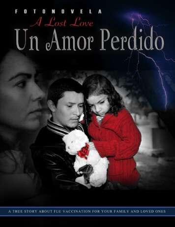
344M. B. Mitrovi}, et al.: Influence of Radiation Dose in Computed Tomography on .Nuclear Technology & Radiation Protection: Year 2017, Vol. 32, No. 4, pp. 342-349ter 7 days. Sacrifice was performed by the decapitationmethod. Immediately following sacrifice, blood samples were collected by exsanguination in order to determine the parameters of the oxidative stress in redblood cells.Dose quantitiesThe dosimetry concept in CT is well-establishedand based on the practical dose quantities: weightedCT dose index (CTDIw), volume weighted CT doseindex (CTDIvol) and dose-length product (DLP). TheCT dose index is an indication of the average dose inthe central part of a scanned region when slices arecontiguous [23]. In addition, it enables comparisonsbetween scanners and scan protocols and can be easilymeasured. All CT vendors are now required to displayCTDI and DLP values on the user interface [24].Prior to data collection, the CTDIw values wereverified by measurements, using a well-establishedprotocol [23]. Values obtained from the CT consolediffered by less than 10 % of the measured values.Data were collected in terms of CTDIw. Data on therabbits and scan protocol were also collected, including length and weight of animals, tube voltage (U),tube current and rotation time product (It), rotationtime (trot), and technique used (acquisition mode,gantry angle, collimation, and pitch).Determination of antioxidantenzyme activityWhole blood samples were taken after decapitation in heparinized vacutainers. Erythrocytes andplasma were immediately separated by centrifugation(10 min, 5000 rpm, 4 C). Aliquots of three-timeswashed erythrocytes with saline (0.9 % ww–1) werelysed in ice-cold distilled water. Antioxidant defenceenzyme activities were measured in lysate. For determination of SOD activity, haemoglobin was removedby the method of Tsuchihashi [25] and values were estimated by the method of Drabkin and Austin [26].at 230 nm. Specific activities were expressed asIU·mg–1 protein.Activity of GSH-Px was measured following thespectrophotometric method [29] based on the NADPHconsumption [i. e. NADPH oxidation by GR, at340 nm]. The reaction mixture consisted of a 50 mMpotassium phosphate buffer (pH 7.0), 1 mM EDTA,1 mM GSH, 1 mM sodium azide, 1 IUmL–1 GR frombaker's yeast, 0.2 mM NADPH and 3 mM t-butyl hydro peroxide (as a substrate and appropriate amountsof the sample). The blank did not contain a sample andthe activity was calculated after subtraction of theblank value. The rate of NADPH oxidation was monitored at 37 C on the basis of the decrease inabsorbance at 340 nm. One unit of enzyme activitywas defined as the amount of enzyme required totransform 1 nmol of substrate [min–1] under the abovedescribed assay conditions. Specific activities are expressed as IUmg–1 protein.Activity of GR was assayed by the method described previously [30] and based on NADPH oxidation concomitant with GSSG reduction. The reactionmixture consisted of 0.5 M sodium phosphate buffer(pH 7.5), 0.1 mM EDTA, 0.1 mM NADPH, 0.1 mMGSSG and appropriate amounts of samples. The rateof NADPH oxidation was monitored at 37 C after thedecrease in absorbance at 340 nm (e340 nm .mmolL–1cm–1)*. The blank did not contain GSSGand the activity was calculated after subtraction of theblank value. Specific activities are expressed asIU·mg–1 protein.Statistical analysisAll values are expressed as mean SEM. Statistical evaluation was calculated by two ways ANOVA(factors: anaesthesia (A) and time (T) as well as dose(D) and time (T)). For all comparisons, p 0.05 wasconsidered as significant.RESULTSRadiation doseEnzyme assaysActivity of SOD was determined using the adrenaline method [27] based on the increase in absorbanceat 480 nm. This method is based on the capacity ofSOD to inhibit the autoxidation of adrenaline to anadrenochrome. The reaction was carried out in a 50mmolL–1 sodium carbonate buffer, pH 10.2, and wasinitiated by the addition of 0.3 mmolL–1 adrenaline.One unit of SOD was defined as the amount of proteinrequired to halve the rate of substrate autooxidation.Activity of CAT was measured spectrophotometrically, according to Beutler [28]. CAT activity was determined by the rate of H2O2 disappearance measuredThe radiation dose in terms of CTDIw for usedscan protocols is presented in tab. 1.Gantry angulation was zero for all examinationprotocols and all image acquisitions were performedusing the axial scanning mode.The effect of anaesthesia on the antioxidantenzymes activity in rabbit erythrocytesThe results indicate that anaesthesia had no influence on the activity of antioxidant enzymes (fig. 1,*e is molar extinction coefficient
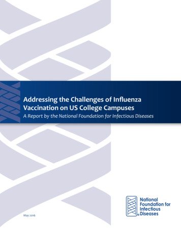
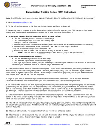
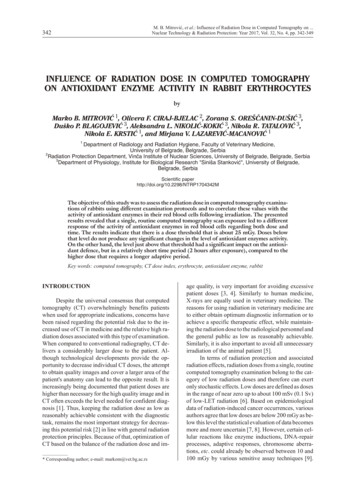
M. B. Mitrovi}, et al.: Influence of Radiation Dose in Computed Tomography on .Nuclear Technology & Radiation Protection: Year 2017, Vol. 32, No. 4, pp. 342-349345Table 1. Scan parameters and assessed CTDIw values fordifferent examination protocolsGroupsI1 and I2II1 and II2III1 and III2IV1 and IV2U [kV]110130110130It [mAs] trot [s] CTDIw [mGy]63117.963125.2105129.8105142.1Figure 3. The effect of different CTDIw values on CATactivity in rabbit erythrocytes measured 2 hours and 7days following computed tomography (a vs. b – statistically significant differences; ab vs. a or b – statisticallynon-significant differences)*n. s. – not significantFigure 1. The effect of anaesthesia on antioxidantenzyme activity in rabbit erythrocytes measured 2 hoursand 7 days after the applicationANOVA, effects of anaesthesia and days, non-significant). Furthermore, the activity of studied antioxidantenzymes in non-treated (NT) animals was similar tovalues registered in anaesthetized animals after 2hours (A1) as well on the 7th day (A2). It can be concluded that the applied anaesthesia had no influenceon the activity of antioxidant enzymes in erythrocytes.The effect of different CTDI values on theantioxidant enzymes activity in rabbiterythrocytes following computed tomographyeffects depend on the dose applied and time of the particular enzyme activity.There is no significant general trend in the levelof SOD activity in erythrocytes after the CT scan (fig.2, ANOVA no significant effect of neither doses nortime), but a very low level of significance betweengeneral groups (interaction D T, p 0.05) was found.However, post hoc Tukey's HSD test showed no significant differences between individual groups.Statistical analysis of the measurements of CATactivity showed no general trend of response after CTscan exposure (fig. 3, ANOVA, no significant effectsof treatment and time). However, there are significantdifferences between individual groups (ANOVA, interaction, p 0.001). There is a significant increase inCAT activity (p 0.001) after 2 hours of exposure of29.8 mGy compared to the 17.9 and 25.2 mGy groups.Moreover, CAT activity in that group decreased after 7days when compared to the other groups.The results showed that the applied treatmentchanged the activity of antioxidant enzymes and theFigure 2. The effect of different CTDIw values on SODactivity in rabbit erythrocytes measured 2 hours and 7days following computed tomographyFigure 4. The effect of different CTDIw values onGSH-Px activity in rabbit erythrocytes measured 2hours and 7 days following computed tomography (a vs.b – statistically significant differences; ab vs. a or b – statistically non-significant differences)
346M. B. Mitrovi}, et al.: Influence of Radiation Dose in Computed Tomography on .Nuclear Technology & Radiation Protection: Year 2017, Vol. 32, No. 4, pp. 342-349Figure 5. The effect of different CTDIw values on GRactivity in rabbit erythrocytes measured 2 hours and 7days following computed tomography (a vs. b –statistically significant differences; ab vs. a or b –statistically non-significant differences)Statistical analyses of GSH-Px activity showedthat, in general, the activity increased after the day 7th(fig. 4, ANOVA, significant day effect, p 0.001, nosignificant changes of dose effect). However, 2 hoursafter exposure GSH-Px activity in the group of 29.8mGy was significantly lower than the control. There isno particular statistically significant individual difference when comparing 2 hours vs. the 7th day, but eachGSH-Px activity on the 7th day was higher to some extent compared to its 2 hours counterpart except controls.Statistical analyses of GR activity showed thatGR activity increased after 2 hours upon exposure to29.8 mGy compared to other groups, but after 7 days,decreased to a level not significantly different to controls and 17.9 and 25.2 mGy groups (fig. 5, ANOVAinteraction, p 0.001). On the other hand, there was anincrease of GR activity in the group exposed to 42.1mGy after 7 days compared to its 2 hours counterpartsand other groups on the 7th day (ANOVA significantdose, p 0.05, and interaction effect, p 0.001).DISCUSSIONNumerous studies show that the radiation dosereceived during CT examination may have adverse effects on living subjects and the International Agencyfor Research on Cancer (IARC) has classified X-raysas carcinogenic to humans on the basis of sufficientevidence for carcinogenicity [31]. The InternationalCommission on Radiological Protection (ICRP) in apublication from 1990 also suggested that a low levelof radiation exposure could result in cancer [32, 33].Contrary to this, some authors suggested that below10 mSv, which is a dose range relevant to radiography,no direct epidemiological data support increased cancer risk. However, this does not mean that this risk isnot present, as even large epidemiological studieswould not have the statistical power to detect increased risk, if present, at a low radiation dose [34].Despite some controversy over the excess cancer riskof low-dose radiation, the linear no-threshold theory iswidely used because an alternative method for assessing the potential risks of low-dose radiation is lacking.In addition, the epidemiological data directly suggestincreased cancer risk in the 10 mSv to 100 mSv range,which is relevant to many CT studies [35]. X-rays andg-rays have also been tested for carcinogenicity at various doses and under various conditions in a range ofanimal species. In adult animals, the incidences of leukaemia and of mammary, lung, and thyroid tumourswere increased in a dose-dependent manner with bothtypes of radiation. Prenatal exposure also gave rise toincreased incidences of various types of tumours [31].Low radiation doses lead to cellular reactions [9] triggering oxidative stress, and changes in the antioxidantenzymes activity were demonstrable when the applieddose ranged from 0,.1 mGy [13, 14] to 50 mGy [12],which corresponds to the doses received during the CTexamination [32].The presented results originate from the firstever performed study on radiation effects during CTexaminations of rabbits. CT scan exposure of rabbitsled to a different response of the activity of antioxidantenzymes regarding both dose and time. The resultsindicate that there is a dose threshold that is about25 mGy despite the fact that changes in the antioxidantenzymes activity were demonstrable when the applieddose ranged from 0.1 mGy [13, 14] to 1 mGy [13].Doses below 25 mGy do not produce any significant changes in the level of antioxidant enzymes activity compared to controls. Higher doses had an impacton antioxidant enzymes, but different components areinvolved.A dose of 29.8 mGy led to changes of CAT andGR activities after 2 hours that decreased to values notsignificantly different compared to the controls andlower dose exposure groups after 7 days. The samegroup (exposed to 29.8 mGy) had lower GSH-Px activity after 2 hours of exposure, but not different compared to the other groups after 7 days. Both CAT andGSH-Px decompose hydrogen peroxide (H2O2), butwith different enzyme kinetics i. e. CAT operates at ahigher H2O2 concentration than GSH-Px. This indicates that in 29.8 mGy, erythrocytes were exposed to ahigh impact of H2O2 after exposure to the CT scan.However, after 7 days the levels of antioxidant enzymes in that group were similar to controls, exceptCAT that was lower compared to other groups. Itseems that GSH-Px activity after 7 days was efficientto in establishing a normal oxidative status and loweramount of H2O2 in this, as well as in other groups.Namely, ANOVA showed that GSH-Px was elevatedto a certain extent in the all the examined groups (except the control), suggesting this enzyme as generallyprotective after 7 days of CT scan exposure.
M. B. Mitrovi}, et al.: Influence of Radiation Dose in Computed Tomography on .Nuclear Technology & Radiation Protection: Year 2017, Vol. 32, No. 4, pp. 342-349On the other hand, exposure to 42 mGy led to theincrease of GR activity after 7 days. This means thatthe reduction of oxidized glutathione was accelerated,suggesting the glutathione mediated way of antioxidant protection as dominant in the group that receivedthe highest dose of irradiation.The presented results suggest that CT exposureled in general to the activation of a protection againsthydrogen peroxide production, which depends on irradiation doses as a determinant of hydrogen peroxideproduction. Moreover, the level just above that threshold had significant impact on the antioxidant defence,but changes were time dependent (2 hours vs. 7 daysafter exposure), suggesting that the higher dose requires a longer adaptive period.Although erythrocytes are not very radiosensitive[36], low radiation doses can trigger the oxidative stressin these cells, resulting in elevated activity of the mainantioxidant enzymes [10, 11]. According to the literaturedata, changes in the antioxidant enzymes activity weredemonstrable when the applied dose was 50 mGy [12] orless, which is confirmed by our results. Despite the factthat the results related to the effect of low-dose radiationon the activity of oxidative enzymes in erythrocytes arepresented in the available literature, they mostly relate tochronic irradiation.Several studies were conducted with the aim ofclarifying the oxidative stress status in radiology workersbut their results are quite contradictory. Recently, someauthors investigated changes of antioxidant enzymes activity in erythrocytes of hospital staff occupationally exposed to ionizing radiation with radiation doses rangingbetween 0.10 and 3.8 mGy per month [37]. Their resultsshowed that activities of erythrocyte CuZn-SOD andSe-GPx, observed for the exposed group, were significantly higher than in the control group. It was also indicated that the activities of antioxidant enzymes were significantly increased in radiation-exposed subjectscompared with control individuals. The activities oferythrocyte CAT levels in the exposed group were foundto be significantly lower than in the control group. Theauthors assume that these findings may be explained interms of low-dose radiation-induced hormesis. According to other authors [17], erythrocyte CAT activities areslightly increased in radiographers when compared to thecontrols.Long-term exposure to low radiation doses inworkers operating X-ray equipment reduced antioxidant enzyme activities (SOD, CAT, and GPx) in erythrocytes as compared to controls [38]. These resultssuggest that long-term exposure to a low radiationdose in workers operating X-ray equipment diminishes their antioxidant defence.Blood samples obtained from the rabbits, beforeand after X-rays irradiation, that was continued for aweek in daily sessions of 100 rad/min until a total doseof 550 rad was reached, were analyzed in order to determine the differences in the content of reduced347glutathione (GSH), and glutathione peroxidase activity (GP). In blood samples that were taken followingirradiation, the activity of GP was increased, whereasthe levels of GSH were decreased [39].As previously stated, there are no available literature data regarding activity of antioxidant enzymes inrabbit erythrocytes following acute low dose X-ray irradiation during CT examination.CONCLUSIONThe presented study investigated the influenceof the radiation dose during CT examination to antioxidant enzyme activity in rabbit erythrocytes. The obtained results show that the antioxidant enzyme response follows dose mediated ROS production.According to this, the highest dose had a lag period foradaptive response compared to the lower one, and athe dose threshold is about 25 mGy. The level justabove that threshold, had significant impact on the antioxidant defence, but in a relatively short time period(2 hours after exposure), compared to the highest dosethat requires a longer adaptive period (7 days). Furthermore, antioxidant protection is established by different antioxidant enzymes and depends on the level ofirradiation.ACKNOWLEDGEMENTSThis work was funded by the Ministry of Education, Science and Technological Development, Republic of Serbia, grant 173014 „Molecular mechanisms of redox signalling in homeostasis: adaptationand ra, W. E., et al., Patient Doses in CT Examinations in 18 Countries: Initial Results from International Atomic Energy Agency Project, Radiation Protection Dosimetry, 136 (2009), 2, pp. 118-126Yu, L., et al., Radiation Dose Reduction in ComputedTomography: Techniques and Future Perspective, Imaging Med., 1 (2009), 1, pp. 65-84Bauhs, J. A., et al., CT Dosimetry: Comparison of Measurement Techniques and Devices, Radiographics, 28(2008), 1, pp. 245-253Rehani, M. M., Limitations of Diagnostic ReferenceLevel (DRL) and Introduction of Acceptable QualityDose (AQD), Br. J. Radiol., 88 (2015), 1045, 20140344***, NCRP – National Council on RadiationProtesction and Measurements, Radiation Protectionin Veterinary Medicine, NCRP Report No. 148, 2004***, BEIR – Committee on the Biological Effects ofIonizing Radiation VII, Phase 2, Health Risks fromExposure to Low Levels of Ionizing Radiation, 2006***, NCRP – National Council on Radiation Protection and Measurements, Influence of Dose and ItsDistribution in Time on Dose-Response Relationshipfor Low LET Radiations, NCRP Report No. 64, 1980
20][21][22][23][24][25][26][27]M. B. Mitrovi}, et al.: Influence of Radiation Dose in Computed Tomography on .Nuclear Technology & Radiation Protection: Year 2017, Vol. 32, No. 4, pp. 342-349***, UNSCEAR – The United Nations Scientific Committee on the Effects of Atomic Radiation, Sources andEffects of Ionizing Radiation, New York, 1994Koteles, G. J., The Lowe Dose Dilemma, CEJOEM, 4(1998), 2, pp. 103-113Buonanno, M., et al., Long-Term Consequence of Radiation-Induced Bystander Effects Depend on Radiation Quality and Dose and Correlate with OxidativeStress, Radiat. Res., 175 (2011), 4, pp. 405-415Azzam, E. I., et al., Ionizing Radiation-Induced Metabolic Oxidative Stress and Prolonged Cell Injury,Cancer Lett., 327 (2012), 1-2, pp. 48-60Emerit, I., et al., Oxidative Stress and Low-Dose Irradiation, in: Low-Doses of Ionizing Radiation: Biological Effects and Regulatory Control, InternationalAtomic Energy Agency, IAEA-TECDOC-976, Vienna, 1997, pp. 1-5Vicker, M. G., et al., Ionizing Radiation at Low DosesInduces Inflammatory Reactions in Human Blood,Radiat. Res., 128 (1991), 3, pp. 251-257Indo, H. P., et al., MnSOD Downregulation Inducedby Extremely Low 0.1 mGy Single and FractionatedX-Rays and Microgravity Treatment in HumanNeuroblastoma Cell Line, NB-1, J. Clin. Biochem.Nutr., 57 (2015), 2, pp. 98-104Yamaoka, K., Applicable Possibility for Treatment ofReactive Oxygen Species-Related Diseases, J. Clin.Biochem. Nutr., 39 (2006), pp. 114-133Focea, R., et al., Low Dose X-Ray Effects on CatalaseActivity in Animal Tissue, J. Phys. Conf. Ser., 398(2012), 11, pp. 1-6Puthran, S. S., et al., Oxidative Stress and low DoseIonizing Radiation, Indian J. Physiol. Pharmacol., 53(2009), 2, pp. 181-184Labruyere, J., Schwarz, T., CT and MRI in VeterinaryPatients: an Update on Recent Advances, BMJ, 35(2013), pp. 546-563Lipmana, N. S., et al., Reversal of Ketamine/XylazineAnaesthesia in the Rabbit with Yohimbine, Lab.Anim. Sci., 37 (1987), 4, pp. 474-477Gnanasekar, R., Vijayalakshmi, R., Response to Oxidative Stress during Surgery under Ketamine/Propofol Anaesthesia in Ace
IN FLU ENCE OF RA DI ATION DOSE IN COM PUTED TO MOG RA PHY ON AN TI OX I DANT EN ZYME AC TIV ITY IN RAB BIT ERYTH RO CYTES by Marko B. MITROVI] 1, Olivera F. CIRAJ-BJELAC 2, Zorana S. OREŠ ANIN-DUŠI] 3, Duško P. BLAGOJEVI] 3, Aleksandra L. NIKOLI]-KOKI] 3, Nikola R. TATALOVI] 3, Nikola E. KRSTI] 1, and Mirjana V. LAZAREVI]-MACANOVI] 1 1 De part m ent of Ra di ol ogy and Ra di a tion Hy .