Transcription
Contents1.AIIMS November 2017 QuestionsExplanations2.AIIMS May 2017 QuestionsExplanations3.AIIMS November 2016 QuestionsExplanations4.AIIMS May 2016 Questions5.AIIMS November 2015 QuestionsExplanations6.AIIMS May 2015 QuestionsExplanations7.AIIMS November 2014 QuestionsExplanations8.AIIMS May 2014 1051273128914171433PREVIEWExplanations117
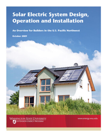
AIIMS NOVEMBER 2017a. Unmyelinatedb. Connects to spinal nervesc. Preganglionicd. Present medial to the white ramus communicans14. Which of the following junctional complexes are notseen in the marked region of the given slide?Multiple Choice Questionsa. Striate cortexb. Orbital cortexc. Hippocampusd. Dentate nucleus5. Development the of heart is from which of the followingmarked structure?a. Gap junction (communicating junctions)b. Zonula occludens (tight junction)c. Fascia adherens (adhering junction)d. Macula adherens (desmosomes)15. Which of the following layer contains abundantdesmosomes?a. Ab. Bc. Cd. D16. Which of the following refers to the lateral semicircularcanal in the specimen of cortical mastoidectomy withposterior tympanostomy?a. Growth hormoneb. Cortisolc. Estrogend. Insulin21. Feed forward control system is employed during theregulation of:a. Blood volumeb. pHc. Temperatured. Blood pressure22. Efferent arteriolar constriction causes all of thefollowing except:a. Decrease in GFRb. Decreasesrenalbloodflowc. Decreases oncotic pressure in peritubular capillariesd. Increases hydrostatic pressure in glomerular capillaries23. Difference in trajectory between inspiratory loop andthe expiratory loop in the curve is due to:a. ab. bc. cd. d11. Identify the marked nerve:a. ac. cb.d.bdPHYSIOLOGYa. ab. bc. cd. d6. Which of the following sinus grows till early adulthood?a. Maxillaryb. Ethmoidalc. Frontald. Sphenoid7. What is the shape of the trapezius muscle?a. Quadrangularb. Triangularc. Strapd. Fusiforma. Abducent nerveb. Spinal accessory nervec. Hypoglossal nerved. Labyrinthine artery12. Whatistherootvalueofcremastericreflex?a. L1-L2b. L2-L3c. L4-L5d. S1-S213. All of the following are true about grey communicansexcept:17. Slow waves are generated by:a. Myenteric neuronsb. Smooth musclec. Interstitial cells of Cajald. Parasympathetic neurons18. Reflex responsible fortachycardia during right atrialdistension is:a. Bezold-Jarischreflex b. Bainbridgereflexc. Cushingreflexd. J-reflexa. Difference in alveolar pressure during inspiration andexpirationb. Difference in concentration of surfactant duringinspiration and expirationc. Difference in airway resistance during inspiration andexpirationd. Inspiration is active and expiration is passive24. Absolute refractory period is due to:a. Opening of calcium channelsb. Closure of potassium channelsc. Closure of active gates of sodium channeld. Closure of inactive gates of sodium channelPREVIEWa. ab. bc. cd. d9. Dense irregularcollagen fibres are found inwhich ofthe following?a. Tendonb. Ligamentc. Dermisd. Lamina propria10. Holocrine cells in the given slide are:19. Identify the stage of sleep from the given picture:a. Stage I NREMb. Stage II NREMc. Stage III NREMd. REM20. Identify the hormone from the picture:8. Which of the marked muscle helps in the opening of jaw?AIIMS ESSENCEANATOMY1. Which of following is not the branch of externalcarotid artery in Kiesselbach’s plexus?a. Anterior and posterior ethmoidalb. Sphenopalatine arteryc. Greater palatine arteryd. Septal branch of superior labial artery2. Structure passing through the central tendon of thediaphragm:a. Esophagusb. Aortac. IVCd. Sympathetic chain3. Talocalcaneonavicular joint is what type of joint?a. Saddleb. Hingec. Ellipsoidd. Ball and socket4. Marked structure in the given image connects whichof the following?AIIMS ESSENCE2
AIIMS ESSENCE182. Ans. c. IVC (Ref: Gray’s 41/e p899, 40/e p1008)Inferior vena cava (IVC) passes through central tendon of diaphragm.Explanations“The vena caval aperture, the highest of the three large openings, lies at about the level of the disc between the eighthand ninth thoracic vertebrae. It is quadrilateral, and located at the junction of the right leaf with the central area of thetendon, and so its margins are aponeurotic. It is traversed by the inferior vena cava, which adheres to the margin of theopening, and by some branches of the right phrenic nerve.”-Gray’s 40/e p1008ANATOMY1. Ans. a. Anterior and posterior ethmoidal (Ref: Gray’s 41/e p563, 40/e p554; Dhingra 7/e p197)Anterior and posterior ethmoidal arteries are branches of ophthalmic artery, which in turn is a branch of internal carotidartery. Sphenopalatine and greater palatine arteries are branches of maxillary artery, which in turn is branch of externalcarotid artery. Superior labial artery is the branch of facial artery, which in turn is branch of external carotid artery.Fig. 2: Openings in diaphragmOpeningVertebral levelPart of diaphragmPassing structureVena CavalT8QCentral tendonQInferior vena cavaQRight phrenic nerveQEsophagealT10QMuscular portion derived from rightcrusQEsophagusQEsophageal branch of left gastric arteryGastric or vagus nerveQAorticT12QOsseoaponeurotic between right &lateral crusQAortaQThoracic ductQAzygous veinQFig. 1: Blood supply of nasal septumSmall Openings in the DiaphragmBlood Supply of Nasal SeptumInternal Carotid SystemExternal Carotid Systeml Anterior & posterior ethmoidal arteryQ l Sphenopalatine arteryQ (branch of maxillary artery Q) gives(Branch of ophthalmic arteryQ)nasopalatine & posterior medial nasal branchesQ.l Septal branch of greater palatine artery Q (branch of maxillaryarteryQ)l Septal branch of superior labial arteryQ (branch of facial arteryQ).OpeningLocationPassing structureMedial lumbocostal ArchBehind medial arcuate ligamentSympathetic chainQLateral lumbocostal archBehind arcuate ligamentSubcostal nerve & vesselsQLarry’s space / Foramen of MorgagniBetween xiphoid & costal origin of Superior epigastric vesselsQ somediaphragmlymphaticsPREVIEWAIIMS ESSENCE“Little’s area: It is situated in the anterior inferior part of nasal septum, just above the vestibule. Four arteries, anteriorethmoidal, septal branch of superior labial, septal branch of sphenopalatine artery and the greater palatine, anastomosehere to form a vascular plexus called “Kiesselbach’s plexus”. This area is exposed to the drying effect of inspiratorycurrent and to finger nail trauma, and is the usual site for epistaxis in children and young adults.”- Dhingra 7/e p197
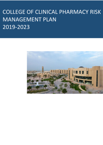
AIIMS MAY 2017AIIMS ESSENCE2186. A patient came with inability to move his 4th and5th digit, cannot hold a pen and he was not able tohold a piece of paper between his fingers. Which ofthe following site given below is the probable cause ofinjury to the nerve in the question?Multiple Choice Questionsa. Alzheimer’s diseasec. Paralysis agitansb. Huntington’s choread. Dementia9. The muscle labeled in the following cross section isresponsible for which movement of the jaw?ANATOMY1. The following coronal section of the abdomen isshowing the relations of epiploic foramen. Which ofthe following structure forms its superior boundary asindicated in the figure below?b. Bd. Db. Duodenumd. Caudate lobe of livera. Ac. Cb. Bd. D7. A 5 years old child presented with absence of thymus,hypoparathyroidism and tetany. Which of the following marked area is defective in this case?a. Ac. Cb. Bd. D8. An area has been marked in the coronal section ofthe brain below. Defect in this area will lead to whatpathology?a. Ac. Cb. Bd. Da. Ac. Cb. Elevationd. Depression10. The arrow marked structure in the given pictureconnects, which of the following structure?b. Bd. D5. The following picture shows various foramina at theskull base. Mandibular nerve passes through which ofthe following foramen?3. Nucleus pulposus of intervertebral disc is a derivativeof which of the following germ layers?a. Protractionc. RetractionPREVIEWa. Lesser omentumc. Inferior vena cava2. The following is the representation of a cervicalvertebra. Which part lies in relation with the thirdpart of vertebral artery?a. Ac. CAIIMS ESSENCEa. Ac. C4. Which of the following part of scapula can be palpatedin the infraclavicular fossa?b. Bd. Da. Hippocampusc. Mammillary bodiesb. Amygdalad. Insular cortex11. The muscle marked in diagram is supplied by thenerve, whose nucleus is situated at the level of:
242AIIMS ESSENCE2. Ans. c. C (Ref: Gray’s 41/e p283)Third part of vertebral artery, which passes over the posterior aspect of atlas vertebra is represented by ‘C’.ExplanationsABCDANATOMYTransverse foramenAnterior arch and articular facet for densGroove for vertebral arteryArticular surface for occipital condyle1. Ans. d. Caudate lobe of liver (Ref: Gray’s 41/e p1107)Caudate lobe of liver forms the superior boundary of epiploic foramen.l Epiploic foramen is the space connecting the greater sac & lesser sac, lying between portal vein & IVC.“The vertebral artery arises from the superoposterior aspect of the first part of the subclavian artery. It passes throughthe foramina in the transverse processes of all of the cervical vertebrae except the seventh, curves medially behind thelateral mass of the atlas and enters the cranium via the foramen magnum. At the lower pontine border it joins its fellowto form the basilar artery. Occasionally it may enter the cervical vertebral column via the fourth, fifth or seventh cervicalvertebra.” —Gray’s 41/e p283“The third part issues medial to rectus capitis lateralis, and curves backwards and medially behind the lateral mass ofthe atlas, with the first cervical ventral spinal ramus lying on its medial side. In this position it lies in a groove on theupper surface of the posterior arch of the atlas, and it enters the vertebral canal below the inferior border of the posterioratlanto-occipital membrane. This part of the artery, covered by semispinalis capitis, lies in the suboccipital triangle.”—Gray’s 41/e p283Boundaries of Epiploic ForamenAnteriorl Hepatoduodenal ligament & portal triad (portal vein, hepatic artery, CBD; cysticduct also present in free edge of lesser omentumQQPosteriorl IVC & right suprarenal glandSuperiorl Caudate process of caudate lobeQ of liver & inferior layer of coronary ligamentInferiorl 1st part of duodenum & transverse part of hepatic arteryQLeft laterall Splenorenal & gastrosplenic ligamentQPringle Maneuver (Total Inflow Occlusion)“The vertebral artery ascends in the neck through the foramina in the transverse processes of the upper six cervicalvertebrae. It passes medially above the posterior arch of the atlas and then ascends through the foramen magnum intothe skull. On reaching the anterior surface of the medulla oblongata of the brain at the level of the lower border of the pons,it joins the vessel of the opposite side to form the basilar artery.” —Snells 9/e p599Vertebral Arteryl Vertebral artery ascends in the neck through foramina in the transverse processes of upper six cervical vertebraeQ.l It passes medially above the posterior arch of atlas & then ascends through foramen magnum into skullQ.l On reaching the anterior surface of medulla oblongata of brain at the level of lower border of pons , it joins the vesselof the opposite side to form the basilar arteryQ.l Total clamping of hepatic pedicle, by placing an atraumatic clamp across the foramen of WinslowQ.l Appropriate-sized vascular clamp or loop snare easily controlshemorrhage from portal vein (effectively) & hepatic arteriesQ.l It doesn’t control bleeding from IVC & hepatic veinsQ.l Inflow occlusion durations of up to 30 minutes can be tolerated safely in cirrhotic livers and possibly up to 60 minutesin early disease.l If prolonged occlusion is required, intermittent clamping can be used with repeated clampings of 10-20 minutesduration, each followed by 5 minutes declamping.Parts of Vertebral ArteryCervical partFirst partl Extends from origin to foramen transversarium of C6 vertebraQ. This partVertebral partSuboccipital partSecond partThird partllIntracranial partFourth partlllies in the scalenovertebral triangle.Lies within foramen transversaria of upper six cervical vertebraeExtends from foramen transversarium of C1 vertebra to the foramenmagnum of skullQ.This part lies within the suboccipital triangleQ.Extends from foramen magnum to the lower border of ponsQ.PREVIEWEpiploic Foramen (Foramen of Winslow, aditus to the lesser sac)AIIMS ESSENCE“The epiploic foramen (foramen of Winslow, aditus to the lesser sac), is a short, vertical slit, usually 3 cm in height inadults, in the upper part of the right border of the lesser sac. It leads into the greater sac. The hepatoduodenal ligament,which is formed by the thickened right edge of the lesser omentum extending from the flexure between the first and secondparts of the duodenum, forms the anterior margin of the foramen. The anterior border contains the common bile duct (onthe right), portal vein (posteriorly) and hepatic artery (on the left) between its two layers. Superiorly the peritoneum of theposterior layer of the hepatoduodenal ligament runs over the caudate lobe of the liver which forms the roof of the epiploicforamen.”-Gray’ 41/e p1107
21.5 cmAIIMS ESSENCE 2017–2014: Volume 1 4th edition YOP: 2018 Pages: 1552By Pritesh Singh Color: ISBN: 9789352704040Available onEduLanche.comPREVIEWA4 size27 cm
AIIMS MAY 2017 AIIMS ESSENCE 218 AIIMS ESSENCE 6. A patient came with inability to move his 4th and 5th digit, cannot hold a pen and he was not able to hold a piece of paper between his fingers. Which of the following site given below is the probable cause of injury to the nerve in the question? a. A b. B c. C d. D 7. A 5 years old child .