Transcription
Vol 443 12 October 2006 doi:10.1038/nature05128LETTERSTransforming the architecture of compound eyesAndrew C. Zelhof1, Robert W. Hardy1, Ann Becker1 & Charles S. Zuker1Eyes differ markedly in the animal kingdom, and are an extremeexample of the evolution of multiple anatomical solutions to lightdetection and image formation. A salient feature of all photoreceptor cells is the presence of a specialized compartment (discouter segments in vertebrates, and microvillar rhabdomeres ininsects), whose primary role is to accommodate the millions oflight receptor molecules required for efficient photon collection.In insects, compound eyes can have very different inner architectures1–3. Fruitflies and houseflies have an open rhabdom system, inwhich the seven rhabdomeres of each ommatidium are separatedfrom each other and function as independent light guides. Incontrast, bees and various mosquitoes and beetle species have aclosed system, in which rhabdomeres within each ommatidiumare fused to each other, thus sharing the same visual axis. Tounderstand the transition between open and closed rhabdomsystems, we isolated and characterized the role of Drosophilagenes involved in rhabdomere assembly. Here we show thatSpacemaker, a secreted protein expressed only in the eyes ofinsects with open rhabdom systems, acts together with Promininand the cell adhesion molecule Chaoptin to choreograph thepartitioning of rhabdomeres into an open system. Furthermore,the complete loss of spacemaker (spam) converts an open rhabdomsystem to a closed one, whereas its targeted expression to photoreceptors of a closed system markedly reorganizes the architectureof the compound eyes to resemble an open system. Our resultsprovide a molecular atlas for the construction of microvillarassemblies and illustrate the critical effect of differences in asingle structural protein in morphogenesis.During ommatidium biogenesis the apical membranes of differentphotoreceptor cells separate from one another and concomitantlyproduce the rhadomeric structure needed for housing the phototransduction machinery. This process involves the precise coordination of membrane–membrane adhesion events both within andbetween rhabdomeres, and culminates in the production of about60,000 microvilli per cell, each 1–2 mm in length and 50 nm indiameter4,5. The generation of the inter-rhabdomeral space (IRS;Fig. 1), by which rhabdomeres of the photoreceptor neurons partition from each other, is an essential event in the transition ofcompound eyes from a closed to an open system. This evolutionarychange resulted in a considerable improvement in angular sensitivity,thus allowing the detection of smaller moving objects. We screenedadult viable ethylmethane sulphonate-mutagenized Drosophila lines6for defects in rhabdomere structure and topology by examiningdeep-pseudopupil phenotypes 7,8 and performing an electronmicroscopic ultrastructural analysis of pupal and adult eyes incandidate lines. Only 2 of 40 different complementation groups,spacemaker (spam) and prominin (prom), had eyes with apparentlynormal microvilli but showing a striking failure of the rhabdomeres to separate from each other, including a marked loss ofIRS, irregular rhabdomere morphology and improper rhabdomeralcontacts (Fig. 1a–d). Interestingly, loss-of-function mutations inspam completely eliminated the IRS, thus generating ommatidia1resembling the compound eyes of animals with closed rhabdomsystems (compare Fig. 1a–c).To understand how spam and prom function, both mutants weremapped and the candidate genes were cloned and tested for rescue ofthe mutant phenotypes by P-element germline transformation (Supplementary Fig. 1c). spam encodes a 2,165-amino-acid polypeptidecontaining several protein motifs commonly found in extracellularmolecules: an amino terminus consisting of seven epidermal growthfactor (EGF)-like repeats, a linker region containing multiple sites forglycosaminoglycan addition, and a carboxy terminus including fouralternating repeats of EGF-like and Laminin G domains (Supplementary Fig. 1a). prom encodes a 910-amino-acid Prominin (Prom)-likemolecule (Supplementary Fig. 1b), a family of evolutionarily conservedtransmembrane proteins often associated with microvilli9 but ofunknown function.What is the role of spam? Sequence analysis of spam indicates thatit might function as an extracellular protein (Supplementary Fig. 1).If Spacemaker (Spam) is a secreted molecule, it should localize to theIRS. Indeed, immunocytochemical studies showed that Spam isselectively localized to the IRS (Fig. 1g, h). However, mosaic analysisreveals that even though Spam is capable of diffusing throughout theommatidium, near wild-type levels of the protein are still required toensure complete rhabdomere separation (Supplementary Fig. 3b,and data not shown). To determine when spam function is required,we placed its transcriptional unit under the control of an inducibleheat-shock promoter (UAS-spacemaker and heat shock (HS)-GAL4)and subjected mutant animals to temperature shifts at various timesduring development. Our results demonstrated that spam expression at36–64 h after puparium formation (APF), a window of time coincidentwith the initiation of rhabdomere biogenesis, is sufficient and necessaryfor complete rescue of the phenotype (Supplementary Fig. 1c). Lateexpression (about 72 h APF), using either HS-GAL4 or a lateexpressing rhodopsin promoter (Rh1-GAL4), leads only to a partialrescue: Spam is still secreted into the IRS and the apical stalkmembranes (the base of the rhabdomeres) are still capable of separating, but the rhabdomeres themselves remain fused in the centre ofthe ommatidium (Supplementary Fig. 1c). Notably, this phenotypeclosely resembles that of prom mutants (Fig. 1d), suggesting that theymight both participate in a common morphogenesis programme.If prom and spam function in the same pathway, we would expectgenetic interactions between these two loci. Indeed, although bothspam and prom are recessive genes, the spam prom double heterozygote shows a failure of the rhabdomeres to separate and the loss of acontinuous IRS (Fig. 1e); these results indicate that the two geneproducts probably act together in the biogenesis of the IRS (seebelow). To explore the function of Prom, we first examined its spatialand temporal expression pattern. Like Spam, Prom is present at thebeginning of rhabdomere biogenesis (48 h APF), when it decoratesthe entire photoreceptor apical surface (Supplementary Fig. 2a). Bythe time of eclosion, however, Prom is selectively localized to the stalkmembrane and the tips of the microvilli; this is best illustrated bysimultaneous labelling with Chaoptin, a photoreceptor adhesionHoward Hughes Medical Institute and Departments of Neurobiology and Neurosciences, University of California at San Diego, La Jolla, California 92093-0649, USA.696 2006 Nature Publishing Group
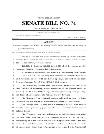
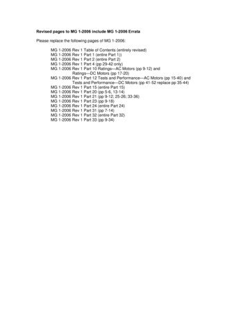
LETTERSNATURE Vol 443 12 October 2006protein expressed throughout the perimeter of microvilli (Supplementary Fig. 2c, f, i). Given that Spam is an IRS-secreted moleculeand Prom is a rhabdomere integral membrane protein, we examinedwhether these two proteins interact. When Drosophila tissue-culturecells are transfected with spam, the protein is secreted into themedium. However, if the same cells are instead transfected togetherwith prom, Spam then localizes selectively to the exterior surface ofthe plasma membrane (Supplementary Fig. 4b). These results indicate that Prom might function as a binding partner for Spam. Todemonstrate that Prom in fact recruits extracellular Spam to theplasma membrane, we performed a mixing experiment: RFP-labelledcells expressing spam were incubated with green fluorescent protein(GFP)-labelled cells transfected with prom. As proposed, secreted Spamnow specifically localizes to the surface of Prom-expressing cells(Supplementary Fig. 4f). Taken together, these findings substantiateProm as a candidate Spam receptor.Figure 1 spam and prom mutants have severe defects in IRS formation.a–f, Electron micrographs showing cross-sections through ommatidia ofDrosophila melanogaster (a), Apis mellifera (b), spam 1 (c), prom 1 (d),spam1 þ/þ prom 1 (e) and flies overexpressing spam (GMR-GAL4,UAS-spam) (f). Asterisks and IRS mark the inter-rhabdomeral space andnumbers label each rhabdomere. Note the absence of IRS in spam and prommutants, and the incomplete rhabdomeric separation in thetransheterozygote (arrow). Scale bar, 1 mm. g, Spam (magenta) expression inwild-type ommatidia; h, spam 1-null ommatidia. Note Spam localization tothe IRS surrounding each of the rhabdomeres (microvilli are labelled greenwith phalloidin).What is the function of prom and spam in orchestrating openrhabdomere development? Microvilli are interconnected by a network of homophilic interactions that ensures their tight and regularpacking within rhabdomeres10,11. However, such an arrangementwould also render microvilli vulnerable to inter-rhabdomericadhesion. We suggest that secretion of Spam into the IRS forces theseparation of the stalk membranes, pushing the rhabdomeres apart,and that the recruitment of Spam to the microvillar surface by thebinding of Prom prevents inter-rhabdomere adhesion. This model isconsistent with the phenotypes of spam and prom mutants, andmakes three significant predictions: first, overexpression of spamshould increase the volume of the IRS by pushing rhabdomeresfurther away from each other; second, in mosaic ommatidia containing prominin mutant cells there should be a significant loss of Spambinding and accumulation surrounding the mutant rhabdomeres;and third, the fused rhabdomeres of prom mutants should be rescuedby independently reducing or abolishing inter-rhabdomere interactions. As predicted, overexpression of spam markedly expands theIRS of wild-type photoreceptors (Fig. 1f), and the presence of Promin mosaic ommatidia promotes the recruitment of Spam selectivelyaround wild-type rhabdomeres (Supplementary Fig. 3c, d). To alterinter-rhabdomere adhesion, we first needed to determine whatmolecule was responsible for the inappropriate fusion of rhabdomeres in prom and spam mutants. We reasoned that Chaoptin, aphotoreceptor-specific glycosylphosphatidylinositol-linked membrane protein expressed very early during photoreceptor morphogenesis and required for crosslinking microvilli by means ofhomophilic interactions, might be a strong candidate10–12. We recognized that the early expression of Chaoptin would ensure the properpacking of microvilli within rhabdomeres and that the subsequentexpression of Spam and Prom would guarantee that individualrhabdomeres remain separated in open rhabdom systems. Indeed,removing just one copy of the gene encoding Chaoptin (chp) in aprom mutant background (prom/prom;chp/þ) is sufficient for aFigure 2 Prom and Spam cooperate to antagonize the adhesive force ofChaoptin. a–c, Electron micrographs showing cross-sections throughprom/prom (a), prom/prom;chp/þ (b) and prom spam/prom þ;chp/þ (c)ommatidia. d, Early expression of Spam produces severely splitrhabdomeres. Asterisks denote the IRS. Scale bar, 1 mm. See SupplementaryFig. 5 for a quantitative analysis of the data. 2006 Nature Publishing Group697
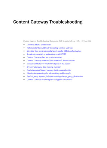
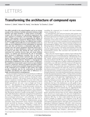
LETTERSNATURE Vol 443 12 October 2006Figure 3 spam is expressed only in insect eyes with an open rhabdomeresystem. a–f, Electron micrographs showing ommatidia of Drosophilamelanogaster (Dm; a), Musca domestica (Md; b), Toxorhynchitesamboinenesis (Ta; c), Apis mellifera (Am; d), Tribolium castaneum (Tc; e)and Anopheles gambiae (Ag; f). Scale bars, 1 mm. g, h, RT–PCR reactionsexamining the expression of spacemaker, prominin, chaoptin and eye Gbetacontrol mRNA in the pupal visual system and bodies of the various species.Multiple Spam-specific primers were designed for each species after thecloning of the corresponding homologues.drastic suppression of the prom mutant phenotype (Fig. 2a, b andSupplementary Fig. 5). These data demonstrate that the capacity ofSpam to push rhabdomeres apart is very sensitive to the amount ofadhesive force available (for example Chaoptin). Given these results,we anticipated that further reducing the levels of spam in thisprom/prom;chp/þ genetic background (that is, bringing the ratio ofspam to chp back to normal levels) should revert the ‘rescuedphenotype’ to the mutant state. As proposed, the rhabdomeres arenow stuck together (Fig. 2c and Supplementary Fig. 5). Thus, in thisgenetic background, there is sufficient Spam to partition the stalkmembranes but not enough to overcome the Chaoptin-dependentadhesiveness between adjacent rhabdomeres. A final prediction ofthese findings is that early expression of Prom and Spam shouldresult in the development of broken-up rhabdomeres, probably asthe Spam/Prom complex outcompetes the adhesive force linkingmicrovilli. The data shown in Fig. 2d soundly validate this postulate.Together, these results demonstrate that Spam, Prom and Chaoptinorchestrate the assembly of microvilli, ensure the structural integrityand the partitioning of rhabdomeres, and guarantee the constructionof an open rhabdom system.Are these molecules responsible for the transformation betweenclosed and open rhabdom systems? To examine this possibility weinvestigated the expression of Spam, Prom and Chaoptin homologuesin other insect species containing open or closed rhabdoms (Fig. 3a–f).First, analysis of Anopheles gambiae eyes, a closed system, showed thatboth Prom and Chaoptin are present but Spam is absent from pupaleyes, suggesting that Spam might be the critical component forpartitioning rhabdomeres (Fig. 3g). To lend further credibility to ourhypothesis, we isolated and examined spam expression in severalother species, two with open rhabdomere systems (the houseflyMusca domestica and the mosquito Toxorhynchites amboinensis)and two with closed ones (the honeybee Apis mellifera and theflour beetle Tribolium castaneum). As predicted, Spam is conspicuously absent from the eyes of insects with closed rhabdomere systems(Fig. 3h), even though spam expression is still detected in the body ofall of these species, probably reflecting an evolutionarily conservedrole13 (Fig. 3g). Taken together, these results substantiate spamexpression as a powerful indicator of visual system architectureand suggest that targeting spam/prom function to a closed rhabdomsystem may well transform it into an open system. To test this finalpostulate, we focused on the ocellar photoreceptors of Drosophila.Ocelli are simple eyes located at the vertex of the fly head andinvolved in navigation14,15. They are composed of 70–90 photoreceptor neurons organized in a near-close rhabdomeric arrangement (Fig. 4a, d; note the lack of an IRS). We engineered flies in which698Figure 4 Expression of Spam induces the formation of an IRS. Electronmicrographs showing cross-sections through wild-type ocelli (a, d), ocellioverexpressing Spam, spam/spam;HS-GAL4/UAS-spam (b, e) and ocellistrongly overexpressing Spam, GMR-GAL4/UAS-spam (c, f). Note thedrastic reorganization of the ocellar visual system with increasing amount ofSpam (see text for details). The bottom panels highlight the IRS (coloured inyellow). Arrows denote the presence of disassembled microvilli withinindividual rhabdomeres, and the dotted lines outline the body of thephotoreceptor cells. Abbreviations: pc, photoreceptor cell; r, rhabdomere;cg, corneagenous cells. 2006 Nature Publishing Group
LETTERSNATURE Vol 443 12 October 2006Spam expression was targeted to the ocelli under the control of eithera weak (HS-GAL4/UAS-spam at 29 8C) or a strong (glass multimerreporter (GMR)-GAL4, UAS-spam) promoter, and examined eyemorphology by electron microscopy. Overexpression of Spam produced a complete reorganization of ocellar eye architecture rangingfrom the creation of a novel IRS surrounding otherwise structurallyintact rhabdomeres (Fig. 4b, e) to the generation of an extensive IRSaccompanied by the disintegration of the rhabdomeres into theirindividual microvilli (GMR–GAL4, UAS-spam) (Fig. 4c, f).Taken together, these studies delineate a vital evolutionary role forSpam in the construction of insect eyes, assign candidate functions tothree important proteins and provide a mechanistic path for theassembly of rhabdomeres. Interestingly, human prominin-1 localizesto the base of the outer segments, a subcellular compartmentresponsible for producing new discs, and frameshift mutations inhProm-1 leads to severe retinal degeneration16. We suggest that,much as in Drosophila, Prom has a key function in the morphogenesis of discs by ensuring their unimpeded folding and stackinginto the outer segment.with DNase and first-strand synthesis was primed with oligo(dT) and completedwith Superscript First Strand Synthesis (Invitrogen). Species-specific primer setswere used for PCR amplification (see Supplementary Table 2).Received 9 May; accepted 28 July 2006.Published online 1 October 2006.1.2.3.4.5.6.7.8.METHODSGenetic stocks/experimental animals. The following stocks were used in thisstudy: cn bw, Heat Shock-GAL4, RH1-GAL4, GMR-GAL4, chp 2, prom 1, andspam 1. FRT Stocks: ey-FLP;GMR-myrGFP FRT40A, ey-FLP;GMR-myrGFPFRT42D, spam FRT40A and prom FRT42D. P-element-mediated germlinetransformation and fly manipulations were performed in accordance withstandard techniques.Electron microscopy, immunofluorescence staining, and imaging. Insectheads were fixed in 4% formaldehyde, 3.5% glutaraldehyde, 100 mM cacodylatebuffer (pH 7.4), 2 mM CaCl for 3 h at 22 8C, and then fixed again overnight at4 8C. Heads were rinsed in 100 mM cacodylate buffer and postfixed in 2%buffered osmium for 1 h at 22 8C. The heads were dehydrated through an ethanolseries, rinsed with propylene oxide and embedded in EMBED-812 for sectioning. After counterstaining, the sections were viewed and photographed with aJeol 1200EX II transmission electron microscope. Developing whole retinas weredissected at the appropriate time and processed as described previously17.Creation and processing of frozen thin sections were performed as described18.The antibodies used were as folows: Prom, rabbit polyclonal antibody producedby injecting rabbits with the C-terminal peptide ARSKGRHRRSDRSP; Spam, inthis study we have shown that the previously unknown antigen for mAb21A6(ref. 19) is Spam; Chaoptin, mAb2A12 (ref. 19). Rhodamine–fluoresceinisothiocyanate (FITC)-conjugated phalloidin (Invitrogen) was used for thedetection of F-actin. FITC-conjugated, Cy5-conjugated and Red-X-conjugatedsecondary antibodies were obtained from Jackson ImmunoResearch Laboratories. All images were captured on a Bio-Rad MRC1024 or Zeiss LSM510confocal microscope and imported into Adobe Photoshop. Antibodies weretested and validated for specificity and selectivity by performing stainingreactions in control and loss-of-function mutant alleles. The anti-Spam, antiProminin and anti-Chaoptin11 staining is completely abolished in null alleles(see Fig. 1h, Supplementary Table 1 and Supplementary Fig. 3a).Transgenic constructs. Complementary DNAs representing spacemaker andprominin were cloned into pUAST20 and transformed into Drosophila. The rescueof spacemaker and prominin was assayed in spam, prom/prom;HS-GAL4/UAS-prom, andprom/prom;RH1-GAL4/UAS-prom and reared at 22 8C. For the temperatureshift experiments white puparia were collected and transferred to 29 8C atvarious times of development and assayed by electron microscopy for rescue.For overexpression of Spacemaker HS-GAL4/UAS-spam larvae were collected,heat shocked for 1 h at 37 8C every 24 h and reared at 29 8C until eclosion.Transfection assays. Drosophila Schneider 2 cells were transfected with Effectene(Qiagen), and 48 h after transfection they were plated on polylysine-coated slidesfor fixing and imaging. Cells were transfected with pUAST–Spacemaker,pUAST–Prominin, pUAST–GFP and pUAST–mRFP and to drive expressionthey were co-transfected with pTub–GAL4 (ref. 21). All fixations and stainingwere performed in the absence of detergent to detect the surface localization ofSpam.RNA isolation and RT–PCR. Extraction of total RNA from insect tissues wasperformed with Trizol (Invitrogen). In addition, all RNA samples were treated9.10.11.12.13.14.15.16.17.18.19.20.21.Land, M. F. in Facets of Vision (eds Stravenga, D. G. & Hardie, R. C.) 90–-111(Springer, Berlin, 1989).Nilsson, D. E. in Facets of Vision (eds Stravenga, D. G. & Hardie, R. C.) 30–-73(Springer, Berlin, 1989).Shaw, S. R. in Facets of Vision (eds Stravenga, D. G. & Hardie, R. C.) 186–-212(Springer, Berlin, 1989).Leonard, D. S., Bowman, V. D., Ready, D. F. & Pak, W. L. Degeneration ofphotoreceptors in rhodopsin mutants of Drosophila. J. Neurobiol. 23, 605–-626(1992).Kumar, J. P. & Ready, D. F. Rhodopsin plays an essential structural role inDrosophila photoreceptor development. Development 121, 4359–-4370 (1995).Koundakjian, E. J., Cowan, D. M., Hardy, R. W. & Becker, A. H. The Zukercollection: a resource for the analysis of autosomal gene function in Drosophilamelanogaster. Genetics 167, 203–-206 (2004).Franceschini, N. & Kirschfeld, K. Pseudopupil phenomena in the Drosophilacompound eye. Kybernetik 9, 159–-182 (1971).Franceschini, N. in Information Processing in the Visual Systems of Arthropods(ed. Wehner, R.) 75–-82 (Springer, Berlin, 1972).Corbeil, D., Roper, K., Fargeas, C. A., Joester, A. & Huttner, W. B. Prominin: astory of cholesterol, plasma membrane protrusions and human pathology.Traffic 2, 82–-91 (2001).Krantz, D. E. & Zipursky, S. L. Drosophila chaoptin, a member of the leucine-richrepeat family, is a photoreceptor cell-specific adhesion molecule. EMBO J. 9,1969–-1977 (1990).Van Vactor, D. Jr, Krantz, D. E., Reinke, R. & Zipursky, S. L. Analysis of mutantsin chaoptin, a photoreceptor cell-specific glycoprotein in Drosophila, reveals itsrole in cellular morphogenesis. Cell 52, 281–-290 (1988).Reinke, R., Krantz, D. E., Yen, D. & Zipursky, S. L. Chaoptin, a cell surfaceglycoprotein required for Drosophila photoreceptor cell morphogenesis,contains a repeat motif found in yeast and human. Cell 52, 291–-301 (1988).Avidor-Reiss, T. et al. Decoding cilia function: defining specialized genesrequired for compartmentalized cilia biogenesis. Cell 117, 527–-539 (2004).Pollock, J. A. & Benzer, S. Transcript localization of four opsin genes in thethree visual organs of Drosophila; RH2 is ocellus specific. Nature 333, 779–-782(1988).Yoon, C. S., Hirosawa, K. & Suzuki, E. Studies on the structure of ocellarphotoreceptor cells of Drosophila melanogaster with special reference tosubrhabdomeric cisternae. Cell Tissue Res. 284, 77–-85 (1996).Maw, M. A. et al. A frameshift mutation in prominin (mouse)-like 1 causeshuman retinal degeneration. Hum. Mol. Genet. 9, 27–-34 (2000).Zelhof, A. C. & Hardy, R. W. WASp is required for the correct temporalmorphogenesis of rhabdomere microvilli. J. Cell Biol. 164, 417–-426 (2004).Tsunoda, S., Sun, Y., Suzuki, E. & Zuker, C. Independent anchoring andassembly mechanisms of INAD signaling complexes in Drosophilaphotoreceptors. J. Neurosci. 21, 150–-158 (2001).Zipursky, S. L., Venkatesh, T. R., Teplow, D. B. & Benzer, S. Neuronaldevelopment in the Drosophila retina: monoclonal antibodies as molecularprobes. Cell 36, 15–-26 (1984).Brand, A. H. & Perrimon, N. Targeted gene expression as a means of alteringcell fates and generating dominant phenotypes. Development 118, 401–-415(1993).Lee, T. & Luo, L. Mosaic analysis with a repressible cell marker for studies ofgene function in neuronal morphogenesis. Neuron 22, 451–-461 (1999).Supplementary Information is linked to the online version of the paper atwww.nature.com/nature.Acknowledgements We thank T. Meerloo for help with cryosections; D. Cowanfor help with P-element injections; Glenn Apiaries, L. McCuiston, R. Denell,S. Brown, T. Shippy, M. Lorenzen, P. Atkinson and A. James for providing variousinsect species; and W. McGinnis, N. Ryba, S. L. Zipursky, K. Kirschfeld andmembers of the Zuker laboratory for comments. This work was supported inpart by a grant from the National Eye Institute. C.S.Z. is an investigator of theHoward Hughes Medical Institute.Author Information The cDNA sequences for spacemaker and prominin aredeposited in GenBank under accession numbers DQ780942 and DQ780943,respectively. Reprints and permissions information is available atwww.nature.com/reprints. The authors declare no competing financial interests.Correspondence and requests for materials should be addressed to C.S.Z.(charles@flyeye.ucsd.edu) or A.C.Z. (azelhof@flyeye.ucsd.edu). 2006 Nature Publishing Group699
Transforming the architecture of compound eyesAndrew C. Zelhof, Robert W. Hardy, Ann Becker and Charles S. ZukerSupplementary Material
Supplementary Figure 1: Deduced genomic and protein organization of spam andprom. a, spacemaker locus. The spam transcript consists of 13 exons spread over 34kb ofDNA. The gene is located at chromosome position 22E1 and includes portions ofpreviously annotated genes CG15388, CG15389, and CG7245. The localization andnature of the spam mutations (spam1-6) are shown on the genomic and protein map. Thesize of the “hash” marked introns are indicated below each intron. Agrin, an extracellularmatrix protein involved in the formation of the neuromuscular junction1, and Perlecan, acomponent of most basement membranes, display prominent domain similarity to the Cterminus of Spacemaker2. b, prominin locus. The prom transcript consists of 11 exons.prom is allelic to eyes closed (eyc)3 and located at chromosome position 60C8.However, CG4556, the mis-identified transcript for eyc is located entirely within the firstintron of the prom transcript. The prom transcript includes portions of previouslyannotated genes CG30166 and CG30164. The localization and nature of the mutations ofprom and eyc are indicated on the genomic map. RT-PCR was used to confirm transcriptsize and organization for both loci. cDNAs encoding the above transcripts rescue thecorresponding mutant phenotypes, c. Scale bar 1 um. Mutants were mapped first usingRFLP, and PLPs strategies4, and then fined mapped using small deficiencies and Pelement insertions. The EMS-induced molecular lesions were identified by sequencinggenomic DNA isolated from homozygous mutant alleles and control cn bwchromosomes.1. Bezakova, G. & Ruegg, M. A. New insights into the roles of agrin. Nat Rev MolCell Biol 4, 295-308 (2003).2. Iozzo, R. V. Matrix proteoglycans: from molecular design to cellular function.Annu Rev Biochem 67, 609-52 (1998).3. Sang, T. K. & Ready, D. F. Eyes closed, a Drosophila p47 homolog, is essentialfor photoreceptor morphogenesis. Development 129, 143-54 (2002).4. Berger, J. et al. Genetic mapping with SNP markers in Drosophila. Nat Genet 29,475-81 (2001).
Supplementary Figure 1a6. BG02208ATG 3 kb10 kb5 kb1. T to A (Y to stop)3.5 kb 1.4 kb1.6 kb4. G to T (G to stop)2. G to A (G to D)1 kbTAG5. C to T (Q to stop)COOH684 aa2165 aa3. T to A (C to S)m AgrinCOOHm PerlecanCOOH100 aa- EGF-like domain- Laminin G domainb- Glycosaminoglycan chainPP1363 eyc21G to A (eyc or prom ) (donor site)CG4556 p47TAAATG1.9 kbG to A (prom1)(acceptor site)1 kb- N Glycosylation sitesTMTMTMTMTMProminin910 aacspam rescueprom rescuespam late rescue**irs***irs
2 hrs APFr396 hrs APFr6a48 hrs APFr3r4C mergecchaoptini1273465Supplementary Figure 2 Prominin and Chaoptin localize to distinct domains duringrhabdomere development. a-c, At 48 hrs APF, Prominin (green) and Chaoptin(magenta) show complete colocalization on the apical surface of photoreceptor cells. d-f,By 72 hrs APF, these two proteins have started to segregate into distinct regions of theapical membrane. g-i, Prior to eclosion, Prominin localization is limited to the apicalregions of microvilli and the apical stalk membranes, while Chaoptin is expressedthroughout the microvillar surface. Dotted lines delineate the boundaries betweenphotoreceptor cells. Left panels show diagrams of rhabdomere morphogenesis at theequivalent times 5 ; red boxes represent adherens junctions between photoreceptor cells, sstalk membrane.
agfp/spamcbgfp/spamdactin/spam** ****Supplementary Figure 3 Mosaic analysis of spam and prom function. a, Mosaiccompound eye, with wildtype prominin photoreceptor cells marked in green (GFP),rhabdomeres in red (phalloidin), and Prom antibody labeling in blue. Note the total lackof Prom staining in mutant cells (prom1 allele). The dotted line outlines the clonalboundary. See Fig. 1h for a corresponding test of the specificity of anti-Spam antibodiesused in this study. b, Mosaic ommatidium, with wildtype Spam-producing photoreceptorcells marked in green. Note diminished Spam (magenta) allocation throughout the IRS.The dotted line outlines a single ommatidium. c, Spam distribution in prom mosaicommatidia. The presence of Prom ensures a rhabdomere is surrounded by a crescent ofSpam (magenta) accumulation; wildtype rhabdomeres are labeled with GFP. d, Panel in bwas labeled with phalloidin to highlight the location of rhabdomeres (blue). Asterisksmark mutant rhabdomeres in which Spam accumulation is either missing or abnormal,and arrows show fused rhabdomeres due to the lack of Prominin.
abcfdeSupplementary Figure 4 Prominin recruits Spacemaker to the plasma membrane.a, Drosophila S2 cell co-transfected with RFP and spam and stained for surface Spamexpression. No Spam is detected on the surface of the cell. b-f, Spam is recruited to thesurface of Prom-expressing cells. S2 cells were co-transfected with Prom GFP andSpam RFP reporters. Spam selectively decorates the surface of Prom expressing cells:b, composite figure, c, RFP staining, d, GFP staining, e, Spam antibody staining (blue).f, When S2 cells expressing RFP (red) and spam are mixed with cells expressing GFP(green) and prom , secreted Spam (blue) selectively accumulates
Vol 443 12 October 2006 doi:10.1038/nature05128 LETTERS . s,