Transcription
Int. J. Med. Sci. 2013, Vol. 10IvyspringInternational Publisher1445International Journal of Medical Sciences2013; 10(11):1445-1450. doi: 10.7150/ijms.4732Research PaperRespiratory Magnetogram Detected with a MEMSDeviceSaul M. Dominguez-Nicolas1, Raul Juarez-Aguirre1, Agustin L. Herrera-May1, Pedro Garcia-Ramirez1,Eduard Figueras2, Edmundo A. Gutierrez-D.3, Jesus A. Tapia4, Argelia Trejo4 and Elias Manjarrez4 1.2.3.4.Research Center for Micro and Nano Technology, Universidad Veracruzana, Calzada Adolfo Ruíz Cortines 455, 94294, Boca del Río,Ver., MexicoMicroelectronics Institute of Barcelona IMB-CNM, CSIC, Bellaterra, 08193, Spain.Department of Electronics, Instituto Nacional de Astrofisica Optica y Electronica, INAOE, Puebla, Pue., Mexico.Institute of Physiology, Benemerita Universidad Autonoma de Puebla, 14 Sur 6301, Colonia San Manuel, Apartado Postal 406, CP 72570,Puebla, Pue., Mexico. Corresponding author: elias.manjarrez@correo.buap.mx or eliasmanjarrez@gmail.com Ivyspring International Publisher. This is an open-access article distributed under the terms of the Creative Commons License ). Reproduction is permitted for personal, noncommercial use, provided that the article is in whole, unmodified, and properly cited.Received: 2012.06.12; Accepted: 2012.11.26; Published: 2013.08.27AbstractMagnetic fields generated by the brain or the heart are very useful in clinical diagnostics. Therefore,magnetic signals produced by other organs are also of considerable interest. Here we show firstevidence that thoracic muscles can produce a strong magnetic flux density during respiratory activity, that we name respiratory magnetogram. We used a small magnetometer based on microelectromechanical systems (MEMS), which was positioned inside the open thoracic cage ofanaesthetized and ventilated rats. With this new MEMS sensor of about 20 nT resolution, werecorded a strong and rhythmic respiratory magnetogram of about 600 nT.Key words: respiration, thoracic muscles, ribs, thoracic cage, magnetocardiogram, magnetometer.IntroductionIn 1963, with a set of copper induction coils,Baule and McFee [1] recorded for the first time amagnetocardiogram (MCG). Although this recordingwas very noisy, the discovery of the MCG stimulatedfuture technological investigations. A subsequentstudy [2] improved the MCG by means of three mutually perpendicular induction coils, coupled by animpedance transformer to a low-noise amplifier.Nevertheless, although this new system of coils improved the MCG signal-to-noise ratio, its resolutionwas not still suitable for real time recordings of the Pand T waves of the cardiac activity.In 1964, in the Ford Research Labs, the superconducting quantum interference device (SQUID) [3]was invented. This new magnetometer allowed arapid development of the MCG research [4-20]. Afterthis invention, several SQUID systems for MCG wereconstructed; for instance, the 67-channels Neuromagsystem, or the 83 channels PTB-Berlin system. Thesemulti-channel systems can be placed on the wholeanterior chest of human subjects to obtain successfulMCG recordings of about 50 pT.However, these systems have been mainly usedfor investigations in humans, and there is scarce information in animal research [16-18]. The aim of thepresent study was to use a MEMS magnetic fieldsensor to detect the magnetic flux density emitted bythe thorax during respiratory activity of animals inexperimental conditions. In a previous report, weperformed several tests for the first prototype of thissensor to detect the magnetic flux density of an artificial neuron [21].Materials and MethodsAnimalsExperiments were performed using male Sprague-Dawley rats (200-350g). Guidelines contained inhttp://www.medsci.org
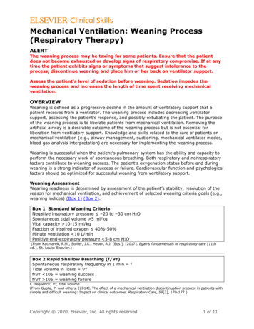
Int. J. Med. Sci. 2013, Vol. 10the National Institutes of Health Guide for the Care andUse of Laboratory Animals (85-23, revised in 1985) andthe “Norma Oficial Mexicana NOM-062-ZOO-1999”,were strictly followed. Anesthesia was induced with amixture of ketamine (100 mg/kg, ip) and xylazine (5.2mg/kg, ip). The level of anesthesia was verifiedthroughout the surgery and the whole experiment bymonitoring arterial blood pressure and by testing forthe lack of withdrawal reflexes and muscle tone.Doses of the ketamine-xylazine mixture were givenwhen necessary. The radial vein was canulated toadminister fluids and the carotid artery to monitorblood pressure. A bicarbonate (100 mM) and glucose(5 %) solution was delivered intravenously throughout the experiment at a rate of 5 mlh-1. Dextran andsaline solutions were given as necessary to maintainblood pressure between 80 and 120 mmHg. Bodytemperature was maintained at 36-37 oC. Rats wereplaced in a supine position with paws taped to theoperating table. With direct visualization of the trachea, an endotracheal tube was inserted and connected to a Harvard rodent volume-cycled ventilatorcycling at physiological speeds, with volume sufficient to adequately expand the lungs. The inflowvalve was supplied with 100 % oxygen. The sternumbone was removed to obtain an open thoracic cagepreparation. We removed tissue to obtain a directvisualization of the heart and the lungs. At the end ofthe experiment each animal was euthanized with anoverdose of pentobarbital. For histological purposesthe thorax with heart and lungs was placed in a solution of 10 % formalin.1446window discriminator, which generated triggerpulses. Then the expiratory activity was detected withthe window discriminator and it was employed foraveraging (32 samples) the signals detected with theMEMS sensor.The MEMS magnetic field sensorOur sensor is a small magnetometer based onMEMS technology developed at the Research Centerfor Micro and Nanotechnology from the UniversidadVeracruzana, Mexico. This research group has publications about this type of MEMS magnetic field sensor[22-24] and some demonstrated applications [21]. Thesensor exploits the Lorentz force and has an easy fabrication process, a small size and a high sensitivity.Figure 1A shows an image of the main elements of theproposed sensor. It has a compact resonant structure(700 600 5 µm) integrated by an array of siliconbeams and uses a piezoresistive sensing system. Asignal conditioning system (Figure 1B), together withthe sensor, was integrated into a PCB in order to makea digital signal processing, and reduce the electromagnetic noise environment below the signal of interest.Electrophysiological recordingsShielded custom-made bipolar electrodes weremade to reduce artifacts. The electrocardiogram(ECG) signal was obtained through standardthree-lead electrodes. Furthermore, other pair of bipolar electrodes was inserted on thoracic muscles toobtain recordings of the electromyogram (EMG)during the respiratory rhythm. The ECG and thethoracic EMG were amplified with GrassP511 amplifiers (Astro-Med, Inc, USA) over a bandwide set between 0.3 Hz and 10 kHz.Recording of respiratory activity in intactanaesthetized ratsIn other series of 3 experiments rats wereanaesthetized with isoflurane via a nose mask. Thethorax was keep intact and the MEMS sensor wasplaced at several positions from the chest surface. Inorder to detect respiratory activity an electret microphone was coupled to the mask. The microphone andcables were shielded. The output of this amplified andfiltered electret signal was send to the input of aFigure 1. A, Image of a magnetic field sensor based on MEMS technology.B, Image of the signal conditioning system implemented in a PCB for theMEMS sensor.http://www.medsci.org
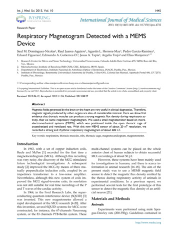
Int. J. Med. Sci. 2013, Vol. 10Data acquisitionData acquisition of the ECG, the thoracic EMG(or the respiratory activity recorded with the electretmicrophone) and the Magnetic signals recorded withthe MEMS sensor was performed at 10K Hz, employing Digidata and Axoscope software (Axon-Instruments). We performed an averaging of thesignals by means of a trigger commanded by a window discriminator (WPI, USA). The ECG or the electret microphone recordings were used as the triggersignal. We averaged 32 samples to obtain clear MCGtraces. The respiratory magnetogram was also recorded in continuous mode.Statistical analysisCoherence between the rectified an integratedEMG and respiratory magnetogram was calculated.The coherence magnitude of a 95 % confidence interval was also obtained. Data were expressed asmean sd. We used the coherence function of theMatlab software (Mathworks) to perform coherenceanalysis.1447ResultsWe performed experiments in 5 anesthetizedand ventilated rats. Our experimental paradigm consisted in the recording of the magnetic flux densityinside or in the proximity of the rat thoracic cage,which was open at the level of the sternum bone(Figure 2A). In order to find the correct position inwhich the emitted magnetic field reaches its maximum value we used a micromanipulator to rotate theMEMS sensor throughout different positions and angles. We found a position dependent magnetic fluxdensity, which is probably given by the compositionof different magnetic fields generated by the heart,lungs, and thoracic muscles. To avoid artifact movements the MEMS sensor did not touch the rat. In allour experiments the thoracic-muscle electromyogramand the electrocardiogram were also simultaneouslyrecorded, and they served as a reference to compareboth magnetic and electric signals generated by activity of heart and thoracic muscles.Figure 2. Sensing magnetic flux density of the thoracic cage with a MEMSdevice during respiratory and cardiac activity. A, Diagram of the experimental arrangement. A MEMS magnetic field sensor was introduced withinthe open thoracic cage of an anesthetized and ventilated rat. B, Electromyogram of the thoracic muscles and magnetic flux density recordingsduring the respiratory activity of the rat (respiratory magnetogram). C andD, Power spectrum density (PSD) calculated from the EMG and from themagnetic flux density, respectively. We found a significant coherence (E)between these rectified and integrated electric and magnetic signals at therespiratory frequency.http://www.medsci.org
Int. J. Med. Sci. 2013, Vol. 10With the MEMS sensor positioned at 0 mm and45 , we recorded a strong magnetic flux density produced by muscles of the thorax during respiration, ofabout 600 104 nT (mean sd, five rats) (Figure 2B), aswell as a MCG of about 98 49 nT (pink box of Figure3B). This angle was the optimal because our MEMSsensor detects the maximal component of the magnetic flux density in parallel direction to its resonantstructure, as illustrated with yellow arrows in Figure1A. This is the first evidence that thoracic muscles canproduce a strong magnetic flux density during res-1448piratory activity (i.e., a respiratory magnetogram).Figure 2B shows continuous recordings of the respiratory magnetogram and electromyogram of the thoracic muscles. A significant coherence of about 1 wasfound between both signals for all the rats; the horizontal line in (Figure 2E) represents the coherencemagnitude of a 95 % confidence interval. Similar results as illustrated in Figures 2 and 3 were obtained in4 other rats. Figure 3A illustrates that the MEMS sensor was displaced in different positions inside andoutside the thoracic cage.Figure 3. A, Diagram of the experimental arrangement, indicating angle (45 ) and positions of the MEMS sensor during the recordings. B, In each box, theupper trace shows the electrocardiogram (ECG) recording; and the lower trace, the magnetic flux density for the respiratory activity (i.e., the respiratorymagnetogram BMEMS). The numbers in millimeters (mm) indicate the different positions in which the sensor was placed.Figure 3B shows the averaged recordings of theelectrocardiogram and the MCG obtained with theMEMS sensor in 5 positions. Because the MCG couldnot be observed in the online recordings we performed and averaging of these magnetic fields bymeans of an ECG triggering averaging. The recordings in Figure 3B were obtained by averaging over 32sample measurements. The sensor was positioned atan angle of 45o. Note that at 4 mm outside the thoraciccage the strength of the magnetic flux density, i.e., theMCG, is close to only about 20 nT. However, there is aresonant position at 2 mm inside the thoracic cage,where the magnetic flux density reaches a maximumvalue close to 100 nT.We employed the same experimental paradigmand the same recording angle of 45 as in Figure 3, butfor the respiratory magnetogram in three other rats.Figure 4B illustrates averaged recordings (32 samples)of this respiratory magnetogram for one rat. Figure 4Bshows that these signals exhibit their maximal amplitude for positions close to the thorax (about -2 mmto 4 mm). We calculated the grand average of the respiratory-magnetogram amplitude for three rats andwe obtained graphs of this grand average versus therecording position (Figure 4D). We found an inverserelationship between the magnitude of this respiratory magnetogram and the recording position.Furthermore, we employed the same experimental paradigm, angle of 45 and positions, as inFigure 4B, but in three other intact rats. Figure 4Cshows averaged recordings (32 samples) of this respiratory magnetogram for one intact rat. We found ahttp://www.medsci.org
Int. J. Med. Sci. 2013, Vol. 10maximal magnitude of this respiratory magnetogramfor positions close to the thorax as illustrated in Figure4C. Furthermore, as illustrated in Figure 4E, we alsofound an inverse relationship between the magnitude1449of this respiratory magnetogram and the recordingposition (results obtained from the grand average ofthe respiratory-magnetogram amplitudes of threeintact rats).Figure 4. A, Diagram of the experimental arrangement, indicating angle (45 ) and positions of the MEMS sensor during the recordings. B, In each box, theupper trace shows the respiratory activity detected with the electret microphone; and the lower trace, the magnetic flux density for the respiratory activity(i.e., the respiratory magnetogram BMEMS). C, The same as B, but for an intact rat. The numbers in millimeters (mm) indicate the different positions in whichthe sensor was placed. D and E, Grand average of the respiratory magnetogram versus the sensor position, for three rats with the open thorax and threeother intact rats, as indicated (mean sd).http://www.medsci.org
Int. J. Med. Sci. 2013, Vol. 10DiscussionWe recorded a strong and rhythmic respiratorymagnetogram of about 600 nT by means of a MEMSsensor. In all the ventilated rats, we observed thatafter an overdose of ketamine the respiratory magnetogram was completely abolished, thus suggestingthat the magnetic signal detected with the MEMSsensor was not produced by a movement artifact dueto the rodent ventilation system. We suggest that asynergistic contraction of the thoracic muscles mainlycontributes to the emission of the magnetic field thatis recorded with the MEMS device.In our experiments, with the open thorax of therat, we also recorded a MCG of about 100 nT which isabout 2,000 or 10,000 times stronger than the typicalMCG detected on the chest of human subjects (50 pT)or rats (10 pT) with the SQUID magnetic detector,respectively. Discrepancies in the magnitude of thesemagnetic flux densities could be explained for differences in the animal preparation employed (i.e., openthorax, in our case versus intact thorax in other studies). An alternative explanation could be the closeproximity to the heart of our MEMS sensor, inside thethoracic cage with an optimal angle, regarding thehorizontal plane of the chest. Other explanation couldbe the method of ECG triggering averaging of theMCG, which exclude uncorrelated signals from theMCG.Fluctuations in strength of the respiratory magnetogram could be employed to analyze the respiratory dynamics in different physiological aspects. Inthis context, our findings could attract the attention ofphysicians, physiologists, biologists and biomedicalresearchers.AcknowledgementsThis work was supported by the followinggrants: FORDECyT-CONACyT 115976 (AH) andCONACyT-84605-and-95111 (PJGR) to develop theMEMS sensor. CONACyT-100028 to purchase aGaussmeter (EG). Cátedra Marcos Moshisky,PROMEP-PIFI-BUAP and CONACyT -Biomedicina”, México. The authors would like to thankPablo Linares for technical assistance in the experiments and John Reid for proof reading the Englishmanuscript.Competing InterestsThe authors have declared that no competinginterest .14.15.16.17.18.19.20.21.22.23.24.Baule G, McFee R. Detection of the magnetic field of the heart. Am HeartJ. 1963; 66: 95-96.Lekkala JO, Malmivuo JAV. Simultaneous measurement of the magneticheart vector components with unipositional lead system. In: Erne SN,Hahlbohm HD, Lubbig H, ed. Biomagnetism, Berlin: de Gruyter; 1980:319-326.Zimmerman JE, Theine P, Harding JT. Design and operation of stablerf-biased superconducting point-contact quantum devices, etc. J ApplPhys. 1970; 41:1572-1580.Cohen D. Measurements of the Magnetic Fields Produced by the HumanHeart, Brain and Lungs. IEEE Trans. Magn. 1970; 2: 694-700.Cohen D, Edelsack E, Zimmerman J. Magnetocardiograms taken inside ashielded room with a superconducting point contact magnetometer.Appl Phys Lett. 1970; 16: 278-280.Cohen D, Edelsack E, Zimmerman J. Magnetocardiograms Taken Insidea Shielded Room with a Superconducting Point Contact Magnetometer.Appl Phys Lett. 1970; 16: 278-280.Cohen, D. et al. MEG versus EEG localization test using implantedsources in the human brain. Ann Neurol. 1990; 28: 811-817.Lekkala JO, Mallmivuo JAV. Multiplexed SQUID vector magnetometerfor biomagnetic research. J Phys E Sci Instrum. 1984; 17: 504.Koch, H. Recent advances in magnetocardiography. J Electrocardiol.2004; 37: 117.Li Z, Xue-Min Z, Li-Hua Z, Xu-Guang H, Yu-Feng R, Geng-Hua C,Qian-Sheng Y, Ji F. An economical magnetocardiogram system based onhigh-T SQUIDs. Chinese Phys Lett. 2006; 23: 2319-2322.Lee YH, Yu KK, Kim JM, Kwon H, Kim K, Park YK. 64-channel second-order axial gradiometer system based on DROS for magnetocardiogram in a thin shielded room. Physica C. 2006; 468: 1942-1945.Lee YK, Kim K, Kwon H, Lee YH. Signal amplitude ratio of magnetocardiogram between sensor assembly and chest surface. IEEE T Magn.2009; 45: 2886-2889.Lim HK, Chung N, Ko YG, Lee YH, Park YK. Magnetocardiogram difference between healthy subjects and ischemic heart disease patients.IEEE T Magn. 2009; 5: 2890-2893.Kang CS, Lee YH, Kim J, Yu KK, Kim JM, Kwon H, Kim IS, Park YK, LimHK, Lee SG. Comparison of magnetocardiograms measured using different SQUID pickup coil configuration. IEEE T Appl Supercon. 2009; 17:835-838.Janawadkar MP, Radhakrishnan TS, Gireesan K, Parasakthi C, Sengottuvel S, Patel R, Sundar CS, Baldev RAJ. SQUID-based measurementof biomagnetic fields. Curr Sci 2010; 99: 36-45.Kim IS, Ahn S, Lee CH, Lee YH. Development of a mouse biomagneticmeasurement system by using a high-T (c) SQUID magnetometer. IEEET Appl Supercon. 2011; 21: 493-496.Kim IS, Kwon H, Ahn S, Song JH. Measurement of rat magnetocardiograms by using a high-T (c) SQUID magnetometer system. IEEE T ApplSupercon. 2011; 21: 497-500.Michael LH, Ballantyne CM, Zachariah JP, Gould KE, Pocius JS, TaffetGE, Hartley CS, Pham TT, Daniel SL, Funk E, Entman ML. Myocardialinfarction and remodelin in mice: effect of reperfusion. Am J Physiol-Heart C. 1999; 277: H660-H668.Maslennikov YV. Magnetocardiographic diagnostic complexes based onthe MAG-SKAN SQUIDs. J Commun Technol El. 2011; 56: 991-999.Leithaeuser B, Jung F, Park JW. Magnetocardiography in clinical cardiology. Status quo and future applications. Postepy kardiologii I JNEJ.2011; 7: 215-222.Tapia JA, Herrera-May AL, Garcia-Ramirez PJ, Martinez-Castillo J,Figueras E, Flores A, Manjarrez E. Sensing magnetic flux density of artificial neurons with a MEMS device. Biomed Microdevices. 2011; 13:303-313.Herrera-May AL, Garcia-Ramirez PJ, Aguilera-Cortes LA, Martinez-Castillo J, Sauceda-Carvajal A, Garcia-Gonzalez L, Figueras-CostaE. A resonant magnetic field microsensor with high quality factor at atmospheric pressure. J Micromech Microeng. 2009; 19: 01516.Herrera-May AL, Garcia-Ramirez PJ, Aguilera-Cortes LA, Figueras E,Martinez-Castillo J, Manjarrez E, Sauceda A, Garcia-Gonzalez L, Juarez-Aguirre R. Mechanical design and characterization of a resonantmagnetic field microsensor with linear response and high resolution.Sensor Actuat A-Phys. 2011; 165: 399-409.Herrera-May AL, Aguilera-Cortes LA, Garcia-Ramirez PJ, Manjarrez E.Resonant Magnetic Field Sensors Based on MEMS Technology. Sensors.2009; 9: 7785-7813.http://www.medsci.org
The ECG and the thoracic EMG were amplified with GrassP511 ampli-fiers (Astro-Med, Inc, USA) over a bandwide set be-tween 0.3 Hz and 10 kHz. Recording of respiratory activity in intact anaesthetized rats In other series of 3 experiments rats were anaesthetized with isoflurane via a nose mask. The thorax was keep intact and the MEMS sensor was