Transcription
1C OPYRIGHT Ó 2014BYT HE J OURNALOFB ONEAND J OINTS URGERY, I NCORPORATEDPosterior Shoulder Instability with a ReverseHill-Sachs Defect: Repair with Use ofCombined Arthroscopic Labral Repairand Fracture DisimpactionA Case ReportSimon A. Euler, MD, Ulrich J.A. Spiegl, MD, and Peter J. Millett, MD, MScInvestigation performed at the Steadman Philippon Research Institute, Vail, ColoradoAcute traumatic posterior glenohumeral dislocations arerare; they are typically caused by high-velocity trauma,epileptic seizures, or electrocution1,2. Anterior impression fractures of the humeral head (reverse Hill-Sachs defect)occur in over 80% of individuals following a posterior dislocation of the shoulder3. The size of reverse Hill-Sachs defectsvaries and has been reported to be as large as 55% of thecartilage surface4. Defects affecting less than 10% of the articular surface can be treated conservatively with closed reduction, and immobilization may produce excellent stability with alow risk for redislocation1. However, traditional sling immobilization in internal rotation puts the shoulder at risk to redislocate. Therefore, surgical treatment is recommended indefects that affect between 10% and 20% of the articular surface; they can be treated either with an arthroscopic approach,open remplissage, bone-grafting, or prosthetic replacement5.Compared with traditional posterior Hill-Sachs defectsthat occur with anterior dislocations, the extent of osseous andcartilaginous destruction of the anterior humeral head isusually more extensive in reverse Hill-Sachs defects6. Therefore, to stabilize the glenohumeral joint and avoid potentialprogressive cartilage destruction, which may lead to early osteoarthritis, reverse Hill-Sachs defects affecting more than 20%of the articular surface are nearly always treated surgically7.Several techniques have been described, including subscapularis transfer with or without the lesser tuberosity into thehumeral head defect, disimpaction and bone-grafting, allograftreconstruction of the humeral articular surface, and arthroplasty8. If the joint becomes unstable, repair of the posteriorcapsulolabral structures can be performed to restore stability, asdescribed for treatment of symptomatic posterior subluxation9,10.Large defects of greater than 40% of the articular surface typicallyrequire initial arthroplasty because of the increased risk of osteoarthritis, humeral head necrosis, and mechanical symptoms orinstability11-13.We present a clinical case of a reverse Hill-Sachs defectthat involved more than 30% of the articular surface. It wassuccessfully treated with combined arthroscopic posterior labralrepair; capsulorrhaphy; open disimpaction of the fracture, whichwas augmented with mineralized subchondral cancellous allograft bone; and internal screw fixation of a humeral head impaction fracture.The patient was informed that data concerning the casewould be submitted for publication, and he provided consent.Case Reportthirty-eight-year-old highly active man sustained a directfall on the right shoulder while snowboarding. The patientsustained a posterior dislocation of the shoulder in combinationwith a reverse Hill-Sachs defect. The shoulder was reduced in theemergency department but it redislocated spontaneously. Afteranother reduction, radiographs (Figs. 1-A, 1-B, and 1-C) andcomputed tomography (CT) (Fig. 1-D) demonstrated the largereverse Hill-Sachs defect. This defect involved approximately33% of the articular surface of the proximal part of the humerus(Fig. 1-D). Because there was instability, the arm was placed inexternal rotation to maintain reduction. Surgical fixation wasrecommended and was performed the following day.ASurgical ProcedureAfter adequate regional and general anesthesia, the patient wasplaced in the beach-chair position. The examination underDisclosure: None of the authors received payment or services, either directly or indirectly (i.e., via his or her institution), from a third party in support of anaspect of this work. One or more of the authors, or his or her institution, has had a financial relationship, in the thirty-six months prior to submission of thiswork, with an entity in the biomedical arena that could be perceived to influence or have the potential to influence what is written in this work. No authorhas had any other relationships, or has engaged in any other activities, that could be perceived to influence or have the potential to influence whatis written in this work. The complete Disclosures of Potential Conflicts of Interest submitted by authors are always provided with the online versionof the article.JBJS Case Connect 15
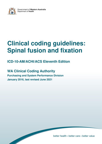
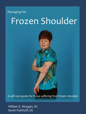
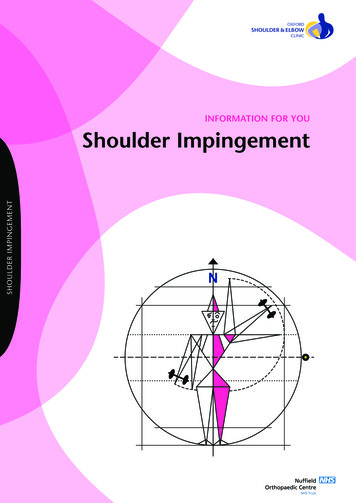
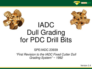
2J BJ S C A S E C O N N E C T O RV O LU M E 4 N U M B E R 3 S E P T E M B E R 24, 2 014ddP O S T E R I O R S H O U L D E R I N S TA B I L I T YA R E V E R S E H I L L -S A C H S D E F E C TWITHFig. 1Figs. 1-A through 1-D Radiographs and CTimaging. Fig. 1-A, 1-B, and 1-C Radiographs of theposterior dislocation. Fig. 1-D CT (axial view) ofthe shoulder. Borders of the articular surface wereset by the red horizontal line; 33% of the articularsurface was involved.anesthesia showed an excessive posterior translation withcrepitation. The humeral head could be easily dislocated posteriorly and locked on the impression fracture of the reverseHill-Sachs defect. The shoulder was manually reduced. Thediagnostic arthroscopic evaluation demonstrated some chondraldebris and hemarthrosis; a posterior labral tear and posteriorcapsular injury also were evident. No other intra-articular pathologies were present.We performed an extensive glenohumeral debridementwith evacuation of the hemarthrosis, a chondroplasty, removalof multiple loose bodies, a partial labrectomy, and a synovectomy. The posterior labrum was then repaired. Three 2.4-mmanchors (BioComposite SutureTak; Arthrex, Naples, Florida)were placed in the posteroinferior glenoid rim, and a crescentsuture shuttle (SutureLasso; Arthrex) was used to pass thesutures around the labrum and the capsule. Sutures were tiedwith use of a sliding, locking Weston knot, and they werebacked up with alternating half-stitches. Upon completion, theposterior capsulolabral complex appeared very stable.The osteochondral impression defect in the anteriorhumeral head was again visualized (Fig. 2). A dynamic arthroscopic examination was performed and showed that evenafter surgical stabilization of the posterior capsulolabral complex, the fracture continued to engage on the posterior glenoidrim when the arm was rotated internally by more than 40 .Therefore, the reverse Hill-Sachs defect fracture was addressed.A deltopectoral approach was performed, and the rotatorinterval was opened. Approximately 1 cm of the upper subscapularis was reflected inferiorly to allow visualization of theFig. 2Arthroscopic view of the defect. The blue arrows indicate the defect’sborder. RC rotator cuff, D defect on the humeral head, CF chondralfragment, and C cartilaginous surface.
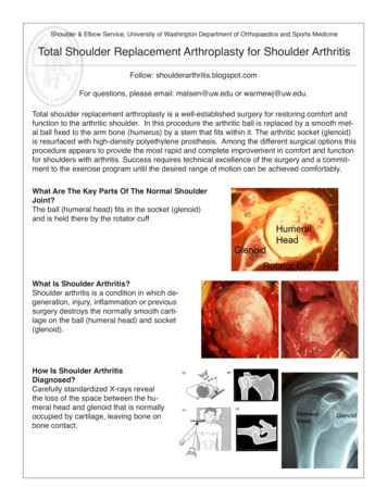
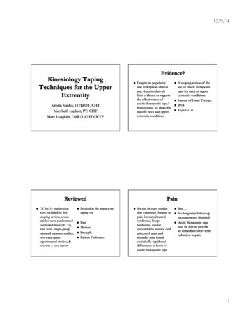
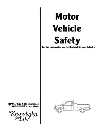
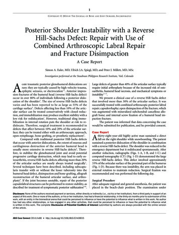
3J BJ S C A S E C O N N E C T O RV O LU M E 4 N U M B E R 3 S E P T E M B E R 24, 2 014ddP O S T E R I O R S H O U L D E R I N S TA B I L I T YA R E V E R S E H I L L -S A C H S D E F E C TWITHFig. 3Figs. 3-A through Fig. 3-D Illustrations of the surgical procedure. Fig. 3-A Access through the major tuberosity with an 8-mm ACL reamer. Fig. 3-BRetrograde reduction of the cartilaginous surface as well as defect filling with osseous chips. Figs. 3-C and 3-D Final setting with the surface reduced; thedefect was filled, and the supportive screws were in place.defect. The defect appeared to be approximately 1 cm deep andwas easily palpable. The arm was internally rotated, and a8-mm cortical viewing portal was created. With use of an8-mm anterior cruciate ligament (ACL) reamer, a tunnel wasmade laterally in the greater tuberosity toward the defect (Fig.3-A). The fracture was disimpacted and reduced anatomicallywith curved bone tamps. Reduction was controlled under direct vision through the rotator interval and with finger pal-pation by rotating the arm externally. The comminutedarticular segments were also placed back into anatomic position. Next, 15 cc of mineralized cancellous allograft bone chips(AlloSource, Centennial, Colorado) were impacted into thebone tunnel to buttress the subchondral fracture (Fig. 3-B).The articular cartilage surface was reduced anatomically andwas controlled manually by finger palpation through the rotator interval. Additionally, to support the subchondral boneFig. 4Intraoperative fluoroscopic views after repair. Two screws can be visualized, leading laterally to medially, supporting the disimpacted region of the anterioraspect of the articular surface.
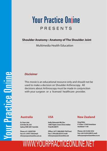
4J BJ S C A S E C O N N E C T O RV O LU M E 4 N U M B E R 3 S E P T E M B E R 24, 2 014ddP O S T E R I O R S H O U L D E R I N S TA B I L I T YA R E V E R S E H I L L -S A C H S D E F E C TWITHFig. 5Postoperative anteroposterior (left) and axial (right) radiographs at forty-three months. The fracture site was well reduced with screws in situ. There were nosigns of osteoarthritis or humeral head necrosis.under the defect, two 4.0-mm fully threaded screws were inserted extra-articularly from the anterolateral direction (Figs.3-C and 3-D). Screw heads were countersunk laterally and thusdid not interfere with rotation.Anteroposterior and axillary fluoroscopic views confirmed an anatomic reduction and correct placement of thescrews anterolaterally to medially, directly supporting the reverse Hill-Sachs defect (Fig. 4). The lesion was now visualizedas flush, and the chondral surface demonstrated restoration toits normal convexity. The patient had an uneventful postoperative course and was discharged the next day.The shoulder was secured in an immobilizing sling forfour weeks. Rehabilitation was initiated on postoperative day1 with pendulums and passive range of motion, limiting external rotation to 30 to protect the repaired upper subscapularis and to restrict internal rotation to the body. Active motionand active-assisted motion were initiated after four weeks.Posterior loading to the shoulder and overhead activities wereavoided for six weeks. The patient was allowed to return to fullsports activities after sixteen weeks.OutcomeAt the time of the final follow-up at forty-three months, thepatient was pain-free and had returned to full activities. He hadunrestricted range of motion (flexion of 170 , external rotationof 60 , and abduction of 120 ; with the arm in abduction to90 : external rotation of 90 and internal rotation of 75 ),which was no different from the uninjured contralateral side.Multiplanar radiographs showed maintenance of the reductionand no signs of osteoarthritis or humeral head necrosis (Fig. 5).The patient had recovered fully from the injury, had no signs ofinstability, and had returned to full activity without limitation.Discussionur patient had an acute traumatic posterior glenohumeraldislocation with a reverse Hill-Sachs defect that affectedmore than 33% of the cartilage surface. The patient was treatedOwith combined arthroscopic posterior labral repair, capsulorrhaphy, and fracture disimpaction of the reverse Hill-Sachsdefect, leading to an excellent outcome.For defects that affect between 20% and 40% of thecartilage surface, a variety of different treatment options havebeen proposed. Banerjee et al. reported excellent results aftertwo years of follow-up in two patients who had sustained reverse Hill-Sachs defects of less than 35% of the articular surface; there was no evidence of osteonecrosis, collapse, orprogression of osteoarthritis14. The defects were filled retrograde with allogenic bone-graft putty, and two screws wereused to support the subchondral zone. However, concerns havebeen raised about the use of an open approach through thesubscapularis tendon, especially with lesions that encompassover 30% of the cartilage surface14.Bock et al. also reported good results for the antegradereconstruction of reverse Hill-Sachs defects after a mean followup of sixty-two and one-half months15. In six cases where 30% to45% of the articular surface had been affected, they used bothautografts and allografts. However, the compacted cartilaginousarea was elevated and completely removed from the shoulder inorder to graft the impacted area. Then, the surface was securedback to the humeral head with anchors, leaving the sutures onthe surface of the cartilaginous joint.Martetschläger et al. described a modified arthroscopicMcLaughlin procedure7. With use of suture anchors, the subscapularis was attached into the reverse Hill-Sachs defect.However, this nonanatomic repair may bear the risk of limitedinternal rotation and might complicate future arthroplasty16. Itmay not be ideal for more centrally located reverse Hill-Sachsdefects or for larger defects because advancing the subscapularis in such settings could certainly affect glenohumeral jointkinematics.Moroder et al. described an all-arthroscopic repair for areverse Hill-Sachs defect that involved over 40% of the humeral head surface in which they used bone-chip allograftingwith cannulated screws17. However, because of humeral head
5J BJ S C A S E C O N N E C T O RV O LU M E 4 N U M B E R 3 S E P T E M B E R 24, 2 014ddnecrosis and partial osseous absorption, the patient required anarthroplasty six months after the initial surgery. Although theprocedure had been technically feasible, the lesion size seemedto limit the biological healing response.In a recent study, Jacquot et al. described a new techniqueof balloon treatment for reverse Hill-Sachs defects18. With fluoroscopic guidance, they percutaneously reduced four reverseHill-Sachs defects in three patients with use of balloon dilatationand cement fixation. The defect sizes were not reported. Aftertwelve months of follow-up, the results were moderate, with amean Constant score19 of 73 points. Nonetheless, informationregarding medium-term to long-term results is still not wellunderstood or available for this technique.To prevent the humeral head from dislocating posteriorly incases of instability, posterior labral repair and capsulorrhaphy arerecommended in order to avoid recurrent (sub)luxation accompanied by potential progressive cartilage wear and osteoarthritis20,21. Our patient received an arthroscopic posterior labral repairand capsulorrhaphy prior to open reduction and internal fixationof the anterior humeral head defect, leading to a stable joint withfull range of motion. To support the subchondral bone, twoscrews were inserted. This screw technique, which has been described by other authors, has been used routinely in other periarticular fractures, including tibial plateau fractures14,17.P O S T E R I O R S H O U L D E R I N S TA B I L I T YA R E V E R S E H I L L -S A C H S D E F E C TWITHIn conclusion, this case report demonstrates the repair ofan acute, impacted, and comminuted reverse Hill-Sachs defectthat involved more than 33% of the articular surface withcombined arthroscopic labral repair and fracture disimpactionof the reverse Hill-Sachs defect with an excellent outcome atalmost four years postoperatively. nNOTE: The authors thank Angelica Wedell, BA, for her work on the illustrations.Simon A. Euler, MDDepartment of Trauma Surgery and Sports Traumatology,Medical University Innsbruck,Anichstrasse 35,6020 Innsbruck, AustriaUlrich J.A. Spiegl, MDDepartment of Trauma and Reconstructive Surgery,University of Leipzig, Liebigstrasse 20,04103 Leipzig, GermanyPeter J. Millett, MD, MScSteadman Philippon Research Institute,181 West Meadow Drive, Suite 1000,Vail, CO 81657.E-mail address: drmillett@thesteadmanclinic.comReferences1. Cicak N. Posterior dislocation of the shoulder. J Bone Joint Surg Br. 2004 Apr;86(3):324-32.2. Robinson CM, Aderinto J. Posterior shoulder dislocations and fracture-dislocations.J Bone Joint Surg Am. 2005 Mar;87(3):639-50.3. Saupe N, White LM, Bleakney R, Schweitzer ME, Recht MP, Jost B, Zanetti M.Acute traumatic posterior shoulder dislocation: MR findings. Radiology. 2008Jul;248(1):185-93. Epub 2008 May 5.4. Gerber C, Lambert SM. Allograft reconstruction of segmental defects of the humeral head for the treatment of chronic locked posterior dislocation of the shoulder.J Bone Joint Surg Am. 1996 Mar;78(3):376-82.5. Paul J, Buchmann S, Beitzel K, Solovyova O, Imhoff AB. Posterior shoulder dislocation: systematic review and treatment algorithm [Review]. Arthroscopy. 2011Nov;27(11):1562-72. Epub 2011 Sep 1.6. Provencher MT, Frank RM, Leclere LE, Metzger PD, Ryu JJ, Bernhardson A, RomeoAA. The Hill-Sachs lesion: diagnosis, classification, and management. J Am AcadOrthop Surg. 2012 Apr;20(4):242-52.7. Martetschläger F, Padalecki JR, Millett PJ. Modified arthroscopic McLaughlinprocedure for treatment of posterior instability of the shoulder with an associatedreverse Hill-Sachs lesion. Knee Surg Sports Traumatol Arthrosc. 2013 Jul;21(7):1642-6. Epub 2012 Oct 7.8. Ponce BA, Warner JP. Management of posterior glenohumeral instabilitywith large humeral head defects. Tech Shoulder Elbow Surg. 2004;5:146-56.9. Bradley JP, Baker CL 3rd, Kline AJ, Armfield DR, Chhabra A. Arthroscopic capsulolabral reconstruction for posterior instability of the shoulder: a prospectivestudy of 100 shoulders. Am J Sports Med. 2006 Jul;34(7):1061-71. Epub 2006Mar 27.10. Savoie FH 3rd, Holt MS, Field LD, Ramsey JR. Arthroscopic management ofposterior instability: evolution of technique and results. Arthroscopy. 2008 Apr;24(4):389-96.11. Engel T, Hepp P, Osterhoff G, Josten C. Arthroscopic reduction and subchondralsupport of reverse Hill-Sachs lesions with a bioabsorbable interference screw. ArchOrthop Trauma Surg. 2009 Aug;129(8):1103-7. Epub 2009 Feb 24.12. Gavriilidis I, Magosch P, Lichtenberg S, Habermeyer P, Kircher J. Chronic lockedposterior shoulder dislocation with severe head involvement. Int Orthop. 2010Feb;34(1):79-84. Epub 2009 Mar 20.13. Hawkins RJ, Neer CS 2nd, Pianta RM, Mendoza FX. Locked posterior dislocationof the shoulder. J Bone Joint Surg Am. 1987 Jan;69(1):9-18.14. Banerjee S, Singh VK, Das AK, Patel VR. Anatomical reconstruction of reversehill-sachs lesions using the underpinning technique. Orthopedics. 2012 May;35(5):e752-7.15. Bock P, Kluger R, Hintermann B. Anatomical reconstruction for Reverse Hill-Sachslesions after posterior locked shoulder dislocation fracture: a case series of six patients. Arch Orthop Trauma Surg. 2007 Sep;127(7):543-8. Epub 2007 May 24.16. Verma NN, Sellards RA, Romeo AA. Arthroscopic reduction and repair of alocked posterior shoulder dislocation. Arthroscopy. 2006 Nov;22(11):1252.e1-5.Epub 2006 Sep 11.17. Moroder P, Resch H, Tauber M. Failed arthroscopic repair of a large reverse HillSachs lesion using bone allograft and cannulated screws: a case report. Arthroscopy. 2012 Jan;28(1):138-44. Epub 2011 Nov 30.18. Jacquot F, Costil V, Werther JR, Atchabahian A, Sautet A, Feron JM, DoursounianL. Balloon treatment of posterior shoulder dislocation with reverse Hill-Sachs injury:description of a new technique. Int Orthop. 2013 Jul;37(7):1291-5. Epub 2013 Apr 9.19. Constant CR, Murley AH. A clinical method of functional assessment of theshoulder. Clin Orthop Relat Res. 1987 Jan;(214):160-4.20. Millett PJ, Clavert P, Hatch GF 3rd, Warner JJ. Recurrent posterior shoulderinstability [Review]. J Am Acad Orthop Surg. 2006 Aug;14(8):464-76.21. Millett PJ, Clavert P, Warner JJ. Arthroscopic management of anterior, posterior,and multidirectional shoulder instability: pearls and pitfalls. Arthroscopy. 2003Dec;19(Suppl 1):86-93.
dislocation with a reverse Hill-Sachs defect that affected ted with combined arthroscopic posterior labral repair, capsulor-rhaphy, and fracture disimpaction of the reverse Hill-Sachs defect, leading to an excellent outcome. For defects that affect between 20% and 40% of the