Transcription
Clinical coding guidelines:Spinal fusion and fixationICD-10-AM/ACHI/ACS Eleventh EditionWA Clinical Coding AuthorityPurchasing and System Performance DivisionJanuary 2016, last revised June 2021
ContentsAnatomy1Types of Spinal Fusion1Interbody Fusion (IF)2Examples of lumbar interbody fusions2Anterior approach2Posterior approach2Posterolateral fusion (PLF)3Spinal fixation3Simple internal fixation3Nonsegmental internal fixation3Segmental internal fixation4Acknowledgement4References4
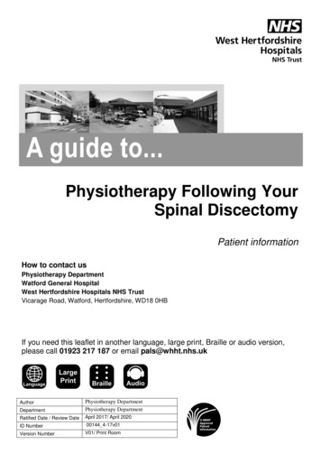
VERSIONDATEAUTHORCOMMENTS1.101/01/2016WA Clinical CodingAuthorityDeveloped1.201/06/2021WA Clinical CodingAuthorityRevision of Spinal fixation page 34.IntroductionFor a general clinical overview on the spine and spinal surgery, nical update - spine - parts a and b.pdfSpinal fusion surgery is intended to alleviate pain by stopping the motion at a painful vertebralsegment. To stop the motion, a bone graft is used to create a fusion.AnatomyAn intervertebral disc is the “cushion” between each vertebrae and acts as a shock absorberduring movement. Each spinal level comprises a disc space at the front and paired facet jointsat the back, which when working together define a motion segment (Ullrich 2012). For example,the lumbar spine has six spinal levels (motion segments), surrounding and in between the fivelumbar vertebrae. Two vertebrae need to be fused together to stop the motion at one segment.A spinal level carries the name of the disc at that level (named by the vertebra above and belowthe disc). For example, the disc space between the L4 and L5 vertebrae is known as the L4-5disc, or the L4-5 level. Therefore an L4-L5 spinal fusion is actually a one level spinal fusion and,an L3-4, L4-5 spinal fusion is a two level spinal fusion. The same nomenclature is applied to theusually associated spinal fixation procedure.Spinal fusion may be done in the cervical, thoracic or (most commonly) lumbar areas of thespine.Examples of conditions treated by spinal fusion are: degenerative disc disease;spondylolisthesis; weak or unstable spine caused by infections, tumors; fractures; scoliosis ordeformity.Types of Spinal FusionThe main types of fusion are: interbody fusionposterolateral fusionSpinal fusion is traditionally performed as an open procedure which involves muscle dissection.There have been advancements in surgical techniques with the development of minimallyinvasive techniques, which involve special instruments, nerve monitoring andradiographic/computer assisted navigation (Cleaver 2013). These techniques involve smallerincisions with use of retractors to dilate/spread (rather than cut) muscles to gain access to thespine. Many spinal procedures can be done using minimally invasive techniques. The surgeondecides the type of fusion procedure/surgical approach needed based on the nature andlocation of the spinal pathology.1
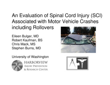
Interbody Fusion (IF)Interbody fusion involves removal of the intervertebral disc (discectomy) and replacement with abone graft and/or a device (spacer or cage) to maintain alignment and disc height. The devicesusually contain bone graft material which facilitates fusion (OrthoInfo 2010).Interbody fusions are categorised in ACHI by their surgical approach i.e. anterior or posterior.Examples of lumbar interbody fusionsAnterior approach Anterior lumbar interbody fusion (ALIF): the disc is approached from an anterior(abdominal) incision. The advantage of this approach is avoidance of cutting muscles ofthe back. The disadvantage is the risk of injury to structures in the abdomen. Transpsoas lumbar interbody fusion (DLIF/direct or XLIF/eXtreme): the disc isapproached through the psoas muscle, from an extreme lateral incision (retroperitoneal)on the patient’s side. The advantage is the avoidance of back muscles and abdominalstructures required in traditional fusion procedures. The disadvantage is that L5-S1 is notaccessible with this procedure (Kleeman 2011). Oblique lumbar interbody fusion (OLIF): the disc is approached from a lateral incisionon the patient’s side. The procedure is done "obliquely" (in front of the iliac crest) whichgives access to L5-S1 (Kleeman 2011).Anterior approach ACHI codes48660-00 [1389] Anterior spinal fusion, 1 level48669-00 [1389] Anterior spinal fusion, 2 levelsPosterior approach Posterior lumbar interbody fusion (PLIF): the disc is approached from a posterior(back) incision. Midline LIF (MidLIF or MLIF): the disc is approached from a posterior (back) incision,however the approach is medial to lateral, rather than lateral to medial (P. Bannan,personal communication October 30, 2013). Transforaminal lumbar interbody fusion (TLIF): the disc is approached from aposterior (back) incision on one side of the spine. This can be done as a traditional“open” TLIF or using minimally invasive techniques (MasTLIF or MisTLIF) wheremultiple small posterior incisions are made further to the side, rather than cutting themuscles near the centre of the back.2
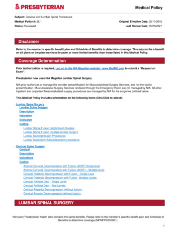
Posterior approach ACHI codes48642-00 [1389] Posterior spinal fusion, 1 or 2 levels48645-00 [1389] Posterior spinal fusion, 3 levels48654-00 [1389] Posterior spinal fusion with laminectomy, 1 level48657-00 [1389] Posterior spinal fusion with laminectomy, 2 levelsPosterolateral fusion (PLF)Posterolateral fusion places the bone graft between the transverse processes (the bonyprotuberances on the vertebrae) rather than the intervertebral disc space, which is left intact.The approach is through a posterior (back) incision, and a laminectomy is typically required togain access. PLF is usually accompanied by fixation.Posterolateral fusion ACHI codes48648-00 [1389] Posterolateral spinal fusion, 1 or 2 levels48651-00 [1389] Posterolateral spinal fusion, 3 levels48654-01 [1389] Posterolateral spinal fusion with laminectomy, 1 level48657-01 [1389] Posterolateral spinal fusion with laminectomy, 2 levelsSpinal fixationIn most cases, spinal fusion is augmented by a process of internal fixation of the spine knownas spinal fixation. This involves the placement of metallic screws, wire loops, hooks, rods, platesor cages to stabilise the vertebrae and facilitate bone fusion. The surgical procedure usuallyconsists of posterior (posterior elements) and anterior (vertebral body) fixation. The fixation isnot removed. The fusion process typically takes 6-12 months after surgery.There are three main types of internal fixation of the spine: simple internal fixationnonsegmental internal fixationsegmental internal fixation.Simple internal fixationFixation of any part of one vertebra (not a spinal/motion segment) and includes parsinterarticularis, spinous process/pedicle or simple interspinous wiring.Nonsegmental internal fixationRods or plates are used to bridge a length of spine at the upper and lower extremity of a multilevel spinal fusion. A multilevel spinal fusion involves three or more vertebrae., The fixation is attwo points only without any intermediate attachment to vertebrae between those two points. The3
attachment to vertebrae at either end is by wire hook or screw fixation. The fixation immobilisesthe spine so that the spinal fusion consolidates.The Harrington rod is an example of nonsegmental internal fixation and was commonly used inScoliosis, for which it was invented.ExampleA rod spanning from the first to fifth lumbar levels is attached to the spine at vertebrae L1 andL5 but not attached to L2, L3, or L4.Segmental internal fixationFixation of a spinal/motion segment with fixation instrumentation, e.g. vertebral body screw,pedicle screw/rod system or hook instrumentation including sublaminar tapes or wires. Aspinal/motion segment includes all anatomical structures between and including the pedicles oftwo adjacent vertebrae. A spinal fixation is segmental when the fixation involves two or morevertebrae attached by the fixation instrumentation, i.e.one or more levels or one or morespinal/motion segments.ExamplesSegmental internal fixation of the spine at one levelA rod attached to second and third thoracic vertebrae and spanning the intervertebral discbetween these.Segmental internal fixation of spine at greater than one levelA rod spanning from the third cervical vertebra to third thoracic vertebra attached at C3 and T3and also attached to at least one other vertebra between these two points (e.g. C6).AcknowledgementThe WA Clinical Coding Authority acknowledges Mr Paul Bannan, Consultant Neurosurgeon atMount Hospital, for the clinical advice provided to produce this document.ReferencesCleaver, Neil. 2013. “Minimally invasive lumbar spine surgery”. Surgery of the Spine, Dr NeilCleaver. Accessed November 22, http://www.cleaverspine.com/miss.htmKleeman, Thomas. 2011. “Oblique lumbar interbody fusion”. Thomas. J Kleeman, MD.Accessed July 17, http://www.tjkleemanmd.com/olif.htmlOrthoInfo. American Academy of Orthopaedic Surgeons. “Anterior lumbar interbody fusion”.2010. Accessed September 11, http://orthoinfo.aaos.org/topic.cfm?topic A00595Pakzaban, Peyman. 2012. “Spinal instability and spinal fusion surgery treatment andmanagement”. Medscape. Accessed September reatment#a1128Ullrich, Peter. 2012. “Lumbar spinal fusion surgery”. Spine Health. Accessed July sion/lumbar-spinal-fusion-surgery4
This document can be made available in alternative formats on request for a person with adisability. Department of Health 2021Copyright to this material is vested in the State of Western Australia unless otherwise indicated. Apartfrom any fair dealing for the purposes of private study, research, criticism or review, as permitted underthe provisions of the Copyright Act 1968, no part may be reproduced or re-used for any purposeswhatsoever without written permission of the State of Western Australia.
3 Posterior approach ACHI codes 48642-00 [1389] Posterior spinal fusion, 1 or 2 levels 48645-00 [1389] Posterior spinal fusion, 3 levels 48654-00 [1389] Posterior spinal fusion with laminectomy, 1 level 48657-00 [1389] Posterior spinal fusion with laminectomy, 2 levels Posterolateral fusion (PLF) Posterolateral fusion places