Transcription
40Will There be a Role for DiscProstheses in Small Animals?Filippo Adamo and Franck ForterreIntroductionSpinal arthroplasty offers an exciting alternative tofusion for the treatment of degenerative disc disease.Currently, the main indication for disc prosthesis insmall animals is the treatment of disc-associatedcervical spondylomyelopathy in dogs, also knownas disc-associated wobbler syndrome (DAWS) (seeChapter 7). Another potential indication is thetreatment of disc-associated lumbosacral stenosis(see Chapter 32). DAWS in dogs shares many similarities with cervical spondylotic myelopathy in people, and the Doberman breed has been proposed as anatural model to study the disease in humans [1].The knowledge gained over the last 15 years inpeople with disc arthroplasty can be applied inveterinary medicine for the treatment of DAWS.However, this information should be interpretedcautiously by veterinarians because of biomechanical differences in dogs such as greater axial forcesand greater amount of coupled motions in dogscompared to people [2, 3].The goal of cervical arthroplasty is to preserveintervertebral mobility while providing distraction,stability, and neural decompression [4–6]. Cervicaldisc arthroplasty involves discectomy, spinal corddecompression, milling of the vertebral end plates,and placement of a device to maintain distraction andpreserve intervertebral mobility at the treated space[4–11]. Maintenance of motion at a decompressedinterspace may result in improved load transfer andreduced stress on the adjacent intervertebral discsand dorsal elements, although this has not yet beendemonstrated conclusively in the canine spine.Comparison of DAWS in peopleand in dogsIn people with cervical myelopathy and radiculopathy secondary to degenerative disc disease,anterior cervical discectomy and fusion (ACDF) isa reliable surgical treatment with a satisfactoryoutcome in 90–95% of patients [12]. However,there is a significant incidence of disc disease atadjacent sites causing recurrence of neurologicsymptoms, termed domino effect [12–14]. Within 5years of surgery, 92% of patients have radiographicAdvances in Intervertebral Disc Disease in Dogs and Cats, First Edition. Edited by James M. Fingeroth and William B. Thomas. 2015 John Wiley & Sons, Inc. Published 2015 by John Wiley & Sons, Inc.2940002203300.INDD 2949/12/2014 4:44:08 PM
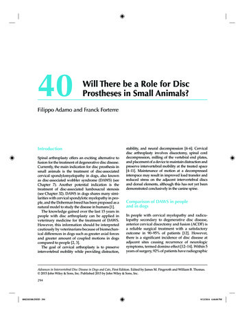
Will There be a Role for Disc Prostheses in Small Animals?evidence degenerative disc disease at adjacentsegments, and by 10 years, about 25% of peoplerequire a second surgery for the same problem atan adjacent space [12, 14]. The cause of thissubsequent disc degeneration remains unknown.One suggested factor is an increase in intradiscalpressure at adjacent discs after fusion [15–18]. Thisincreased pressure blocks the diffusion of nutrients from the end plate and is the most significantcause of disc degeneration [19].Clinical studies using dynamic radiographyshow increased motion at adjacent segmentsabove and below the level of cervical fusion, andthis is also a factor associated with deterioration[20, 21]. After arthroplasty, the range of motion isincreased or maintained in the surgically treatedsegment and mildly decreased at adjacent levels.However, this reduction is compensated by themovement of the artificial disc itself [22].An interesting dilemma regarding dominolesions is whether such lesions are the naturalprogress of an underlying similar process at theadjacent vertebral motion units or if they are anaccelerated degenerative process influenced bythe biomechanical effect of fusion [21–23]. Motionpreservation at the surgery site may reduce therate of adjacent-level cervical disc disease [24–28].295The use of a cervical disc prosthesis in humanpatients was first reported by Ferstron in 1966 [29].Over the last decade, clinical experience with manydifferent types of artificial discs has been reported,including Cummins artificial cervical joint [30](which evolved into Prestige [31], ProDisc-C [32],Bryan Cervical Disc [33], Discover [34, 35]), andseveral clinical trials are currently underway [28].some are used in conjunction with supplementalvertebral body screw fixation [36]. Certain designspromote biological bone ingrowth at the disc-endplate interface. In terms of motion, artificial discsmay be constrained, semiconstrained, or unconstrained [36]. Devices are considered constrained incertain planes if they restrict motion to less than thatseen physiologically. Devices are considered semiconstrained in certain planes if they allow motionsimilar to that seen physiologically. Devices are considered nonconstrained in certain planes of motion ifthere is no mechanical stop to the motion and if theyare reliant on the perispinal soft tissue and theinherent compression across the disc space to provide restraint to extremes of motion [37].The distribution of force and subsidence ispossibly the most important biomechanicalconsideration for an artificial disc. The idea is todistribute the forces involved as uniformly aspossible over a large area [37].Intradiscal replacement of the nucleus pulposusrepresents an alternative to total disc replacementand spinal fusion procedures. The aim is to reconstruct the nucleus pulposus primarily while preserving the biomechanics of the annulus fibrosusand cartilaginous end plate. Nucleus pulposusimplants are designed to provide stable motion,increase disc space width, relieve or lessen transmission of shear forces on the remaining annulus,and stabilize ligaments [38]. Nucleus pulposusreplacement devices can be categorized into twogroups: the intradiscal implants and in situ curablepolymers. Intradiscal implants are biomechanically more similar to the native nucleus pulposus,whereas in situ curable polymers consist of compounds that harden after implantation. Theseimplants are currently at different stages of preclinical and clinical investigations in veterinary medicine [39; F. Forterre, personal communication].Classification of artificial discsCervical disc prosthesis in dogsCurrently, artificial discs are classified into threetypes: non-, uni-, and biarticulating. The implantmay consist of a metal-on-metal design, metal-onpolymer (ultrahigh-molecular-weight polyethylene),and less commonly ceramic-on-polymer or aceramic-on-ceramic design [36]. The disc is eithermodular (having replaceable components) or nonmodular (without replaceable components, andThe first cervical prosthesis specifically for thecanine cervical spine1 was designed and biomechanically tested in vivo in 2007 [7]. This prosthesisis made of a titanium alloy (Ti-6AI-4 V-ELI) andconsists of two end plates, with a range ofmovement of 30o between the plates (Figure 40.1).Titanium alloy was selected because it is resistant,is relatively inexpensive, has a good corrosionCervical disc prosthesis in people0002203300.INDD 2959/12/2014 4:44:08 PM
296 Advances in Intervertebral Disc Disease in Dogs and Cats(A)(B)(C)(D)Ball in PEEK(E)Figure 40.1 Adamo spinal disc . US Patent # 8,496,707 B2. The prosthesis is made of a titanium alloy, and the concavity andconvexity of the central aspects of the end-plate surfaces result in a ball-and-socket type of connection between the end plates.The external surface of each end plate is convex, resembling the concavity of the caudal end plate of the most caudal cervicalvertebrae. The outer surface of each end plate has concentric grooves to allow bone ingrowth into the implant and is pretreatedwith a dual acid etch bath to promote bone ingrowth into the prosthesis (A). The convexity of the central aspect of theend-plate surface is made of polyether ether ketone (PEEK) (B). Two end-threaded stainless steel pins screwed into each piece ofthe implant and a barrel holder are used to hold together the two pieces of the implant during placement within the disc space(C and D). Six standard sizes of prosthesis are available, labeled S1, S2, and S3 and M1, M2, and M3. However, since eachmale piece of the prosthesis has to be assembled with a separate female piece, this makes a total of 12 different sizes when thedisc is assembled in all possible combinations (E).resistance compared to other materials, and is idealfor follow-up MRI studies in the event of postsurgical worsening and to evaluate domino lesions.This prosthesis is rotationally unconstrained, following the ball-and-socket principle, and does notrequire supplemental fixation. Unconstrainedcervical prostheses designed for people usuallyallow about 11 of freedom [24]. The higher degreeof freedom in this prosthesis was arbitrarily chosenconsidering that heterotopic ossification at thetreated site observed in people significantlydecreases the range of motion at the site of0002203300.INDD 296implantation over time. The outer surface of eachend plate is convex to avoid prosthesis migrationand has concentric grooves to allow bone ingrowthinto the prosthesis. In the in vitro study, it was concluded that cervical spine specimens with theimplanted prosthesis have biomechanical behaviors more similar to an intact spine compared tospinal specimens with ventral slot and PMMAfusion [7]. This was the prerequisite for thesubsequent clinical investigation [8–10].A pilot long-term clinical study using this prosthesis in two dogs affected by DAWS showed that9/12/2014 4:44:11 PM
Will There be a Role for Disc Prostheses in Small Animals?cervical arthroplasty was well tolerated with excellent outcome in both dogs. Mobility at the treatedsite was lost and distraction decreased over time inboth dogs, without affecting the clinical outcome.Furthermore, the presence of the prosthesis did notaffect the ability to reassess the area via MRI at afollow-up 18 months later, and domino lesionswere not observed in either dogs [9].A multicenter study evaluating the short andintermediate clinical and radiological results(average 15 months) using the same prosthesis in 12dogs with single- and multilevel lesions has been(A)(B)(C)297reported [10]. In this study, the external surface ofthe prosthesis was treated with dual acid bath etch[40, 41] to promote bone–implant incorporationat the implant–vertebral end-plate interface, anddedicated surgical tools were created to facilitateimplantation (Figure 40.2). To improve visualassessment, a high-powered headlamp , magnification loupes, and a Gaspar cervical distractor wereused as in the previous study (Figure 40.3). All 12dogs had immediate postoperative recovery withgood degree of distraction in the immediate postoperative radiographs (Figure 40.4). All dogs in this(F)(D)(E)Figure 40.2 Dedicated surgical tools and other accessories. US Patent # 8,491,655 B2. Barrel holder. (A) After the two piecesof the implant are assembled, the two pins are inserted into the barrel holder. (B) The barrel holder has the double function ofholding the prosthesis assembled with one extremity and to unscrew the end-threaded pins after the prosthesis is placed in theslot with the other extremity. Custom-made burrs. (C) Two dedicated custom-made burrs are used. The small burr head is usedto enlarge the discectomy and to create enough space for the insertion of larger burr. The large head burr resembles theexternal convexity of the implant, and it is used for the final burring to create the specular concavity to accommodate theimplant into the disc space. Twenty degree angle attachment. (D) A 20 angle attachment for the high-speed air drill is used tofacilitate burring at an angle parallel to the disc space. Caspar cervical distractor. (E) This instrument is used to distract the twocervical vertebrae at the affected disc space. It allows better visualization of the dorsal aspect of the discectomy, facilitates theremoval of the bulging disc, improves visualization of the dorsal longitudinal ligament, and facilitates the inspection of thevertebral canal for adequate spinal cord decompression. Sizing probe. (F) This instrument resembles at each extremity theshape of the S or M disc size, respectively it is used to probe the discectomy between burring (in place of the implant), and toassess the congruity of the discectomy to the implant. This instrument shortens the total surgery time by avoiding multiple timesof checking of the discectomy with the actual implant, and it also facilitates proper selection of the size of the implant after thediscectomy is completed.0002203300.INDD 2979/12/2014 4:44:15 PM
298 Advances in Intervertebral Disc Disease in Dogs and Cats(A)(B)(C)(D)(E)(F)(G)(H)(I)Figure 40.3 Surgical technique. After routine surgical preparation, the ventral aspect of the affected vertebral bodies isaccessed via a standard approach as for a ventral slot procedure. A discectomy is performed across the intervertebral space.The self-retaining Caspar distractor is placed and maintained distracted (A) to allow the final cleaning of the disc space and thesubsequent burring with the two dedicated burrs (B). Burring is done following the direction of the intervertebral space, whichis facilitated by the 20 angle attachment. Burring should be kept on the midline of the disc space and the widest part of theburr should be centered at a depth of approximately 50% of the total disc space depth. Burring of the end plate of the cranialvertebra is minimal and limited to the removal of the final debris of the annulus fibrosus. The cranial end plate does not requireexcessive burring because it already has a natural concavity, which accommodates the convexity of the implant. Burring of theend plate of the caudal vertebra is limited in the central surface. Burring is also extended to the lateral edge of the discectomyas needed to allow the insertion of the sizing probe and later of the prosthesis. If the MRI study is suggestive of static spinalcord compression, the dorsal longitudinal ligament should be incised to visualize the spinal cord and explore the vertebralcanal (C). In between burring, the sizing probe (which simulates the prosthesis) is used for pretesting how the actual prosthesiswill be firmly seated in place (D). The prosthesis is inserted while maintaining the Caspar distractor in maximal distraction.Gentle pressure is applied to force the implant into the slot, and an audible click is usually heard when the convex area of theimplant slips past the edge of the slot (E). To ensure that the implant is seated as much as possible on the midline, the barrelholder is rotated until the fissure of its proximal end is aligned with the long axis of the vertebrae. After implantation, thedistraction is released, allowing the two vertebral end plates to collapse on the prosthesis (F). After assessing that the prosthesisis correctly seated in position, in order to ensure that the implant is firmly in position, the two pins still screwed in theprosthesis are grasped with a needle holder, and upward traction is applied in an attempt to dislocate the prosthesis from theslot (G). Once the surgeon is satisfied that the prosthesis is held snuggly between the vertebrae, the two pins are unscrewedfrom the two prosthetic end plates (I), and the two Caspar distractor pins are removed from the vertebral bodies. Bone wax orgel foam is placed to stop the bleeding that might occur at the holes created by the two Caspar distractor pins. The longus colli,sternohyoideus, and sternocephalicus muscles and subcutaneous and subcuticular tissues are then closed in a routine manner.0002203300.INDD 2989/12/2014 4:44:21 PM
(A)(B)(C)(D)(E)(F)Figure 40.4 Postoperative radiographs. Immediate postoperative radiographs. German shepherd, 9 years old. Correctpositioning of the implant on the lateral (A) and ventrodorsal projection (B) at the level of C5–C6. The implant used is M1,which provides adequate distraction. Two weeks post-op (same dog). The lateral (C) and ventrodorsal (D) views show that theimplant is still well in place providing distraction at the treated site. The extension (E) and the flexion (F) views are performed toevaluate the degree of mobility at the treated site. The opening of the angle between the two implant end plates, as shown inthe flexion view in this patient, indicates that mobility is maintained. At the reevaluation 6 months later, the radiographicfindings were unchanged.0002203300.INDD 2999/12/2014 4:44:23 PM
300 Advances in Intervertebral Disc Disease in Dogs and Catsstudy, except one in which the technique wasimproperly performed, showed improvement inthe neurological status during the observationperiod. In the majority of dogs, the distraction wasmoderately lost, and mobility at the treated sitesdecreased or became undetectable over time(Figure 40.5). A subsequent study using a redesigned thinner disc (2nd-generation disc) with theinternal convex surface replaced with polyether(A)(B)(D)(H)ether ketone (PEEK) and an additional tool (sizingprobe) to probe/test the disc space during burringbefore final disc implantation showed similarresults but with the advantages that the surgerywas facilitated by less burring and less implantmanipulation and the implant accommodatedeasier along the natural angle of the disc space [11].A recent study evaluating 50 disc spaces in 33 dogstreated with CDA for DAWS showed that on the(E)(I)(C)(F)(G)(J)Figure 40.5 Two-level cervical disc replacement. Intraoperative photo (A) and immediate postoperative radiographs (B and C)of disc-associated wobbler syndrome (DAWS) at C3–C4 and C5–C6, with a suspected congenital vertebral fusion at C4–C5.Multiple cervical disc arthroplasty was recommended to avoid additional fusion at the affected spaces, which, in addition tothe preexisting one, could have had predisposed the patient to a domino lesion at C6–C7. Nine-month postoperativeradiographs with the neck in lateral (D), extension (E), flexion (F), and ventrolateral (G) positioning showed mild decrease of theoriginal vertebral distraction, no heterotopic ossifications, and retention of mobility at both treated spaces. This dog hadpresented with a 4 month history of progressive ataxia/tetraparesis and regained full neurological function. MRI T2 weighted of6-year-old Doberman with a 2-year history of ataxia/tetraparesis treated with NSAID who presented acutely tetraplegic; theMRI showed multilevel DAWS lesions at C5–C6 and C6–C7 (H). The dog was treated with double cervical disc replacementand the immediate postoperative radiograph showed adequate disc placement at both treated sites (I). At 7-month follow-up,recheck radiographs showed decreased vertebral distraction and heterotopic ossification at both treated spaces (J). Mobility wasnot detectable at C6–C7 and significantly decreased at C5–C6. However, this did not affect the clinical outcome. The dogbecame ambulatory 2 weeks postsurgery and neurologically normal 4 months later. He continued to maintain his normalneurological status without any adjuvant anti-inflammatory or analgesic medication.0002203300.INDD 3009/12/2014 4:44:24 PM
Will There be a Role for Disc Prostheses in Small Animals?immediate postoperative radiographs, 15 sites wereoverdistracted, 34 sites were adequately distracted,and only 1 site was underdistracted. Overdistractionwas mostly observed with the 1st-generation(thicker) implant, while the adequate distraction was mostly observed with the 2nd-generation(thinner) implant. On serial radiographic evaluation, the distraction was gradually decreased compared to the immediate postoperative radiographsand subsidence (defined as reduction of the widthof the disc space equivalent to or less than the widthof the preoperative radiographs) was seen in 7/19sites (37%) at 6 months postsurgery and progressedto all the five sites evaluated at 2 years postsurgery.Subsidence, except for one dog, was seen onlywhen the disc spaces were overdistracted, and itwas more pronounced with the 1st-generation(thicker) and narrower implants and less pronounced with the 2nd-generation (thinner) andwider implants (Adamo, personal observation).Mobility was not detectable in 8/36 treated sites(22%) 2 weeks after surgery in 24 dogs examinedand was lost or not detectable in 17/22 sites (77%)at 6 months after surgery in 14 dogs examined. Noimplant migration or implant infections wereobserved on serial radiographs or on MRI whenavailable. In this study, the mean and median followup time was 16 and 12 months, respectively (range,2 weeks to 42 months), and the outcome was considered good to excellent in 30 out of 33 dogs (91%).The three dogs with poor or unsatisfactory outcomewere presented with over 2 months of nonambulatory tetraparesis and severe extensor rigidity inboth thoracic limbs. Among these, one dog(12.4-year-old sheltie mix weighing 9 kg [19.5 lb])was euthanized 2 weeks postsurgery because of acompressive fracture of C6 with ventral displacement of the implant, and another dog (13.5-year-oldchow mix) was euthanized 8 months after surgerybecause of lack of significant improvement. Twodogs had recurrence of the neurological signs 18months after surgery secondary to suspected osteophytes (noted on MRI) causing ventral spinal cordcompression at the treated sites. Ventral osteophytes were seen at six treated sites, in four dogs onpostoperative radiographs, causing bridging spondylarthrosis and ankylosis at three disc spaces intwo dogs. No neurologic domino lesion adjacent totreated sites was seen in any of the dogs during theobservation period. Median postoperative hospitalization time was 1 day (range 0–5 days), and 5 dogs0002203300.INDD 301301were discharged on the day of surgery. In this study,it was concluded that correct patient selection(dog’s weight not less than 20 kg [44 lb]), neurological status at presentation, size of the implant, endplate preparation, and applied distraction duringsurgery were important factors that may influencethe outcome (Adamo, personal observation).These studies showed that cervical disc arthroplasty is well tolerated and might be a valuablemethod to treat DAWS [9–11]. Furthermore, thesestudies showed that the cervical cast used in theprevious study was not needed.Benefits of cervical disc arthroplastyCervical arthroplasty in people is still an area ofactive research, debate, and controversy, and a5-year follow-up has been recommended to assessthe long-term functionality of the prosthesis andits influence at adjacent levels [42]. A technologyoverview from the American Academy of OrthopaedicSurgeons analyzed the results of multiple clinicalstudies and concluded that artificial disc arthroplasty is more beneficial in the short term compared to ACDF, although its long-term benefitover standard ACDF remains unclear [43]. Theimpact of cervical disc replacement on adjacentsegment degeneration and the degree of heterotopic ossifications (HOs) of the treated segmentsremain a subject of intensive investigation. HO is apathologic condition that leads to the developmentof bone within nonosseous soft tissues [44]. Thebone that forms is believed to develop throughstimulation by cellular mediators and altered neurovascular signaling [44]. Although the precisecause of HO remains unclear, it is certain that itincreases with time, it may occur at the four cornerof the disc space (most commonly anterosuperioror posterosuperior), and it has been reported withan incidence as high as 60.3% [45].A prospective, randomized, multicenter studycomparing cervical arthroplasty with ACDF with aminimum of 2-year follow-up concluded thatcervical arthroplasty was associated with significantly greater overall success rate than ACDF.Furthermore, there were significantly fewerpatients in the cervical arthroplasty group showingsevere adjacent-level radiographic changes at the2-year follow-up [46]. Another study with anaverage follow-up of 49.4 months evaluated9/12/2014 4:44:24 PM
302 Advances in Intervertebral Disc Disease in Dogs and Catscervical disc replacement and its effects on adjacentsegment discs and found satisfactory clinical andradiographic outcome. In this study, progression ofadjacent segment degeneration was observed in23% of patients, but this did not affect the clinicaloutcome [47]. A study of cervical arthroplasty withup to 2-year follow-up showed motion preservation at the treated site in 85% of patients, HOs in4.5% of the treated levels, and radiological signs ofadjacent-level degeneration in 9.1% patients. Thisstudy confirmed the efficacy and safety of the technique and concluded that the presence of HOsdoes not alter the clinical outcomes [48]. In anotherstudy, HOs was detected in 27.7% of treated segments but did not affect the clinical outcomes, andno specific risk factors for HOs were identified [49].Fitting of the implants to end plates has been identified as a factor to reduce the development of HO[45, 49]. Finally, multilevel cervical disc replacementwith contiguous and noncontiguous implants hasbeen reported to be a safe and effective alternativeto fusion. However, the impact of multilevel arthroplasty, especially on the adjacent segments, remainsto be evaluated [50–52].In dogs, studies comparing long-term resultsof cervical disc arthroplasty with other surgical(A)(B)(C)(D)Figure 40.6 Three-level cervical disc replacement. Immediate postoperative radiographs of a 13-year-old dalmatian affectedby disc-associated wobbler syndrome at C3–C4, C4–C5, and C5–C6. Correct positioning of the implant on the lateral (A) andventrodorsal projection (B). The implant used is S1, which provides adequate distraction at all sites. Same dog 3 weekspostsurgery (C and D). Distraction and mobility are maintained at all treated sites. Immediate postoperative radiographs of a14-year-old chow–lab mixed affected by disc-associated wobbler syndrome at C2–C3, C5–C6, and C6–C7. Correct positioningof the implant on the lateral (E) and ventrodorsal projection (F). The implants used in this patient are M1 at C5–C6 and C6–C7and S1 at C2–C3, which provide adequate distraction at all sites.0002203300.INDD 3029/12/2014 4:44:25 PM
Will There be a Role for Disc Prostheses in Small Animals?techniques are still lacking. However, there areseveral benefits of this technique includingminimal invasiveness, quick recovery, spinal corddecompression, distraction with immediate reliefof radicular pain and vascular compression at theintervertebral foramina, treatment of multilevellesions at adjacent or nonadjacent sites (Figure 40.6),and ability to reassess the spine with MRI inthe event of complications and for long-termassessment of domino lesions. Furthermore,because cervical disc arthroplasty is well toleratedand the neurological status does not worsen in theimmediate postoperative phase, dogs that areambulatory prior to surgery can be potentiallytreated as outpatients.The advantage of cervical disc arthroplasty overa ventral slot procedure is that the prosthetic discacts as a spacer preventing early collapse of theintervertebral disc space as may occur with aventral slot alone. Collapse of the disc space cancompress the nerve roots and vasculature at theintervertebral foramina, causing cervical hyperesthesia and focal spinal cord ischemia, which in turnmay cause an immediate worsening from thepreoperative neurological status. Ventral slot alonemay also be ineffective in completely decompressing the spinal cord [53–56]. Clinical effectiveness ofthe ventral slot procedures is typically evident overtime, and any instability may be alleviated becausethese disc spaces may eventually fuse [57–59].An advantage of cervical disc arthroplasty overother distraction and stabilization techniques isthat the prosthetic disc is retained in the slotwithout the use of additional fixation [8]. Thiseliminates complications associated with impingement on neurovascular structures, plate fractures,screws pulling out, and delayed graft incorporation [10, 57, 58, 60, 61]. An additional advantage isthat cervical disc arthroplasty may preventdomino lesions [10]. However, long-term follow-up studies in a large number of dogs areneeded to investigate this potential benefit.Prophylactic treatment of mildly affected adjacent disc spaces has been suggested to reduce theincidence of domino lesions [57, 58]. Because thecervical prosthesis is relatively easy to implant, iscost-effective (its cost may be equivalent to thecost of the pins and PMMA), and does not requirespecial instrumentation for plating, it could alsobe used to other mildly affected disc spaces inconjunction with disc replacement at the affected0002203300.INDD 303303space [10, 11]. This could be particularly indicatedwhen at the suspected disc space there is a mildevidence of spinal cord signal hyperintensity onMRI T2-weighted images, which may be suggestive of “incipient lesions” (P.F. Adamo, personalcommunication).Complications with cervical discarthroplastyIn people, the most common complications includeimplant or end-plate subsidence (the penetrationand collapse of the implant into the adjacentvertebral bone) [62, 63], splitting of the vertebralbody during implantation [64], HOs and ankylosisat the treated site [65–67], adjacent disc degeneration including new formation or enlargement ofosteophytes [65, 68], and device migration [69].Less common complications include delayedhyperreactivity to metal ions and subsidencesecondary to osteoporosis [62, 70].In dogs, possible complications associated withthis type of prosthesis may be similar to thosereported in human patients. Among these, devicesmigrating out, infections, and subsidence would bethe most serious. Although subsidence, HOs, andankylosis have been observed, devices migratingout and infection have not been reported [9–11].While canine disc arthroplasty may offer benefits over arthrodesis, it also requires that the surgeon acquire new operative techniques, and newcomplications might be introduced during thislearning curve. Since the key to success in any surgical procedure is correct patient selection,treatment criteria also have to be determined inthe future. Many patients
Cervical disc prosthesis in dogs The first cervical prosthesis specifically for the canine cervical spine1 was designed and biome-chanically tested in vivo in 2007 [7]. This prosthesis is made of a titanium alloy (Ti-6AI-4 V-ELI) and consists of two end plates, with a range of movement of 30o between the plates (Figure 40.1).