Transcription
cancersArticleAmino Acids Regulate Cisplatin Insensitivityin NeuroblastomaVenugopal Gunda 1 , Anup S. Pathania 1 , Srinivas Chava 1 , Philip Prathipati 2 ,Nagendra K. Chaturvedi 3 , Don W. Coulter 3 , Manoj K. Pandey 4 , Donald L. Durden 5,6and Kishore B. Challagundla 1,7, *1234567*Department of Biochemistry and Molecular Biology & The Fred and Pamela Buffett Cancer Center,University of Nebraska Medical Center, Omaha, NE 68198, USA; venu.gunda@unmc.edu (V.G.);anup.pathania@unmc.edu (A.S.P.); srinivas.chava@unmc.edu (S.C.)Laboratory of Bioinformatics, National Institutes of Biomedical Innovation, Health and Nutrition,7-6-8 Saito-Asagi, Ibaraki City, Osaka 567-0085, Japan; philip.prathipati@gmail.comDepartment of Pediatrics, Division of Hematology/Oncology, University of Nebraska Medical Center,Omaha, NE 68198, USA; nchaturvedi@unmc.edu (N.K.C.); dwcoulter@unmc.edu (D.W.C.)Department of Biomedical Sciences, Cooper Medical School of Rowan University, 401 South Broadway,Camden, NJ 08103, USA; pandey@rowan.eduDivision of Pediatric Hematology and Oncology, Department of Pediatrics, Moores Cancer Center,University of California, San Diego, 3855 Health Science Drive, MC-0815, La Jolla, CA 92093, USA;ddurden@health.ucsd.eduSignalRx Pharmaceuticals, Inc. 8330, Loveland Drive, Omaha, NE 68124, USAThe Children’s Health Research Institute, University of Nebraska Medical Center, Omaha, NE 68198, USACorrespondence: kishore.challagundla@unmc.edu; Tel.: 1-402-559-9032Received: 29 July 2020; Accepted: 8 September 2020; Published: 10 September 2020 Simple Summary: Neuroblastomas mostly show poor response to the Cisplatin therapy. Amino acidsserve as building blocks for proteins, which are acquired either through diet or protein breakdown.Our study reveals high amino acid pools and dependence of Cisplatin-tolerant neuroblastomas cellson amino acids for their survival, especially, in drug treated conditions. Our study also demonstratesthat response of neuroblastomas to Cisplatin can be improved by decreasing cellular amino acid levelseither by reducing amino acid supplements or by applying autophagy inhibitor, Hydroxychloroquine.Thus, our findings establish that neuroblastomas can be sensitized to Cisplatin by targeting aminoacid metabolism.Abstract: Neuroblastoma are pediatric, extracranial malignancies showing alarming survival prognosisoutcomes due to their resilience to current aggressive treatment regimens, including chemotherapies withcisplatin (CDDP) provided in the first line of therapy regimens. Metabolic deregulation supports tumorcell survival in drug-treated conditions. However, metabolic pathways underlying cisplatin-resistance areleast studied in neuroblastoma. Our metabolomics analysis revealed that cisplatin-insensitive cells altertheir metabolism; especially, the metabolism of amino acids was upregulated in cisplatin-insensitive cellscompared to the cisplatin-sensitive neuroblastoma cell line. A significant increase in amino acid levels incisplatin-insensitive cells led us to hypothesize that the mechanisms upregulating intracellular amino acidpools facilitate insensitivity in neuroblastoma. We hereby report that amino acid depletion reduces cellsurvival and cisplatin-insensitivity in neuroblastoma cells. Since cells regulate their amino acids levelsthrough processes, such as autophagy, we evaluated the effects of hydroxychloroquine (HCQ), a terminalautophagy inhibitor, on the survival and amino acid metabolism of cisplatin-insensitive neuroblastomacells. Our results demonstrate that combining HCQ with CDDP abrogated the amino acid metabolism incisplatin-insensitive cells and sensitized neuroblastoma cells to sub-lethal doses of cisplatin. Our resultssuggest that targeting of amino acid replenishing mechanisms could be considered as a potential approachin developing combination therapies for treating neuroblastomas.Cancers 2020, 12, 2576; ncers
Cancers 2020, 12, 25762 of 17Keywords: neuroblastoma; MYCN; cisplatin-sensitivity; metabolism; amino acids; hydroxychloroquine1. IntroductionNeuroblastomas are extracranial tumors arising in adrenal glands, most commonly found ininfants and young children. Rarely, children older than ten years are diagnosed with these extracranialneuroblastoma tumors that originate from neuroblasts [1,2]. Overall, five-year survival outcomes havebeen alarming, with around 40% or less survival observed in children diagnosed with neuroblastoma.This is an indicative of high mortality, especially in neuroblastoma patients diagnosed with high-risktumors who are predicted to have poor survival outcomes due to the aggressive growth and therapyresilience prevalent in high-risk patient tumors [3–5]. Standard therapies prescribed for neuroblastomapatients include a combination of chemotherapy, radiation therapy and immunotherapies, along withstem cell therapies and surgery being mostly opted for high-risk neuroblastoma patients [6,7].Despite the strategic application of clinically available chemotherapy agents in combination therapeuticapproaches, neuroblastoma patients often show disease recurrence due to therapy resistance whichis commonly evident in the poor responders [8,9]. Thus, a deep understanding of the resistancemechanisms which counteract the therapeutic outcomes would enable in developing alternativestrategies to improve the therapeutic outcomes in neuroblastoma patients [10].Cisplatin (CDDP) is applied as a frontline chemotherapy agent for neuroblastoma patients,including the high-risk cases [11]. Like other platinum-based chemotherapeutic agents, CDPP’s efficacyrelies on DNA damaging effects, which often fails due to the innate and acquired resistance prevalentamong tumor cells [12]. Cell line models of neuroblastoma exhibit heterogeneity in their CDDP responsedue to the resistance mechanisms underlying their drug sensitivity. Availability of the sensitive andresistant, neuroblastoma cell lines models for CDDP response have enabled in deciphering the role ofCDDP-resistance mechanisms in neuroblastoma. Elaboration of CDDP resistance mechanisms usingtumor cell lines has revealed that survival of tumor cells in cisplatin-treated conditions primarilyrelies on adaptive mechanisms that enable tumor cells in switching to oncogenic mode which enableevasion of cytotoxicity in drug treated conditions [13]. Thus, utilizing cell lines as models mechanismsmediated through intra-tumoral oncogenes, mutations and signaling cascades, as well as extra-tumoralfactors, such as stromal components and non-cellular components, have been identified to affect CDDPsensitivity [10,14–17]. Multiple mechanisms mediated by oncogenes, such as exosomal-microRNAs andcell signaling pathways, were identified to promote CDDP-insensitivity in neuroblastoma [10,18–20].The oncogenes, including v-myc avian myelocytomatosis viral oncogene neuroblastoma derivedhomolog (MYCN) and Hypoxia-inducible factor (HIF)-1 , were shown to regulate metabolism andmediate CDDP-resistance in neuroblastoma [21–23].Metabolic rewiring is an effective phenomenon opted by tumor cells in evading therapyresponse. Recent studies have demonstrated that oncogene-mediated metabolism augments tumorgrowth in neuroblastoma [24]. Direct metabolic dependencies of tumor cells are being explored toexploit these dependencies as novel therapeutic targets for sensitizing neuroblastoma to therapeuticagents [25]. In similar lines, the pathological role of metabolic pathways, including purine metabolism,sphingolipid metabolism, and glutathione metabolism, has been enumerated in regulating neuroblastomas’response to chemotherapeutic agents, such as Thioguanines [26], fenretinide [27], and Etoposide [28],respectively. Metabolic pathways involving amino acids also play key role in promoting cancer [29].Metabolism of Serine, Glycine, Glutamine, and Tryptophan metabolites have been shown to promotecellular growth in neuroblastoma models [24,30,31]. Tumor cells maintain their amino pools throughuptake of exogenous amino acids which is under tight regulation through cellular receptor expressionand mechanistic Target of Rapamycin kinase (mTOR) [32]. In addition to the uptake of amino acids,endogenous protein degradation through autophagy, especially in stress conditions, such as drug treatments,enables cancer cells in maintaining their amino acid pools [33]. Identifying such precise, metabolic readouts
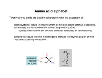
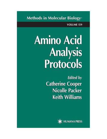
Cancers 2020, 12, 25763 of 17in drug-resistant tumor cells facilitates targeting of metabolic pathways, especially in drug-induced scenarios,such as CDDP-induced resistance. In the current study, we applied a metabolomics-based approach todecipher the role of amino acid metabolism in regulating cisplatin-insensitivity of neuroblastoma using cellline models. Our study establishes the reliance of CDDP-insensitive cells on amino acids for surviving incisplatin-treated conditions.2. Results2.1. Cisplatin-Insensitive Neuroblastoma Cells Exhibit Metabolic PlasticityIn order to sort out the CDDP-sensitive and -insensitive neuroblastoma cell line models,we evaluated the survival response of different neuroblastoma cells, including SK-N-BE(2)C, SK-N-DZ,CHLA-255, SK-N-AS, and SK-N-BE(2)Cres —a CDDP-resistant model developed in our laboratory—,and murine NB-975 cells, upon treatment with various concentrations of CDDP. As shown inFigure 1A, SK-N-BE(2)Cres , SK-N-AS, and murine NB-975 cells showed less sensitivity to CDDP,whereas CHLA-255, SK-N-BE(2)C, and SK-N-DZ showed higher sensitivity to CDDP as evident fromthe differences in cell survival upon drug treatments. We confirmed the difference in sensitivitiesof our model cell lines by analyzing percentage of the late and early apoptotic cells detected afterCDDP-treatments using Annexin-V staining and flow cytometric analyses. As shown in Figure 1B,cisplatin-sensitive CHLA-255 and SK-N-BE(2)C cells showed significantly high percentage of apoptoticcells in CDDP-treatments, in comparison to the vehicle treated conditions. In contrast to sensitive cells,apoptotic phenotype was not altered in CDDP-treated SK-N-AS cells in comparison to the vehicletreatments. Intriguingly, the percentage of apoptotic cells was low in CDDP-treated SK-N-BE(2)Crescells in comparison to their vehicle treated counterparts (Figure 1B). Hereafter we referred SK-N-AScells as CDDP-insensitive and CHLA-255 as CDDP-sensitive cells throughout the rest of the manuscript.To characterize the metabolic changes upon CDDP-treatment, we used SK-N-AS (CDDP-insensitive)and CHLA-255 (CDDP-sensitive) cell lines as innately insensitive and sensitive CDDP-cell lines modelsfor polar metabolomic analysis. We present the metabolite differences between control and CDDPtreated cells in the form of the Principal Component Analysis (PCA) plot. As evident from the PCAplot in Figure 1C, CDDP-sensitive CHLA-255 cells and CDDP-insensitive, SK-N-AS cells separated onthe PC1 axis, indicating metabolic differences among these cells. Furthermore, the segregation of thevehicle and CDDP-treated, SK-N-AS and CHLA-255 cells was different along the PC2 axis. The vehicleand CDDP-treated, cisplatin-sensitive CHLA-255 cells showed relatively minor change in their polarmetabolic profile, indicated by the close clustering of the vehicle and CDDP-treated groups on thePCA plot (Figure 1C). In contrast to the sensitive cells, vehicle and CDDP-treated cisplatin insensitive,SK-N-AS cells showed higher separation along the PC2 axis (Figure 1C), indicating significant contrastin the metabolite pools of SK-N-AS cells upon CDDP-treatment. In addition to the PCA analysis,the metabolite changes presented in the form of heatmap analysis also indicated that sensitive andinsensitive neuroblastoma cells exhibited a contrasting trend in metabolic profiles upon CDDP-treatment(Figure 1D). Based on metabolic profile in the heatmap, vehicle-treated, sensitive cells and insensitivecells clustered closely with their respective CDDP-treated groups. Further, heatmap analysis revealedthat the relative alterations in the individual metabolite levels were more drastic in SK-N-AS cells upontreatment with CDDP.
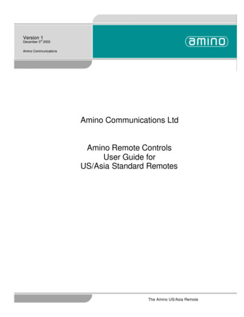
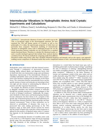
Cancers 2020, 12, 2576Cancers 2020, 12, x FOR PEER REVIEW4 of 174 of 17Figure 1. Cisplatin (CDDP) treatment alters metabolism in neuroblastoma. (A) The half-maximalFigure 1.Cisplatin (CDDP) treatment alters metabolism in neuroblastoma. (A) The half-maximalinhibitory concentration (IC50) curves showing different survival response in neuroblastoma cellsinhibitory concentration (IC50 ) curves showing different survival response in neuroblastoma cellsupon cisplatin (CDDP) treatment. Each data point represents mean SE values from three biologicalupon cisplatin(CDDP)Eachpercentagedata pointofrepresentsmean SEvalues fromthree24biologicalreplicates.(B) Bartreatment.graphs showingtotal (late andearly)apoptoticcells afterhreplicates.(B) Bargraphsshowingpercentageof intotal(late and cells.early)apoptoticafter 24 htreatmentswitheither vehicleor CDDPtreatmentsneuroblastoma* and** indicatecellsp 0.05treatmentseithervehicleor CDDPtreatmentsneuroblastomacells. * t-test.and **and pwith 0.01obtainedfrom threebiologicalreplicate indataderived using Student’s(C)indicatePrincipalp 0.05Analysisshowingreplicatethe segregationof vehicleand Student’scisplatin (CDDP)and p Component0.01 obtainedfrom(PCA)three plotbiologicaldata derivedusingt-test.treated(C) Principalcells basedon theirmetaboliteprofiles. Eachcircle representsbiologicalreplicateof the treatedComponentAnalysis(PCA)plot showingthe coloredsegregationof vehiclea andcisplatin(CDDP)treatment condition. (D) Heatmap showing the clustering of vehicle and cisplatin (CDDP) treatedcells based on their metabolite profiles. Each colored circle represents a biological replicate of thegroups. Colored cells in each row represent metabolites, and each column represents a sample. Thetreatmentcondition. (D) Heatmap showing the clustering of vehicle and cisplatin (CDDP) treatedscale for the heatmap is shown under the heatmap.groups. Colored cells in each row represent metabolites, and each column represents a sample. The scaleAminoAcid Metabolism in Drug Insensitive Neuroblastoma Cellsfor 2.2.the CisplatinheatmapTreatmentis shownAltersunderthe heatmap.validated the key differences in metabolic pathways altered upon drug treatment in CDDP2.2. CisplatinWeTreatmentAlters Amino Acid Metabolism in Drug Insensitive Neuroblastoma Cellsinsensitive and sensitive neuroblastoma cells. Pathway enrichment analysis using MetaboAnalystthe top50 metabolicpathways inalteredin tedthekey differencesmetabolicpathwaysalteredSK-N-ASupon anddrugtreatment e and sensitive neuroblastoma cells. Pathway enrichment analysis using MetaboAnalystencompassing amino acids, vitamins, carbohydrates (like Glycolysis, Warburg effect), sugar andrevealed the top 50 metabolic pathways altered in CDDP-treated insensitive, SK-N-AS and sensitive,CHLA-255 cells compared to their respective vehicle-treatments (Figure 2). Metabolic pathwaysencompassing amino acids, vitamins, carbohydrates (like Glycolysis, Warburg effect), sugar and nucleotidemetabolism, mitochondrial metabolism (including branched amino acid metabolism, Citric acid cycle,and β-oxidation), and peroxisomal metabolism were significantly altered in SK-N-AS cells upon treatmentwith CDDP. Metabolic pathways in particular central carbon and nucleotide related (Pentose phosphate
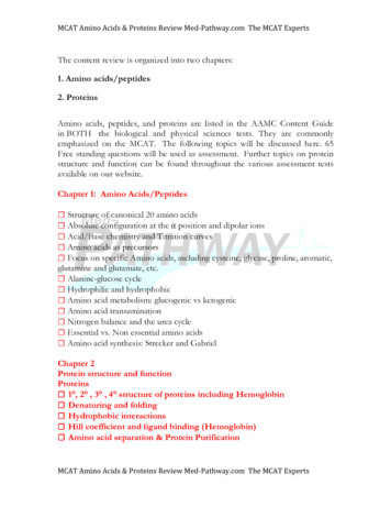
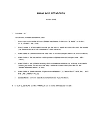
Cancers 2020, 12, x FOR PEER REVIEW5 of 17nucleotide metabolism, mitochondrial metabolism (including branched amino acid metabolism,5 of 17Citric acid cycle, and β-oxidation), and peroxisomal metabolism were significantly altered in SK-NAS cells upon treatment with CDDP. Metabolic pathways in particular central carbon and nucleotiderelated Warburg(Pentoseeffect,phosphatepathway, Warburgeffect,gluconeogenesis,and mannose,pathway,gluconeogenesis,fructose andmannose,pyrimidinefructoseand pyruvatemetabolicpyrimidine and pyruvate metabolic pathways), and lipid metabolic pathways (inositol, bile salts,pathways), and lipid metabolic pathways (inositol, bile salts, glycolipid, sphingolipid, and phospholipidglycolipid, sphingolipid, and phospholipid metabolic pathways) were among the most significantlymetabolic pathways) were among the most significantly altered pathways in CHLA-255 cells upon CDDPaltered pathways in CHLA-255 cells upon CDDP treatment. Thus, alterations in metabolic pathwaystreatment. Thus, alterations in metabolic pathways observed in CDDP-treated, sensitive, CHLA-255 cellsobserved in CDDP-treated, sensitive, CHLA-255 cells were distinct from CDDP-treated, insensitive,were distinct from CDDP-treated, insensitive, SK-N-AS cells based on the differences in the mostSK-N-AS cells based on the differences in the most significantly affected pathways. Among the topsignificantly affected pathways. Among the top 50 hits, there was a high frequency of alterations in50 hits, there was a high frequency of alterations in amino-acid metabolic pathways in CDDPamino-acidmetabolicpathwaysin CDDP-insensitive,cells. Therefore,we concludethat theinsensitive,SK-N-AScells. Therefore,we conclude SK-N-ASthat the metabolismof Methionine,Tyrosine,metabolismof Methionine,Lysine,Valine, Lysine, onine,Histidine,and tedwerein ive,SK-N-AScellsuponsignificantly altered in insensitive, SK-N-AS cells upon CDDP-treatment,CDDP-treatment,werenot noticedin sensitive,which were notwhichnoticedin sensitive,CHLA-255cellsCHLA-255(Figure 2). cells (Figure 2).Cancers 2020, 12, 2576FigureCisplatin treatmentmetabolicpathwaysin neuroblastomacells. ThepathwayimpactFigure2. sin neuroblastomacells.The din(A)cisplatin-insensitive,SK-N-ASandimpact analyses showing top 50 metabolic pathways altered in (A) cisplatin-insensitive, oncisplatintreatment.Threebiologicalreplicateseach condition.condition. Theplot,whereasthethecolorin eachThe ,whereascolorscale adjacent to the respective plot indicates p-values for the impacted pathways.scale adjacent to the respective plot indicates p-values for the impacted astomaCellsCellsUpregulateUpregulate Amino Acid2.3.2.3.Cisplatin-InsensitiveAcid etabolitelevelsin idualaminoacidmetabolitelevelsin nupregulationeightcisplatin as presented in Figure 3. Our comparative analysis revealed an upregulation of eightofessentialessentialamino acids(Histidine,Isoleucine,Leucine, Lysine,Methionine,Phenylalanine,aminoacids (Histidine,Isoleucine,Leucine,Lysine, Methionine,Phenylalanine,Tryptophan,and Valine)in CDDP-insensitive, SK-N-AS cells upon treatment with CDDP (Figure 3). The upregulation ofessential amino acids, and their derived metabolites was noticed in only few metabolites in CDDPtreated, sensitive CHLA-255 cells. Thus, the frequency of upregulation in amino acids and theirmetabolites was relatively low in sensitive cells compared to SK-N-AS cells (Figure 3). We also observed
Cancers 2020, 12, x FOR PEER REVIEW6 of 17Tryptophan, and Valine) in CDDP-insensitive, SK-N-AS cells upon treatment with CDDP (Figure 3).The upregulation of essential amino acids, and their derived metabolites was noticed in only fewCancers2020, 12,in25766 of 17metabolitesCDDP treated, sensitive CHLA-255 cells. Thus, the frequency of upregulation in aminoacids and their metabolites was relatively low in sensitive cells compared to SK-N-AS cells (Figure3). We also observed that Tryptophan levels were significantly higher in both sensitive andthat Tryptophan levels were significantly higher in both sensitive and insensitive neuroblastoma cells.insensitive neuroblastoma cells. However, Tryptophan derived metabolites Serotonin,However, Tryptophan derived metabolites Serotonin, Aminobutyrate, and 5-Hydroxyindole aceticAminobutyrate, and 5-Hydroxyindole acetic acid (HIAA) were significantly upregulated only inacid (HIAA) were significantly upregulated only in CDDP-treated, insensitive but not in CHLA-255CDDP-treated, insensitive but not in CHLA-255 cells. Similarly, the relative increase of Leucine,cells. Similarly, the relative increase of Leucine, Histidine, Lysine, Isoleucine, Homocysteine, Serine,Histidine, Lysine, Isoleucine, Homocysteine, Serine, and Aspartate levels was also higher in CDDPand Aspartate levels was also higher in CDDP-treated, insensitive, SK-N-AS cells compared totreated, insensitive, SK-N-AS cells compared to CDDP-treated, sensitive cells. In conclusion, out ofCDDP-treated, sensitive cells. In conclusion, out of 16 amino acids that were altered, we observed16 amino acids that were altered, we observed only three amino acids (Glycine, Asparagine, andonly three amino acids (Glycine, Asparagine, and Valine) to be relatively enhanced in CDDP treatedValine) to be relatively enhanced in CDDP treated sensitive cells compared to CDDP-treatedsensitive cells compared to CDDP-treated insensitive cells (Figure 3).insensitive cells (Figure 3).Figure3.3. Aminoin eAmino vehicleandcisplatin-treatedinsensitiveBar graphs showing relative quantification of amino acid levels in vehicle and cisplatin-treatedSK-N-AS andsensitivecells. Eachcells.bar representsmean SEmeanof itive CHLA-255Each bar represents SEof datafromobtainedbiological*, ** and ****, indicatep indicate0.05, p p 0.01andp p 0.010.001,which werefromthree replicates.biological replicates.** and *** 0.05,andrespectively,p 0.001, respectively,which were obtained through Student’s t-test by comparing the relative metabolite of the treated cellline with its respective vehicle, control value.
Cancers 2020, 12, 25767 of 172.4. Amino Acid Deprivation Abrogates Survival and Cisplatin-Sensitivity of Neuroblastoma CellsUpregulation of amino acids in CDDP-insensitive cells prompted us to evaluate the role ofamino acids in the survival response of neuroblastoma cells. We prepared cell culture mediawith or without (w/o) individual essential amino acids, including Arginine (R), Cysteine (C),Isoleucine (I), Leucine (L), Lysine (K), Methionine (M), Phenylalanine (F), Tryptophan (W), Tyrosine (Y),and Tryptophan (W). Cell survival data derived from neuroblastoma cells cultured in either regular ormedium-lacking individual amino acids are presented in Figure 4A. Deprivation of most of the essentialamino acids resulted in significant reduction of cell survival in CDDP-sensitive (CHLA-255) andCDDP-resistant [SK-N-B(E)2Cres ] cells (Figure 4A). We also evaluated the survival of cisplatin-resistant,human SK-N-BE(2)Cres and murine NB-975 cells in Tryptophan-catabolite supplemented media,since Tryptophan derived metabolites were upregulated in CDDP-insensitive cells (Figure 3). Both thecisplatin-insensitive, SK-N-BE(2)Cres and murine NB-975 neuroblastoma cells showed reduction incell survival in 72 h and ten-day cultures performed in Tryptophan-deprived media (Figure 4B).Interestingly, culturing SK-N-BE(2)Cres cells in Tryptophan catabolites supplemented media reducedthe survival of these neuroblastoma cells in the long-term (10 days) cultures, indicating a negativeeffect of Tryptophan catabolites on cell survival (Figure 4B). From the experimental data we concludedthat the reduction in cell survival observed in neuroblastoma cells as evident in our amino aciddeprived conditions was a cumulative effect of a decrease in multiple amino acid derivatives ratherthan individual catabolites of amino acids.Further, we evaluated the role of amino acids on CDDP-sensitivity through cell survival analysisusing two cisplatin-insensitive cell lines; SK-N-AS that we applied for metabolomic analysis andSK-N-BE(2)Cres cells. We observed that deprivation of specific amino acids sensitized CDP-insensitive,SK-N-AS and SK-N-BE(2)Cres neuroblastoma cells to CDDP (Figure 4C). Culturing both theCDDP-insensitive cells with a fixed dosage of CDDP concentration less than the IC50 for eachcell line, in media deprived of individual amino acids, M, S, T, F, P, W, Y, and V reduced the survival ofthese two cell lines in a significant manner as indicated by the significant differences in survival valueswith p 0.001(***) obtained through One-way ANOVA and Sidak’s multiple comparisons test shownin Figure 4C.2.5. Hydroxychloroquine Suppresses Amino Acid Metabolism in Cisplatin-Insensitive CellsOur cell survival analyses indicated that exogenous amino acids were regulating survival, as wellas CDDP-sensitivity, in neuroblastoma cells (Figure 4C). In addition to exogenous amino acids,autophagy-mediated recycling of amino acids also facilitates the maintenance of amino acids incells subjected to stress conditions. Therefore, we applied hydroxychloroquine (HCQ) for inhibitingamino acid metabolism in cisplatin-insensitive neuroblastoma cells based on the premise that HCQinhibits terminal phase of autophagy, recycling of cellular receptors involved in nutrient uptake,as well as mTOR pathways. For identifying the effect of HCQ on metabolism of drug insensitive cells,we compared the metabolite profiles of SK-N-AS cells treated with CDDP alone and in combinationwith HCQ. The heatmap derived from metabolite pools of CDDP and CDDP HCQ treated cellsindicated that the metabolite profile of CDDP treated SK-N-AS cells were different from HCQ CDDPtreated cells (Figure 5A). HCQ and CDDP combined treatment also affected the metabolic pathwayscompared to CDDP treatment alone, in CDDP-insensitive cells as shown in Figure 5B. We analyzed therelative levels of individual amino acids in CDDP alone with HCQ CDDP treated SK-N-AS cells.Our analyses showed that the relative levels of amino acids and the metabolites derived through aminoacid metabolism were reduced significantly in HCQ CDDP treated conditions compared to CDDPalone treated conditions (Figure 5C). Thus, we concluded that HCQ suppressed amino metabolism inCDDP treated, insensitive, SK-N-AS cells.
Cancers 2020, 12, 2576Cancers 2020, 12, x FOR PEER REVIEW8 of 178 of 17Figure 4. Amino acids regulate cisplatin-sensitivity. (A) Bar graphs showing relative change in cellFigure 4. Amino acids regulate cisplatin-sensitivity. (A) Bar graphs showing relative change in cellsurvival of cisplatin-sensitive (CHLA-255) and cisplatin-insensitive (SK-N-AS) and cells cultured insurvival -insensitive(SK-N-AS)andcontrolcells cultured inand amino-acid-deprivedmedia for72 h.Statistical comparisons weremade betweenindividual amino acid deprivedbar representsmean SE ofdata obtainedfromcontrol andandamino-acid-deprivedmediaconditions.for 72 h.EachStatisticalcomparisonsweremade betweencontrolthree aminobiologicalreplicates.*, ** and*** indicateEachp 0.05, 0.01 and p mean0.001, respectively,whichand individualaciddeprivedconditions.barp representsSE of dataobtained fromwere obtained through Student’s t-test. (B) Left panel: Relative change in survival of cells cultured forthree biological replicates. *, ** and *** indicate p 0.05, p 0.01 and p 0.001, respectively, which were72 h in media constituted either with or without Tryptophan and Tryptophan metabolites. Each barobtained through Student’s t-test. (B) Left panel: Relative change in survival of cells cultured for 72 h inmedia constituted either with or without Tryptophan and Tryptophan metabolites. Each bar representsmean SE of data obtained from three biological replicates. *** indicates p 0.001 obtained throughStudent’s t-test by comparing survival in control media with the survival in modified media as shownin legend. Right panel: Images of cells cultured for 10 days according to conditions maintained in leftpanel of (B). (C) Bar graphs showing changes in cell survival of cisplatin-insensitive (SK-N-BE(2)Cresand SK-N-AS) cells treated with cisplatin for 72 h in control and amino acid deprived media. Each barin the graph represents mean SE of data obtained from either three biological replicates. *** indicatep 0.001 obtained through One-way ANOVA and Sidak’s multiple comparisons test. Standard singleletter amino acid codes were used to represent the amino acid labels in (A,C).
as shown in legend. Right panel: Images of cells cultured for 10 days according to conditionsmaintained in left panel of (B). (C) Bar graphs showing changes in cell survival of cisplatin-insensitive(SK-N-BE(2)Cres and SK-N-AS) cells treated with cisplatin for 72 h in control and amino acid deprivedmedia. Each bar in the graph represents mean SE of data obtained from either three biologicalreplicates. *** indicate p 0.001 obtained through One-way ANOVA and Sidak’s multipleCancers2020, 12, 2576comparisonstest. Standard single letter amino acid codes were used to represent the amino acid labels 9 of 17in (A,C).Figure 5. Hydroxychloroquine (HCQ) inhibits cisplatin-induced amino acid metabolism. (A) Heatmapshowing alterations in metabolic pathways in cispl
Cell survival assays performed for 72 h indicated that treatment with HCQ (2.5 M) reduced cell survival in neuroblastoma cells when treated with HCQ alone or in combination with CDDP (Figure6B). Treatment of CDDP-insensitive cells with HCQ (1.25 M) for ten days also reduced the survival of SK-N-BE(2)Cres and NB-975 cells. Furthermore .