Transcription
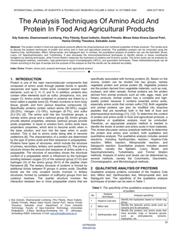
INTERNATIONAL JOURNAL OF SCIENTIFIC & TECHNOLOGY RESEARCH VOLUME 9, ISSUE 10, OCTOBER 2020ISSN 2277-8616The Analysis Techniques Of Amino Acid AndProtein In Food And Agricultural ProductsEdy Subroto, Elazmanawati Lembong, Fitry Filianty, Rossi Indiarto, Gisella Primalia, Miswa Salza Kirana Zaenal Putri,Hanna Christy Theodora, Salsabila JunarAbstract: The protein content in food and agricultural products affects the physicochemical and nutritional properties of these products. This review aimsto discuss the analysis techniques of protein and amino acid in food and agricultural products. The qualitative analysis can be conducted using theHopkins-Cole, Xanthoproteic, Millon, Nitroprusside, and Sakaguchi test. In contrast, the quantitative analysis of proteins can use the Kjehldahl, Biuret,Lowry, UV Spectrophotometry, and Turbidimetry. It also discussed the immunohistochemical techniques to identify cellular or tissue constituents(antigens) by staining techniques, while Formol titration measures the hydrolysis of proteins and N-amino quickly. The amino acids can be analyzed bymicrobiological methods, colorimetric, high-performance liquid chromatography (HPLC), and gravimetric techniques. These methods/techniques can bechosen according to the type of sample and the purpose of the analysis so that the results can be obtained accurately.Index Terms: Protein, amino acid, analysis technique, food, agricultural product—————————— ——————————1. INTRODUCTIONProtein is one of the main macromolecular components thatcomposed of amino acids through peptide bonds in specificsequences and types. Amino acids contained several mainelements, such as C, H, O, and N. In addition, proteins alsocontain other elements such as sulfur, phosphorus, iron, andcopper [1]. Amino acids contained in proteins are linked by abond called a peptide bond [2]. Protein functions to form bodytissue, growth, and form various bioactive compounds [3].Specifically, proteins contain carbon atoms (50-55%), oxygen(20-23%), nitrogen (12-19%), hydrogen (6-7%), and sulfur(0.2-3%) [4]. The amino acid has two functional groups, anamely amino group and a carboxyl group [5]. Amino groupsprovide alkaline properties, whereas carboxyl groups provideacidic properties. In liquid or solution form, amino acids haveamphoteric properties, which tend to become acidic when inthe base solution, and turn into the base when in acidicsolution. This is due to amino acids being able to becomezwitterions [6]. The characteristics of a protein are determinedby the type of amino acids and their sequence in polypeptides.Proteins have types of structures, which include the structureof primary, secondary, tertiary, and quaternary [7]. The primarystructure shows the amount and sequence of amino acids in apolypeptide. The structure of secondary shows the structuralcontent of a polypeptide which is influenced by the hydrogenbonding between oxygen (O) of the carbonyl group (C O) andhydrogen (H) of the amino group (N-H) of the peptide chainframework [8]. The tertiary structure of proteins is formed byadditional bonds between R groups in amino acids. Disulfidebonds are the only covalent bonds involved in tertiarystructures, formed by oxidation of sulfhydryl groups from twocysteinyl residues. The quarter structure involves theinteraction between two or more polypeptide chains that arespecifically associated with forming proteins [2]. Based on thesource, protein can be divided into two groups, namelyvegetable protein and animal protein [9]. Vegetable proteinsare the protein derived from vegetable materials, such as nuts,soybean, and other cereals. Animal proteins are the proteinderived from animal products such as milk, eggs, meat, andfishery products. Animal protein has a complete and highquality protein because it contains essential amino acids,especially amino acids that contain sulfur [10]. Both vegetableand animal proteins can also be modified into bioactivepeptides that are beneficial both in the field of food andpharmaceutical technology [11], [12]. To determine the contentof protein and amino acids in food and agricultural products, aquantitative or qualitative analysis must be conducted.Therefore, an appropriate analysis method is needed toidentify the levels of protein and amino acids in food materials.This review discusses various analytical methods to determinethe protein and amino acid content, both qualitative andquantitative analysis. The qualitative analysis includes severalreactions, including Xanthoproteic reaction, Hopkins-Colereaction, Millon reaction, Nitroprusside reaction, andSakaguchi reaction. Quantitative analysis includes severalmethods, namely the Kjeldahl, Lowry, Biuret, andSpectrophotometry, Turbidimetry, and Formol titrationmethods. Analysis of amino acid levels can be determined byseveral methods, namely the Colorimetric, Gravimetric,Chromatographic, and Microbiological methods.2 QUALITATIVE ANALYSIS OF PROTEINSQualitative analysis proteins consisted of the Hopkins Coletest, Millon test, Xanthoproteic test, Nitroprusside test, andSakaguchi test. The specificity of the qualitative analysistechniques of protein can be seen in Table 1.Tabel 1. The specificity of the qualitative analysis techniquesof —— Edy Subroto, Elazmanawati Lembong, Fitry Filianty, Rossi Indiarto,Gisella Primalia, Miswa Salza Kirana Zaenal Putri, Hanna ChristyTheodora, Salsabila Junar: Department of Food IndustrialTechnology, Faculty of Agro-Industrial Technology, UniversitasPadjadjaran, Jl.Raya Bandung-Sumedang Km. 21, Jatinangor,Sumedang 40600, Indonesia. E-mail: s Cole test2Millon test3Xanthoproteic testAnalysis specificityIdentify the tryptophan based on Indole ringgroup.Identify the phenolic amino acids such astyrosine and its derivatives.Identify the proteins and amino acids thathave aromatic rings or benzene groupssuchasphenylalanine,tyrosine,tryptophan, etc.29IJSTR 2020www.ijstr.org
INTERNATIONAL JOURNAL OF SCIENTIFIC & TECHNOLOGY RESEARCH VOLUME 9, ISSUE 10, OCTOBER 20204Nitroprusside test5Sakaguchi testDetect the presence of cysteine aminoacids.Identify the amino acid arginine based onguanidine group in its side chain.2.1 Hopkins Cole testHopkins Cole Test is one of the qualitative test methods todetermine differences in types of proteins and amino acids.This test is used to identify the presence of the amino acidtryptophan, which is the only amino acid that has an indolering group [13]. Tryptophan belongs to the group of essentialamino acids. Tryptophan is a precursor of niacin vitamin andan introduction to the serotonin nerve. Tryptophan functions tomaximize the use of vitamin B complex, improve nerve health,stabilize emotions, increase feelings of calm, preventinsomnia, and increase the release of growth hormone. One ofthese amino acids can be found in egg whites. Hopkins Colereagents which contain glyoxylic acid (C2H2O3) react withsulfuric acid. Tryptophan in the protein solution will becondensed with the aldehyde group on glyoxylic acid with thehelp of strong oxidizing sulfuric acid. Positive results areindicated by the formation of purple rings between 2 separatelayers [13], [14]. According to Elzagheid [15], the principle oftesting tryptophan by the Hopkins Cole test method is byadding 1 mL of Hopkins Cole reagent into a test tube thatalready contains a 2% protein solution, then the concentratedsulfuric acid solution is added as much as 1-2 mL. Thesolution is homogeneous with vortex, and then color changesare formed.2.2 Millon testMillon test is one of the qualitative test methods to determinedifferences in types of proteins and amino acids, namely thetype of phenolic amino or amino acids that have phenolgroups such as tyrosine and its derivatives [13]. Tyrosine is anon-essential amino acid that has a phenyl group and is aweak acid. One of these amino acids can be found in milkcasein. Millon test is not a specific test, because this testidentifies all types of phenol compounds, so to ensure thattesting is needed by other means [16]. In this test, Millonreagents are used, which are solutions containing mercury(Hg) dissolved in nitric acid [17]. These mercury compoundsbind with hydroxyphenyl groups to produce a white precipitatein a protein solution [18]. Tyrosine in protein solution will forma solution or reddish-brown sediment when heated. Theprinciple of testing tyrosine with the Millon test method is tohomogenate 1-3 drops of Millon reagents into a test tube thatalready contains a 2% protein solution. After that, the solutionis heated using a water bath and observed the changes incolor and the formed deposits [15].2.3 Xanthoproteic testXanthoproteic test aims to determine differences in types ofproteins and amino acids that have aromatic rings or benzenegroups such as phenylalanine, tyrosine, tryptophan, etc. [13].In this test used a solution of concentrated nitric acid and abase in the form of ammonia or sodium hydroxide (NaOH).Phenylalanine, tyrosine, and tryptophan will form whitedeposits that can turn yellow when reacting with nitric acid inthe presence of heat [17]. This yellow color is called Xanthoprotein. Addition of a basic solution such as HNO3 or NaOHwill create a very alkaline so that the nitro compound that hasbeen previously formed can be ionized and the solutionchanges color to dark yellow or orange. The visible colorISSN 2277-8616appears based on the nitration reaction on the aromatic ring ofamino acids [18]. The principle of testing phenylalanine,tyrosine, and tryptophan by the Xanthoproteic test method isby homogenizing 1 mL of HNO3 solution that already containsa 2% protein solution. After that, the sample is heated using awater bath for 15-20 minutes or on fire for 2 minutes, then achange in the color of the solution is observed. The test tube isincubated at room temperature until the temperaturedecreases then NH3 solution or 20-40% NaOH is added to it,and a change in the color of the formed solution is observedagain [15].2.4 Nitroprusside testNitroprusside test is a test used to detect the presence ofcysteine amino acids. This test uses sodium nitroprussidereagents and ammonia solution. The testing principle is thatthe free -SH group called thiol or mercapto, which is owned bythe amino acid cysteine will react with nitroprusside in thecase of excess ammonia to form red compounds. The S-Sgroup in cysteine will give positive results in nitroprussidetesting if it is reduced first. Nitroprusside testing is conductedby entering 0.5 mL of sodium nitroprusside 1% into the testtube then add 2 mL of the sample to be tested, then add 0.5mL of NH4OH solution. The sodium nitroprusside solution usedmust be in a new condition and made just before the test [19].Nitroprusside reactions can be applied in various tests, one ofwhich is to detect the content of cysteine in urine clinically [20].The test was carried out by adding of sodium nitroprusside20% (w/v), then the color intensity was measured using aspectrophotometer at a wavelength of 521 nm in 1 minute, andthe cysteine concentration was calculated using a standardcurve [21].2.5 Sakaguchi TestThe Sakaguchi test is a chemical test that includes acolorimetric reaction for the identification and quantification ofthe amino acid arginine. Arginine is an amino acid that has aguanidine group in its side chain, which is the C atom thatbinds N2 with a single bond and binds N with a double bond.Sakaguchi reaction is carried out using naphthol and sodiumhypobromite or sodium hypochlorite reagents. The guanidinegroup in arginine, which is oxidized by sodium hypochlorite,will react with alpha-naphthol and produce red compoundsunder alkaline conditions; the absorption spectrum producedby the Sakaguchi reaction at a wavelength of 520 nm [22]. Ingeneral, Sakaguchi testing is carried out by inserting about 2mL of the sample into a test tube then adding two drops of 1%α-naphthol in alcohol, 4% sodium hydroxide and 8-10 drops ofbromine water. Testing gives positive results if red complexesare formed [23]. Adding hypobromite to the arginine solutionthat has been given α-naphthol in an alkaline state willproduce a red complex immediately, followed by rapid fadingof the color [24].3 QUANTITATIVE ANALYSIS OF PROTEINQuantitative analysis proteins consisted of the Kjeldahl, unohistochemistry, Formol titration. The specificity of thequantitative analysis techniques of protein can be seen inTable 2.30IJSTR 2020www.ijstr.org
INTERNATIONAL JOURNAL OF SCIENTIFIC & TECHNOLOGY RESEARCH VOLUME 9, ISSUE 10, OCTOBER 2020Tabel 2. The specificity of the quantitative analysis techniquesof protein.NoAnalysis ry5Turbidimetry6Immunohistochemistry7Formol titrationAnalysis specificityDetermine the total protein basedon total nitrogen.Determine the soluble proteincontent based on peptide bonds.Determine the soluble proteincontent in small amounts basedon peptide bonds. More sensitivethan BiuretDetermine the soluble proteinbased on the interaction of thesample with UV light.Determine the protein based onthemeasurementoflightscattering in a cloudy proteinsolution.Determine the cellular antigens(protein) through antigen-antibodyinteractionsDetermine the protein hydrolysisto show N-amino levels3.1 Kjeldahl methodDetermination of total protein in various fields such as biology,pharmacy, environment, and food is generally carried out bythe Kjeldahl method. This method is still recognized by AOACInternational as the official method for protein analysis. Theprinciple of the Kjeldahl method is to measure the total proteinbased on the nitrogen content, which represents the protein inthe materials [25]. In general, the Kjeldahl method is carriedout through 3 stages, namely destruction, distillation, andtitration. The destruction stage involves the destruction oforganic compounds in the sample using sulfuric acid orpotassium sulfate with a catalyst to convert nitrogen in proteininto ammonium sulfate quantitatively. Then the solution isdistilled using excess sodium hydroxide to free ammonia andthen absorb it in boric acid [26]. Ammonia distillate in boricacid is then titrated using hydrochloric acid to measurenitrogen in ammonia that reacts with acids [27]. Nitrogenlevels obtained from the titration results represent the amountof crude protein present in the sample [28]. In general, theconversion factor used in determining total protein using theKjeldahl method is 6.25, which is based on the assumptionthat the general nitrogen content is 16% in food proteins, andall nitrogen in food is bound to protein. This assumption is afairly crude and inaccurate assumption because of therelatively variable nitrogen content between the varying proteinand amino acids content of food products. The presence ofvarious other compounds containing non-protein nitrogen suchas free amino acids, nucleic acids, urea, ammonia, nitrate,chlorophyll, and alkaloids in food products is one proof that theconversion factor 6.25 is less accurate in determining totalprotein. Specific conversion factors for various types of foodhave been made to overcome this problem so that now thecalculation of the conversion of nitrogen into protein is moreprecise [29].3.2 Biuret methodBiuret method is a method used to determine the proteincontent based on peptide bonds in the material being tested.Peptide bonds obtained indicate that the protein containedbecause amino acids bind to other amino acids throughpeptide bonds. The reagents used in this method are NaOHand CuSO4 [30]. The principle of the biuret method is that theISSN 2277-8616protein solution is converted into alkalis with NaOH, then aCuSO4 solution is added so that the protein reacts with Cu 2 toform a blue-purple complex under alkaline conditions. Themore or the longer the peptide bonds contained in the protein,the stronger the purple color produced [31]. Biuret reagentscan be prepared by dissolving 150 mg of copper (II) sulfate(CuSO4.5H2O) and potassium tartrate (KNaC4H4O6.4H2O) in50 mL of distilled water. The next step, add 30 mL of 10%sodium hydroxide while shaken, then add distilled water to thelimit of the measuring flask line [32]. According to Janairo et al.[33], testing by the Biuret method can be conducted by takinga few samples of dissolved protein, such as albumin. Albuminneeds to be precipitated before being used by addingcrystalline ammonium sulfate until it approaches the saturationof ammonium sulfate in solution. The precipitating protein isseparated by centrifuge at 11,000 rpm for 10 minutes. Theprecipitate, which is a protein, is then dissolved with aceticacid (pH 5) of 10 mL. The solution is then taken and biuretreagent added and incubated for 10 minutes. The solutionthen reads its absorbance at a wavelength of 550 nm to theblank containing the biuret reagent and acetate pH 5. Dilutionfactors must be considered, and the absorbance of the sampleshould be within the absorbance range of the standard curve.3.3 Lowry methodLowry method is a development of the Biuret method. Theprinciple of the Lowry method is the reaction that occursbetween Cu2 with peptide bonds and the reduction reaction ofphosphotungstic acid and phosphomolybdic acid bytryptophan and tyrosine contained in a protein [34]. Protein inthe alkaline condition then added phosphomolybdic andphosphotungstic acid to produce a blue color whose thicknessdepends on the concentration of protein contained in thematerials. The resulting bluish color can be measured atwavelengths of 500-750 nm [29]. There are two kinds ofreagents used in the Lowry method, namely solution A andsolution B. Solution A is a mixture of phosphotungstic acid andphosphomolybdic acid in a ratio of (1:1). Solution B can bemade by mixing 2% sodium carbonate in 100 mL of 0.1 Nsodium hydroxide, then adding with 1 mL of 1% copper (II)sulfate and 1 mL of potassium sodium tartrate 2% [35].Determination of protein content by the Lowry method requiresa standard curve that describes the relationship betweenprotein concentration and optical density (OD) [35]. A standardcurve can be made by preparing a bovine serum albumin(BSA) solution with a concentration of 300 µg/ mL. Next, add 8mL of Lowry B reagent into each of the tubes with differentconcentrations and leave it for 10 minutes, then add 1 mL ofLowry A reagent, shake and let stand for 20 minutes. Thesolution obtained was then measured for its absorbance at awavelength of 600 nm against the blank [29]. The test step ofthe Lowry method is almost the same as the protein testingstep of the Biuret method, the difference at the end isdetermined by the addition of 8 mL of Lowry A reagent and soon as in the standard curve.3.4 UV-Spectrophotometry methodDetermination of protein content by the UV Spectrophotometrymethod is based on the interaction of the sample with UV light[36]. UV light has a wavelength of around 100-400 nm. UVrays cannot be seen in the human eyes; therefore, compoundsthat can absorb UV rays are compounds that have a clear andtransparent color. The principle of the UV spectrophotometer31IJSTR 2020www.ijstr.org
INTERNATIONAL JOURNAL OF SCIENTIFIC & TECHNOLOGY RESEARCH VOLUME 9, ISSUE 10, OCTOBER 2020method is that the tested sample must be clear and completelydissolved; there are no colloidal particles or suspensions [37].Tryptophan, tyrosine, and phenylalanine are amino acidsmaking up proteins that have aromatic groups. Tryptophan hasa maximum absorption at 280 nm [38]. Tyrosine has amaximum absorption at 278 nm [39]. Phenylalanine has lessstrong light absorption and absorption at shorter wavelengths.Estimation of protein concentration in a test solution can beseen absorbance at 280 nm. For more accurate results, it isnecessary to correct possible contamination of nucleic acidcontent at 260 nm. The absorption ratio of 280/260 nm can beused to determine the correction factor [40].3.5 Turbidimetry methodTurbidimetry is the analysis method based on themeasurement of light scattering in solution. The method ofmeasurement is by reducing the intensity of the light afterpassing through the suspension solution. Turbidimeters applya continuous scattering of light or 180 , in contrast to anephelometer that uses light scattering with an angle of 90 [41]. Basically, turbidimetry measures the ratio between theintensity of the light that is passed on with the intensity of theinitial beam. The continued measurement of light intensity as afunction of concentration is a basic principle of turbidimeterequipment. Protein analysis using the turbidimetry method canuse benzethonium chloride (BZ) from cationic detergents. Theresults showed that turbidity is dependent on pH and isreversible. However, when BZ concentrations in sediment arelow, turbidity decreases with increasing pH at a higher pHrange. Turbidity formation by aromatic organic acids such assulfosalicylic acid is reversible and can only be observed in thelower pH range [42]. The formation of turbidity in theturbidimetry method uses BZ because the positively chargedcationic detergent will bind to the negatively charged protein inthe pH range of 5-6 with the dissociated and complex carboxylgroups produced together by their respective intermolecularforces. However, when the BZ concentration is low, theprinciple of reaction that has been reported is not enough toexplain the phenomenon that turbidity decreases withincreasing pH in a higher pH range. According to Boumaza etal. [43], the turbidimetry method is suitable to be implementedto evaluate protein from milk and cereal wastewater. This hasproven to be effective in terms of analysis time and precision,so as to avoid the disadvantages of using other classicalmethods to determine proteins that usually take a long timeand are expensive.3.6 Immunohistochemistry methodImmunohistochemistry (IHC) is the method that can be usedfor identifying cellular antigens through antigen-antibodyinteractions, where binding of antibodies are identified bydirect labeling of antibodies or by using a secondary labelingmethod [44]. Immunohistochemical staining methods relatedto the use of antibodies labeled enzymes (immunoperoxidase)and labeled fluorophore (immunofluorescence) to identifyproteins in cells. In general, the mechanism of theimmunohistochemical method begins with deparaffinization.Then proceed with the collection of antigens. The unspecifiedbinding site is blocked and primary antibody bound. Then, thebiotinylated secondary antibody is bound as well. In thismethod, detection uses the peroxidization-anti peroxidasemethod, the biotin-avidin conjugate, the peroxidation complexor the two-step polymer labeling method, which is more widelyISSN 2277-8616used. Then, additional chromogen substrate, usually DAB, andthe final stage is counterstaining and dehydration. There aretwo basic methods of using immunohistochemistry to identifyantigens in tissues, namely the direct method and the indirectmethod. The principle of the direct method is the use oflabeled primary antibodies so that they will bind directly to theantigen directly. Meanwhile, the principle of the indirectmethod is the use of primary antibodies that are not labeled,but this method also uses secondary antibodies that alreadyhave a label and will react with Immunoglobulin G (IgG) fromthe primary antibody. The advantages of immunohistochemicalmethods are the location and distribution of visible proteins,which can be detected in the biopsy of small and large tissuesas well as fixed tissues, and validation of other high yieldstudies (DNA microarrays). While the shortcomings of thismethod are limited ability to measure protein content,problems with antibody types, limited ability to detect proteinmodification, limited or less evidence-based criteria, nomethod of normalizing, the results are limited, and limitedcapacity for creating clinical biomarker profiles (only withtissue microarrays) [45].3.7 Formol titration methodFormol titration is a titration that is usually conducted todetermine the protein content in milk quickly. In addition,formol titration can also be used to measure protein hydrolysisto show N-amino levels in a processing or storage process.The principle of the formol titration method is to neutralize thesolution with a NaOH added with formaldehyde in which theamino group is bound and does not affect the acid-basereaction of NaOH to form Dimethylol. The indicator commonlyused is phenolphthalein (PP), and the color change reactionturns pink at the end of the titration [46]. In general, thedetermination of protein content by formol titration isconducted by weighing a sample that has been mashed about10 g, then dissolved in distilled water and shaken out with astirrer for 15 minutes. The filtrate is filtered and diluted withdistilled water, then taken about 10 mL to be added withdistilled water, potassium oxalate, and PP indicator. Thereaction mixture is then titrated with 0.1 N NaOH until it turnspink. The titrated sample added 2 mL of 40% Formaldehyde,and the PP indicator was added. After that, it was titratedagain using 0.1 N NaOH, then calculate the protein content[47].4 AMINO ACID ANALYSIS4.1 Colorimetric MethodThe colorimetric is a chemical analysis that is included inphotometric analysis. The principle of the analysis is tocompare the intensity of the color between the sample solutionmade using a Nessler tube or Dubosque colorimeter with thecolor of the solution whose concentration is known or is calleda standard solution, using polychromatic light as a light sourceand the eye as a detector. The colorimetric method is widelyused in the amino acid analysis because of the low costsincurred, minimal equipment used, the discoloration can beobserved easily even at very low concentrations [48]. Colorduplication in the analysis using the colorimetric method isconducted by using two solutions in an upright position in thedirection of the light or visualization tool that has the samesubstance in the column with the same cross-sectionaerometer capabilities. There are several reagents used in32IJSTR 2020www.ijstr.org
INTERNATIONAL JOURNAL OF SCIENTIFIC & TECHNOLOGY RESEARCH VOLUME 9, ISSUE 10, OCTOBER 2020amino acid analysis using the colorimetric method, amongwhich the most common is ninhydrin and several otherreagents such as 2,4-dinitrofluorobenzene and theirderivatives and sodium nitroprusside [49]. The colorimetricmethod with the ninhydrin reagent is widely used for theanalysis of compounds containing amino groups in thepharmaceutical products and food industry [50]. Amino acidanalysis using the colorimetric method can be conducted byadding 1 mL of the ninhydrin reagent to 5 mL of the sampleand reacted at 80-100 C for 4-7 minutes, then cooled to roomtemperature. The sample is then absorbed using aspectrophotometer and compared with a standard solution.4.2 High-performance liquid chromatography (HPLC)methodHPLC is one method that is often used to separate heatresistant compounds such as amino acids [51]. HPLC is aspecial type of column chromatography. This method uses ahigh-pressure liquid as the mobile phase [52]. The principle ofamino acid analysis using this method is freeing amino acidsfrom protein through hydrolysis by HCl 6 N. Hydrolyzate isthen dissolved with a sodium citrate buffer which then theseamino acids will be separated by HPLC [53]. Analysis of aminoacids by the HPLC method can be conducted using OPAreagents [54]. Provision of OPA reagents can be conducted bypreparing 50 mg of OPA, 4 mL of methanol, 0.025 mL ofmercaptoethanol, 0.050 mL of 30% Brij-30, and borate buffer0.5 M, pH 10.4. The second step is to prepare mobile phase A,which consists of 2 g Na-acetate hydrate, 90 mL methanol, 0.5g Na-EDTA, and 10 mL THF. The reagent mixture was addedwith water up to 1 liter in a measuring flask; then the pH wasadjusted to 6.5 by NaOH. Furthermore, mobile phase Bconsisting of 95% methanol is made. Both phases werefiltered with a 0.45 µl membrane filter. The next step is to dosample preparation. First, insert a sample containing 3 mg ofprotein into the screw tube; then add 1 mL of HCl 6 N.Hydrolysis by heating the tube in an oven at 110 C for 24hours, then cooled. The sample was filtered with sinteredglass, rinsed several times with 0.01 N HCl then dried with avacuum evaporator. Next, re-dissolve the dried sample with 5ml of 0.01 N HCl. The sample is ready to be injected intoHPLC [55]. Injection of the sample at HPLC is conducted bypreparing a sample, then adding with potassium borate in aratio of 1: 1. A total of 1 µl of the sample is put into an emptyvial, then 25 µl of OPA reagent is added, leave it for 1 minuteso that the derivatization is fully flawed. Furthermore, inject asample of 5 µl into the HPLC, wait until the amino acidseparation is complete, the time required is approximately 30minutes [56].4.3 Microbiological methodsMicrobiological method is one of the quantitative test methodsfor amino acid content in a material [57]. The implementationof amino acid testing using microbiological methods is notmore widely used compared to other methods. This is becauseto determine accurate results, and this testing requires a longtime and adequate expertise. But in addition to theseshortcomings, this method has very good sensitivity to aminoacids, and the process does not cost a lot. The use ofmicrobiology and biotechnology continues to grow rapidly bothfor analysis and bioprocess purposes [58], [59]. Utilization ofmicroorganism growth is used in microbiological test methods.Some of the most commonly used bacteria are LactobacillusISSN 2277-8616arabinosus for leucine, isoleucine, valine, and tryptophan;Leuconostoc mesenteroides for histidine, phenylalanine, andmethionine; and Streptococcus faecalis for arginine andthreonine. Apart from microorganisms, other importantcomponents involved in the microbiological test method arethe medium in which th
Identify the amino acid arginine based on guanidine group in its side chain. 3 2.1 Hopkins Cole test Hopkins Cole Test is one of the qualitative test methods to determine differences in types of proteins and amino acids. This test is used to identify the presence of the amino acid tryptophan, which is the only amino acid that has an indole