Transcription
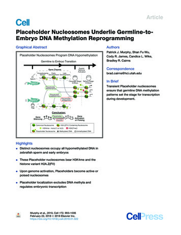
ArticlePlaceholder Nucleosomes Underlie Germline-toEmbryo DNA Methylation ReprogrammingGraphical AbstractAuthorsPatrick J. Murphy, Shan Fu Wu,Cody R. James, Candice L. Wike,Bradley R. CairnsCorrespondencebrad.cairns@hci.utah.eduIn BriefTransient Placeholder nucleosomesensure that germline DNA methylationpatterns set the stage for transcriptionduring development.HighlightsdDistinct nucleosomes occupy all hypomethylated DNA inzebrafish sperm and early embryosdThese Placeholder nucleosomes bear H3K4me and thehistone variant H2A.Z(FV)dUpon genome activation, Placeholders become active orpoised nucleosomesdPlaceholder localization excludes DNA methyla andregulates embryonic transcriptionMurphy et al., 2018, Cell 172, 993–1006February 22, 2018 ª 2018 Elsevier Inc.https://doi.org/10.1016/j.cell.2018.01.022
ArticlePlaceholder Nucleosomes UnderlieGermline-to-Embryo DNA Methylation ReprogrammingPatrick J. Murphy,1 Shan Fu Wu,1 Cody R. James,1 Candice L. Wike,1 and Bradley R. Cairns1,2,*1Howard Hughes Medical Institute, Department of Oncological Sciences and Huntsman Cancer Institute, University of Utah School ofMedicine, Salt Lake City, UT 84112, USA2Lead Contact*Correspondence: cell.2018.01.022SUMMARYThe fate and function of epigenetic marks duringthe germline-to-embryo transition is a key issue indevelopmental biology, with relevance to stem cellprogramming and transgenerational inheritance.In zebrafish, DNA methylation patterns are programmed in transcriptionally quiescent cleavage embryos; paternally inherited patterns are maintained,whereas maternal patterns are reprogrammed tomatch the paternal. Here, we provide the mechanismby demonstrating that ‘‘Placeholder’’ nucleosomes,containing histone H2A variant H2A.Z(FV) andH3K4me1, virtually occupy all regions lacking DNAmethylation in both sperm and cleavage embryosand reside at promoters encoding housekeepingand early embryonic transcription factors. Upongenome-wide transcriptional onset, genes withPlaceholder become either active (H3K4me3) orsilent (H3K4me3/K27me3). Notably, perturbationscausing Placeholder loss confer DNA methylationaccumulation, whereas acquisition/expansion ofPlaceholder confers DNA hypomethylation andimproper gene activation. Thus, during transcriptionally quiescent gametic and embryonic stages, anH2A.Z(FV)/H3K4me1-containing Placeholder nucleosome deters DNA methylation, poising parentalgenes for either gene-specific activation or facultative repression.INTRODUCTIONA major issue in early development is how chromatin packagingof the parental genomes in embryos helps ensure proper generegulation during and after initial genome-wide transcriptionalactivation. Chromatin packaging greatly differs between maturesperm and eggs, yet these differences are largely harmonized(imprinted loci excepted) via chromatin reprogramming in earlyvertebrate embryos (Akkers et al., 2009; Dahl et al., 2016;Hammoud et al., 2009; Liu et al., 2016; Vastenhouw et al.,2010; Wu et al., 2011; Zhang et al., 2016). The zebrafish (Daniorerio) model system offers an opportunity to study this reprog-ramming process in detail because of the abundant availabilityof sperm, eggs, and embryos and key properties of zebrafishcleavage-stage embryos. Unlike mammals, prior to initiatingzygotic genome transcriptional activation (ZGA), zebrafish undergo 10 rapid cell divisions during cleavage phase, resultingin a transcriptionally active 4,000-cell largely pluripotent embryo at 4 hr post-fertilization (4hpf) (Lee et al., 2013).In zebrafish sperm and eggs, both the paternal and maternalgenomes are solely packaged in histones, whereas mammaliansperm utilizes primarily protamine (Wu et al., 2011; Zhang et al.,2016). However, similar to mammals, the zebrafish spermgenome appears ‘‘poised’’ by chromatin for the future activationof particular gene categories. For example, zebrafish sperm lackDNA methylation at the promoters of housekeeping genes(H3K4me3-marked) and also at bivalent genes (H3K4me3 andH3K27me3-marked; Bernstein et al., 2006) that encode transcription factors (TFs) for guiding somatic development (e.g.,hox-family, gata-family factors), pluripotency factors (e.g., nanogand pou5f3 - zebrafish OCT4 ortholog), and genes for germlineidentity/function (e.g., dazl, piwil1) (Wu et al., 2011, Potoket al., 2013). Thus, the paternally inherited DNA methylationpatterns of zebrafish sperm are compatible with achievingpluripotency and with the derivation of germline. Likewise,zebrafish oocytes lack DNA methylation at housekeeping genesand certain developmental genes; however, they possess DNAmethylation at several hundred loci important for development,pluripotency, and germline identity/function (e.g., many hoxfamily genes, nanog, dazl). Whereas the paternal DNA methylation patterns are virtually identical to those in pluripotentembryos at 4hpf, the maternal genome is gradually reprogrammed to the paternal pattern during cleavage (Jiang et al.,2013; Potok et al., 2013). Presently, the mechanisms that enablemaintenance of paternal DNA methylation and reprogramming ofmaternal DNA methylation in zebrafish embryos are unknownand may inform similar issues in mammals.In other systems, achieving DNA hypomethylation(hypoDNAme) occurs via a host of mechanisms; however,many of these cannot account for DNA methylation reprogramingin early zebrafish embryos. For example, H3K4-methylation and/or H3K27me3 antagonize DNA methylation (Brinkman et al.,2012; Ooi et al., 2007; Zhang et al., 2010), yet cleavage-stagezebrafish embryos display strong focal hypoDNAme in theabsence of high H3K4me3 and/or H3K27me3, which aredepleted during cleavage (Lindeman et al., 2010; Vastenhouwet al., 2010; Zhang et al., 2014). Likewise, bivalent regions areCell 172, 993–1006, February 22, 2018 ª 2018 Elsevier Inc. 993
ACPlaceholderNucleosomeMethylated CG1.010-million regions(representative)TsDNMUnmethylated CGMetagene EnrichmentSperm ChIP-Seq10X Zoom0.0-1kbTSSH3K27me3H3K4me3 1kb 2kbH2AFVH3K4me1DNAmeH3K4me3TTS 1kbH3K27acH3K14acH3K27me323ChIP Enrichment Z-Scores or DNAme Fraction 0H3K4me1H2AFVH3K14ac1H3K27acAll PromotersD1130K regions(representative)NormalizedEnrichmentBGenome WideK4me3 K27me3 K4me1 H2AFV K14ac K27ac DNAmeDNMTs-1.5kbTSSChIP Enrichment or DNAme Fraction 1.5kbFEH3K4me301.0All K27acH3K27me3H2AFVH3K4me1Genome VH3K4me3H3K27acDNAmeWGBS Coveragehibabhbhoxa13bhoxa10bPearsonR Values1.0-0.5hoxa2bGH2AFVH3K4me1DNAmeDNAme rg1Figure 1. Identification of Placeholder Features in Zebrafish Sperm Chromatin(A) A model for ‘‘Placeholder’’ nucleosome blocking DNA methyltransferases (DNMTs) to facilitate focal hypoDNAme.(B) Metagene analysis of normalized ChIP-seq enrichment. Epigenetic features are enriched at the transcription start site (TSS) and depleted over the gene body.(C) K-means clustering of epigenetic features in sperm. Top: representative view of the whole genome. Bottom: 103 zoom for higher resolution. Z scores aredisplayed for ChIP-seq data and fraction methylation for DNA methylation (DNAme). Regions are 200 bp.(legend continued on next page)994 Cell 172, 993–1006, February 22, 2018
also largely absent in early mouse embryos (Liu et al., 2016;Zhang et al., 2016). Thus, alternative factors or modificationsmay be needed in zebrafish to maintain focal hypoDNAme in earlyembryos. Moreover, the use of TET-family enzymes and 5hmC(Hackett et al., 2013; Huang et al., 2014; Tahiliani et al., 2009) isexcluded in cleavage-stage zebrafish (Potok et al., 2013) and, et al., 2016).instead, utilized at/after gastrulation (BogdanovicHere, we provide a mechanism for establishing and maintainingfocal hypoDNAme in cleavage-stage embryos, involving a nucleosome bearing the histone H2A variant H2A.Z(FV) and H3K4me1.The histone H2A variant H2A.Z (termed H2AFV in zebrafish) issimilar to canonical H2A, but differs along its amino terminal‘‘tail’’ and at key residues in the globular domain and C terminus.Precise genomic incorporation of H2A.Z occurs by the nucleosome remodeler SRCAP (Snf2-related CREBBP activator protein) complex (Kobor et al., 2004; Krogan et al., 2003; Wanget al., 2016). H2A.Z installation is antagonized by its chaperoneAnp32e, which promotes H2A.Z removal (Mao et al., 2014;Obri et al., 2014). In vertebrates, H2A.Z-containing nucleosomesreside at most gene promoters, including developmentallyregulated genes (Hu et al., 2013; Ku et al., 2012), and H2A.Zhas roles in differentiation, nucleosome stability, transcriptionalregulation, and Polycomb complex recruitment (Chen et al.,2014; Creyghton et al., 2008; Hu et al., 2013; Jin et al., 2009;Ku et al., 2012). H2A.Z and DNA methylation are antagonistic,both in plants (Arabidopsis thaliana) and in human cancercells (Conerly et al., 2010; Zilberman et al., 2008); however,whether H2A.Z helps maintain DNA hypomethylation duringthe germline-to-embryo transition is unexplored.Here, we utilized zebrafish to address a central question invertebrate germline-to-embryo epigenetics: how are paternalDNA methylation patterns maintained and maternal DNAmethylation patterns reprogrammed during a transcriptionallyquiescent cleavage phase, during which H3K4me3-markedand bivalent chromatin is absent? Our work provides evidencefor ‘‘Placeholder’’ nucleosomes, which consist of zebrafishH2A.Z (H2AFV) and H3K4me1, to ‘‘hold in place’’ hypoDNAmeregions, which then resolve at ZGA into either transcriptionallyactive or poised chromatin. Through a series of gain-of-functionand loss-of-function approaches, we demonstrate the molecularutility of Placeholder in programming in vivo DNA methylationpatterns and in the regulation of transcriptional activation duringearly zebrafish development.RESULTSSpecific Chromatin Features Mark DNA HypomethylatedRegions in SpermDNA hypomethylated regions in sperm and in early embryos arestriking in their sharp boundaries (Jiang et al., 2013; Potok et al.,2013). To account for this precision of focal DNA methylationhypomethylation during zebrafish cleavage stage, we reasonedthat a ‘‘Placeholder’’ type of nucleosome/chromatin bearingparticular histone variants and/or modifications might existto antagonize DNA methylation in both sperm and transcriptionally quiescent embryos (Figure 1A). We identified candidate‘‘Placeholder’’ marks/features by genome-wide chromatinimmunoprecipitation sequencing (ChIP-seq) to profile knownpromoter (H3K4me3, H3K27me3, H3K14ac, and H2A.Z/FV)and enhancer features (H3K4me1 and H3K27ac) in zebrafishsperm. These marks showed the expected profiles at transcription start sites (TSS) and gene body regions (Figures 1Band S1A). K-means clustering of these data revealed low DNAmethylation overlapping with high levels of these marks (Figures1C, cluster 2, and S1B) (DNA methylation data from Potok et al.[2013]). However, DNA methylation appeared compatible withrobust levels of H3K4me1 and low-moderate H3K4me3 (cluster1), indicating their insufficiency (alone, at those levels) for deterring DNA methylation in sperm. In counter-distinction, virtuallyall regions with high H2AFV lacked DNA methylation and wereoften associated with high H3K4me1 and moderate H3K14ac(cluster 2; Figure S1C).Similarly, the majority (55%) of sperm promoters lacked DNAmethylation and were marked with one or more of these modifications, most prominently high H2AFV or H3K4me1/3 (Figure 1D). Promoters with high H3K27me3 and H3K4me3 (bivalent)also contained H2AFV and lacked DNA methylation; these represented only a small fraction ( 5%) of total promoters (Figure 1D,asterisk) but included many key developmental genes (e.g.,hox clusters; Figure 1E). Pearson correlational analyses revealedH3K4me3, H2AFV, and H3K4me1 strongly anti-correlated withDNA methylation both genome-wide and at promoters (Figure 1F), a result clearly evident in browser snapshots (Figure 1G).DNA methylation is anti-correlated with CpG density in mammalsand zebrafish (Potok et al., 2013). Interestingly, nearly all spermloci marked by H2AFV lack DNA methylation, regardless of promoter/intergenic context or CpG density (Figures S1D and S1E).Taken together, H2AFV is strikingly anti-correlated with DNAmethylation and is frequently found with H3K4me1.Acquisition of H2AFV Confers Loss of DNA Methylationin SpermFunctional tests for H2AFV regulation of DNA methylation byh2afv knockout approaches were deterred due to the ubiquitousexpression and separate paralogs of h2afv. Therefore, we disrupted the single-copy gene, srcap, which encodes theconserved catalytic subunit of the chromatin remodeler SRCAPcomplex (Kobor et al., 2004; Krogan et al., 2003; Wang et al.,2016). Our TALEN-mediated disruption (Dahlem et al., 2012) ofsrcap was successful; however, homozygous null animals failed(D) H3K4me3, H3K4me1, H2AFV, and H3K14ac mark hypoDNAme promoters in sperm. Heatmaps of Z scores are displayed for ChIP-seq data and fractionmethylation for DNAme. *, bivalent loci.(E) A genome browser snapshot of the hoxA locus. log10 fold enrichment (FE) values (0–1) for ChIP-seq data and DNA methylation fraction (0–1).(F) H3K4me3, H3K4me1, H2AVF, and H3K14ac are correlated with one another and anti-correlated with DNA methylation in sperm. Clustering of pairwisePearson correlation R values are displayed as a heatmap.(G) A gallery of browser snapshots for three genomic loci depicting the anticorrelation of H2AFV and H3K4me1 with DNAme.See also Figure S1.Cell 172, 993–1006, February 22, 2018 995
FOR1anp32eREVSAla03501TgC-/-STOP SDFOR2anp32e -/-Wt18hpfD5dpf40%10%-1.5kbn 200Wild TypeEH3K4me1anp32eH2AFVFH3K4me1121,430 DVRsH2AFV-3-5-76hpf embryosH2AFVWild Typeyypp20%-1H3K4me1anp32epWild Typeyp anp32eypp60,725 Regions6hpfChIP Enrichment 1.5kbWild TypeH2AFVanp32e-H3K4me1H2AFVH3K4me1DNAmeChange1.0 1.00.0-1.0123ChIPEnrichmentChIP Enrichment 01.0GO Analysis:Cluster 1: Regulation of transcription (p 10-3)Cluster 2: Transcription regulation (p 10-21)Cluster 3: DNA binding (p 10-14)WT DNAmeHDVR CpGsinc.87%dec.WT SpermDNAme (0-1)JH2AFV Change0.6Log10MutantDNAme (0-1)DecreasedDNAmeanp32e-/- DNAme (0-1)G7,340 Regions (DVRs with sufficient RRBS coverage)0.0DecreasedDNAme LociAll LociCpG DensityDistance to TSS1.5Observed/Expected RatioBFigure 2. A Null Mutation for anp32e CausesIncreased Genomic H2AFV and DecreasedDNA Methylation in Spermkrt4 rpl32 anp32e1Log2FC in anp32e-/-A10kb8kb1.06kb4kb0.52kb00.0INucleosome AffinityKPercentile ScoreWT H2AFVanp32e- H2AFVWT H3K4me1anp32e- H3K4me1WT DNAmeWt RRBS Coverageanp32e- DNAmeanp32e- RRBS Coverage0.80.40.0tpst2-1.5kb0 1.5kbDistance to H2AFV Peak Centernlgn4a(exon3)to survive to sexual maturity (phenotypes described later).Also, transplantation of srcap / germline stem cells into wildtype (WT) surrogates solely yielded males that lacked sperm,consistent with a necessity for germline stem cell survival. Therefore, as an alternative strategy, we isolated zebrafish mutantslacking Anp32e, the histone chaperone known to removeH2A.Z in mice (Obri et al., 2014).Zebrafish anp32e null mutants (if viable) would offer the potential to study the impact of H2A.Z mislocalization in gametesand maternal zygotic Anp32e-deficient embryos. To proceed,we obtained the available heterozygous anp32e null mutation(transgenic la013501 insertion; Figures 2A and S2A) (Varshney996 Cell 172, 993–1006, February 22, 2018(A) Scheme for the anp32e mutant allele. A gene trapcassette (la013501Tg) within the anp32e gene causes premature translation termination. PCR primersare indicated.(B) anp32e / homozygotes display a variety ofphenotypes. Lethality occurs at the displayedstages. 30% are viable and fertile.(C) anp32e mRNA in the anp32e mutants. An earlyzygotically transcribed gene (krt4) and a housekeepinggene (rpl32) are unaffected. Error bars represent SD.(D) H2AFV enrichment regions become more broadin anp32e sperm. Heatmaps for H2AFV andH3K4me1 (normalized enrichment) are displayed atall WT sperm H2AFV peaks.(E) anp32e sperm display ectopic H2AFV localization.H2AFV and H3K4me1 enrichment (Z scores) is displayed as a heatmap. Differential variant regions (DVRs)were analyzed and segregate into 3 clusters byK-means clustering. Results of GO analysis for affectedgene categories is shown below.(F) Heatmaps indicate DNA methylation decreases atthe majority of DVRs. Displayed regions are onlythose with sufficient read depth from RRBS-seqassays (see the STAR Methods).(G) Individual CpGs within DVRs indicate decreasedDNAme. Scatterplot of mutant versus control isdisplayed with a bar graph quantifying changingCpGs ( 20% absolute change), and the percentagedecreasing DNA methylation is shown.(H) H2AFV increases more at regions at which DNAmethylation decreases. Boxplots of log10FE valuesare shown. *** p 0.001.(I) Browser snapshots are displayed for WT andanp32e sperm. Regions with sufficient coveragefor DNA methylation data are indicated with barsbeneath each track. Ectopic H2AFV accumulation ishighlighted with red dashed boxes. log10FE data arefrom 0–1 for ChIP-seq and DNA methylation fractionfrom 0–1.(J) Analysis of CpG density (left) and neighboringgene proximity distance (right) for clusters identifiedin (E). t tests: ** p 0.01, *** p 0.001. Boxplots aredisplayed.(K) Analysis of predicted nucleosome affinity atDVRs. Regions from (E) were grouped by cluster andcolored like in (J).See also Figure S2.et al., 2013). Homozygous mutants displayed phenotypic variationwith lethality occurring either at shield stage ( 40%), early somitestage ( 20%), or 5 days post-fertilization (dpf; 10%) (Figure 2B).However, 30% of the embryos survived to adulthood, genotyped as anp32e / , lacked detectable anp32e RNA at shieldstage (unlike WT) (Figure 2C; logFC log fold change), and werefertile, providing a source of anp32e sperm and oocytes.We first examined H3K4me1 and H2AFV localization followingAnp32e loss. Notably, we observed a clear expansion of H2AFVin anp32e mutant sperm, which primarily occurred at intergenicregions (Figure S2B) and often involved H2AFV peak spreadinginto the surrounding H3K4me1-marked region (Figure 2D).
AFigure 3. Maintenance of H2AFV andH3K4me1 at DNA Hypomethylated Regionsin Zebrafish EmbryosBGenome Wide Pearson CorrelationpreZGA DNAmeSpermpostZGA DNAmepreZGApostZGASperm DNAmeSperm H3K14acpostZGA H2AFVH3K4me1preZGA H2AFVpreZGA H3K4me1Sperm H3K4me1H2AFVSperm H2AFVpostZGA H3K4me1postZGA H3K14acH3K14acpreZGA H3K14acCR Values -0.5All Dynamic LociH2AFVSppreZ0K4me1postZ Sp preZ postZ0.51.0K14acSppreZ postZK27acSpK4me3preZ postZ SpK27me3postZ SppostZDNAmeSppreZpostZ32,000 regions (5% sample)All PromotersChIP Enrichment Z-Scores or DNAme Fraction 01(A) Correlation of Placeholder candidates in spermand embryos. Pearson correlation R values aredisplayed, and the order was determined by theclustering in Figure S3C.(B) Maintenance of Placeholder candidates duringsperm-to-embryo transition. Venn diagrams indicate overlap of called peaks comparing sperm(blue), preZGA (green), and postZGA (red) samples.(C) Heatmaps for epigenetic marks in sperm andembryos. Displayed are dynamic loci at whichepigenetic marks change during development(top) and promoter regions (bottom).(D) Haploid parthenogenesis by UV irradiation.Top: in vitro fertilization resulted in phenotypicallyWT or haploid embryos dependent on UV treatment. Bottom: 2-day-old haploid embryos wereshorter with reduced intestinal length, as indicatedby the black bars.(E) DNAme reprogramming is normal in gynogenetic haploids. Genome-wide RRBS revealedDNA methylation in haploids is similar to sperm.Displayed is a heatmap for Pearson correlations.(F) H2AFV patterns in gynogenetic embryos arenormal. Haploid H2AFV at WT H2AFV peaks ishighly similar to normal embryos. Displayed is aheatmap of normalized enrichment.31,953 regionsectopic H2AFV lost DNA methylation(Figure 2F). Although most regions disEggSpermSpermEggEggplayed a modest reduction ( 7.4%Parthenote Sphereaverage) in fraction DNA methylationSpermand few gain DNA methylation (FigDiploid Sphereure 2G), the cluster which received theR Values 0.6Haploid ParthenoteControl Diploidlargest increase in H2AFV (cluster 1)0.81.0Sphere StageSphere StageDiploidParthenoteshowed a striking reduction in meanSphereH2AFVSphereH2AFVF2dpf Haploid ParthenoteDNA methylation (50% to 25%). Reciprocally, regions that decreased in DNAmethylation gained significant H2AFV(Figure 2H), with examples of affectedloci in Figure 2I. (Note: transcription of-1.5kb 1.5kbChIPEnrichment2dpf Diploid ControlDNMTs was not affected; Figure S2C.)DVR clusters differed in CpG contentWe further identified 120,000 de novo ectopic H2AFV regions and location: cluster 1 was low CpG and intergenic, whereasin anp32e mutant sperm, defined as differential variant regions clusters 2 and 3 were CpG rich and promoter proximal(DVRs; see the STAR Methods). Relationships between H2AFV (Figure 2J). Thus, CpG-rich promoter-proximal regions thatand H3K4me1 at DVRs were revealed through K-means clus- were marked by H3K4me1 but lacked H2AFV in WT sperm,tering, which yielded three clusters (Figure 2E): cluster 1 loci gained H2AFV in anp32e mutant sperm whereas at CpGcontain H2AFV but lack H3K4me1, loci within clusters 2 and 3 poor distal intergenic regions that gained H2AFV in anp32ebear moderate-high H2AFV and H3K4me1, and cluster 3 bears mutant sperm lacked H3K4me1 in WT sperm. CpG densitythe highest H3K4me1. Thus, a portion of the sites that acquire and GC richness can affect nucleosome stability, andH2AFV elevate H3K4me1. Notably, examination of gene cate- sequences that stabilize or destabilize nucleosomes can begories (via GO analysis) that acquire ectopic H2AFV yielded predicted in silico (Kaplan et al., 2009). Here, we observed aenrichment at genes encoding DNA-binding transcriptional high nucleosome affinity score at the center of ectopicH2AVF peaks in all three clusters (Figure 2K). Taken together,regulators (Figure 2E, bottom).Next, we quantified the impact of altering H2AFV on DNA Anp32e loss causes ectopic acquisition of H2AFV in sperm atmethylation via reduced representation bisulfite sequencing regions of high nucleosome affinity and attendant DNA(RRBS-seq). The vast majority (77%) of regions that acquired methylation loss.UVECorrelation of Genome Wide DNAmeWT H2AFV peaks(29,590 regions)DCell 172, 993–1006, February 22, 2018 997
AH2A.Z NucleosomeH2A.Z C-terminalcontacts with H3Anp32e bound H2A.Zsuperimposed with H3H3Anp32e15oH2A.Z55oH2BBCAnp32eControlDAnp32e InjectedBSAControlAnp32e InjectedDAPI75kD30kD14hpf embryos0.40.2Soluble H2AFVn 34H2AFV4hpf ControlAnp32e InjectedControlReprogrammedHypoDNAme LociIRNA-Seq in 6hpfembryos (FPKM)***Fraction DNAme1.0dec.WT SphereDNAmeAnp32eInjectedJWT 6hpftotal RNAAnp32e Injectedtotal RNA2931foxc1aWT 6hpfAnp32e Injectedtotal RNAWT 6hpf4hpf ControlAnp32e Injectedcdx4total RNA31110.04hpf embryos76%inc.Actinanp32e InjectedHIncreasedDNAmeDecreasedDNAmeWT DNAme200.0GH2AFV:GFP**0.66MutantDNAme (0-1)Fn 20Anp32e InjectedFraction DNAmeCytoplasmic H2AFVNormalized H2AFVFraction Cytoplasmic GFPSignal at 4hpfE 2 Fold Change, 0.01 p-valuetbx6lFigure 4. Anp32e Protein Injection Causes Reduced Nuclear H2AFV and Increased DNA Methylation(A) Anp32e binding inhibits H2A.Z within the nucleosome. Left: crystal structure for the nucleosome octamer containing mouse H2A.Z (yellow) (Suto et al., 2000).Middle: zoomed-in and rotated view of the H2A.Z nucleosome octamer (dashed box from left), where the C terminus of H2A.Z interacts with histone H3. Right:the same view as middle panel, with the superimposed H2A.Z bound to human ANP32E (Obri et al., 2014).(B) Purification of recombinant zebrafish Anp32e protein. A comparison to BSA and Coomassie staining to demonstrate purity.(C) Phenotype of early somite-staged zebrafish following Anp32e protein injection.(D) H2AFV signal is depleted in nuclei following Anp32e injection in embryos. DAPI and H2AFV:GFP localization in cells from sphere-staged (4hpf) embryos.Injection was into 1-cell embryos.(E) Microscopy-based quantification of H2AFV following Anp32e injection. Fraction H2AFV:GFP signal in control and injected embryos like in (D). t tests:** p 0.01(legend continued on next page)998 Cell 172, 993–1006, February 22, 2018
Similar approaches with oocytes (either WT or anp32e ) wereprevented, as H2AFV levels were barely detectable, and we wereunable to identify H2AFV-enriched loci in oocytes by ChIP. Inkeeping, H2AFV stains weakly and diffusely in oocytes (Paulset al., 2001), suggesting minimal chromatin-bound Placeholder.Instead, we find focal hypoDNAme in oocytes co-incident withhigh H3K4me3 and present at housekeeping gene promotersbut absent at loci for developmental TFs (e.g., nanog; Figure S3Aand S3B). Thus, oocytes appear to utilize focal H3K4me3 toachieve focal hypoDNAme and reserve the use of Placeholderfor maternal genome DNA methylation reprogramming duringcleavage (this is explored below).Locations of H2AFV in Sperm Are Largely Maintained inEmbryosNext, we compared Placeholder locations in sperm to embryosby performing genome-wide ChIP-seq in embryos for H2AFV,H3K4me1, and H3K14ac and additional modifications (Figure S3C). We performed ChIP-seq at two time points, prior toZGA (2.5hpf) or just after ZGA (4hpf), and utilized publishedH3K4me1, H3K4me3, and H3K27me3 postZGA datasets(Bogdanovic et al., 2012; Zhang et al., 2014). Furthermore, weattempted ChIP-seq for H3K4me3 and H3K27me3 in preZGAsamples, but, in keeping with prior observations (Vastenhouwet al., 2010; Zhang et al., 2014), these marks were very weakprior to ZGA and lacked clear ChIP-seq enrichment.Interestingly, we find that H2AFV and H3K4me1 locations insperm and preZGA embryos correlated and were strongly anticorrelated with embryonic DNA methylation (Figure 3A). Next,we directly compared the embryonic peak localization forH3K4me1, H2AFV, and H3K14ac to their respective peaklocations in sperm. For H3K4me1, H2AFV, and H3K14ac, weobserved 31%, 84%, and 7% of peak locations were maintainedfrom sperm to preZGA embryos, respectively (Figure 3B). Here,most H3K4me1 reductions occurred at sites lacking H2AFV, andmost H2AFV gains involved H2AFV spreading from existing sitesinto flanking H3K4me1-marked regions in embryos (Figure S3D).Most notably, the regions bound by both H2AFV and H3K4me1in preZGA embryos lacked DNA methylation, whereas H3K14accoincided with moderate DNA methylation (Figure 3C), reinforcing H2AFV and H3K4me1 as the consistent Placeholder marks.Supporting their co-incidence on individual nucleosomes in embryos, we co-precipitated H3K4me1 and H2AFV from mononucleosomes that were purified from preZGA embryo chromatin(2.5hpf) (Figure S3E).Although DNA methylation patterns are nearly identical insperm and early embryos, paternal DNA is dispensable formaternal DNA methylation reprogramming (Potok et al., 2013).Here, to determine whether embryonic H2AFV localization isalso independent of paternal DNA, we generated embryoslacking sperm DNA contribution (maternal haploids) (Figure 3D,top). Our parthenogenesis procedure was highly efficient atgenerating phenotypically haploid 2-day-old embryos (30 of30) (Figures 3D, bottom, and S3F) (Heier et al., 2015). As reportedpreviously, the DNA methylation pattern of haploid parthenotes(4hpf) closely matched the sperm, and was distinct from theegg pattern (Figure 3E). Notably, H2AFV enrichment in haploidparthenotes was also highly similar to the sphere stage WT control (Figure 3F), consistent with a role for Placeholder nucleosomes in reprogramming the maternal chromatin and DNAmethylation without using the paternal genome as a template,suggesting that one function of Placeholder is to establish achromatin and DNA methylation ‘‘state’’ that is identical onboth parental genomes.Depletion of Nuclear H2AFV Causes Increased DNAMethylation in EmbryosTo test whether Placeholder antagonizes DNA methylation inembryos, we injected translation-blocking morpholinos targeting srcap mRNA into fertilized 1-cell embryos or oocytes, followed by in vitro fertilization (IVF). Srcap morphants from injected1-cell embryos died at 3 dpf, with phenotypes resembling ourgenetic srcap / mutants: an enlarged heart, a recessed jaw,and smaller retinas than WT embryos (Figures S4A and S4B).Oocyte injections improved morpholino efficacy, and conferredan earlier arrest, but only partly reduced Srcap protein in preZGAembryos (Figures S4C and S4D). This partial reduction conferreda moderate and significant increase in DNA methylation atregions normally marked by H2AFV, with higher levels at locithat normally undergo cleavage-phase loss of DNA methylation(Figure S4E). Thus, Srcap attenuation had a greater effect atregions that undergo both DNA demethylation and H2AFV acquisition in the maternal genome. Although these results supportH2AFV in deterring DNA methylation, we sought an orthogonalloss-of-function approach.The eukaryotic chaperone Anp32e antagonizes H2A.Z(FV)incorporation by binding and preventing the H2A.Z C terminusfrom interacting with H3 (Mao et al., 2014; Obri et al., 2014) (Figure 4A). Here, we depleted H2AFV from chromatin via ectopicelevation of Anp32e protein in embryos by injecting purifiedrecombinant Anp32e into 1-cell embryos (Figure 4B). Thisapproach conferred arrest at or prior to early somite stagesof development in all injected animals (Figure 4C). Utilizing atransgenic H2AFV:GFP tracer line (Pauls et al., 2001), Anp32e(F) Quantification of H2AFV in cellular fractions resulting from Anp32e injection. Western blotting for soluble H2AFV indicates that Anp32e injection causesincreased cytoplasmic H2AFV. t tests: *** p 0.001.(E and F) Error bars represent SD.(G) Anp32e injection causes increased DNAme. Scatterplot of CpGs for treatment versus control is displayed, with a bar graph quantifying changingCpGs ( 20% absolute chang
rerio) model system offers an opportunity to study this reprog-ramming process in detail because of the abundant availability of sperm, eggs, and embryos and key properties of zebrafish . bryo at 4 hr post-fertilization (4hpf) (Lee et al., 2013). In zebrafish sperm and eggs, both the paternal and maternal